
THE 6 ZONES IN IMMEDIATE ESTHETIC IMPLANTOLOGY
6 zones immediate esthetic implantology dental implant
immediate implant placement esthetic zone six zones
PMID: 41090894
Sala Zufia "zones immediate esthetic implantology" periodontium peri-implant 6 zones
The 6 Zones in Immediate Esthetic Implantology
Context
The 6 Zones
Intra-Alveolar Zones (inside the socket)
| Zone | Name | Key Role |
|---|---|---|
| Zone A | Anchorage Zone | Primary implant stability - the apical or palatal bone where the implant fixture engages for initial mechanical fixation |
| Zone B | Bone Compartment | The buccal and interproximal alveolar bone walls surrounding the implant; bone volume here determines long-term peri-implant bone support and contour |
| Zone C | Clot Compartment | The jump gap (gap between implant surface and socket wall) filled with a blood clot; critical for bone regeneration and soft tissue healing |
Extra-Alveolar Zones (coronal to/outside the socket)
| Zone | Name | Key Role |
|---|---|---|
| Zone D | Dermal Compartment | The soft tissue envelope - gingival and mucosal architecture around the emergence profile; drives papilla preservation and marginal tissue height |
| Zone E | Esthetic Zone | The visible clinical zone of gingival/mucosal display; the area the patient perceives as the esthetic result (pink esthetics) |
| Zone F | Fixed Restoration | The provisional and definitive crown; further subdivided into F1, F2, and F3 to address the subgingival, junctional, and supragingival portions of the restoration |
Zone F Subdivision
- F1 - Subgingival/emergence profile portion: shapes the soft tissue tunnel and supports gingival architecture
- F2 - Junctional/transmucosal portion: interacts with the biologic width and mucosal seal
- F3 - Supragingival/visible portion: the esthetically visible crown form
Clinical Significance
- Zones A, B, C are surgically managed: implant position, bone grafting of the jump gap, and socket management all target these zones.
- Zones D, E, F are prosthetically and biologically managed: soft tissue support, emergence profile, and provisional restoration design target these zones.
- The zones concept provides a structured framework for diagnosis, surgical planning, and prosthetic design - ensuring each area receives specific attention to maximize the esthetic and biologic outcome.
- The authors emphasize that IIPIP success depends on the integration of planning, surgical intervention, and prosthetic restoration as a unified approach across all 6 zones.
This zone classification was published in 2025 and represents current best-practice thinking for immediate implant therapy in the esthetic region. The framework helps clinicians understand why immediate implant therapy works - and where failures occur - by linking each zone to a specific tissue response.
Can i get this article as pdf
pubmed_fulltext tool has a budget of 0 for this session, so I cannot download the full text directly. However, I can point you to where you can access it:-
PubMed direct link - Check if a free full text is available: https://pubmed.ncbi.nlm.nih.gov/41090894/
-
Publisher (Quintessence / Int J Esthet Dent) - The journal is published by Quintessence. Visit their website and search the article title directly.
-
ResearchGate - Authors often self-upload their papers. Search the exact title on researchgate.net and you can request the PDF from the authors directly if it is not freely available.
-
Unpaywall browser extension - Install the Unpaywall extension in Chrome/Firefox - it automatically finds legal free PDF versions of papers as you browse PubMed.
-
Your institution's library - If you have access to a university or hospital library, they can retrieve it via interlibrary loan.
-
Email the corresponding author - Leticia Sala is listed as first author. A brief email requesting a reprint is almost always honored by authors.
Make 6 zones picture
A detailed cross-sectional anatomical diagram of a dental implant in an anterior tooth socket showing 6 labeled zones for immediate esthetic implantology. The diagram shows a sagittal/cross-section view through the jaw bone and gum tissue with a dental implant placed in the socket. Label each zone clearly: Zone A (Anchorage Zone) - at the apical tip of the implant in the bone, shown in deep blue Zone B (Bone Compartment) - the buccal and surrounding alveolar bone walls, shown in light orange/bone color Zone C (Clot Compartment) - the jump gap space between implant and socket wall, shown in red/pink Zone D (Dermal Compartment) - the soft gingival tissue envelope around the emergence, shown in pink Zone E (Esthetic Zone) - the visible coronal gingival display area, shown in light pink Zone F (Fixed Restoration) - the crown/provisional restoration above, subdivided into F1 (subgingival), F2 (junctional), F3 (supragingival), shown in white/tooth color with dashed subdivision lines Include a clear legend on the side. The intra-alveolar zones (A, B, C) should be grouped with a bracket labeled "Intra-Alveolar" and the extra-alveolar zones (D, E, F) grouped with a bracket labeled "Extra-Alveolar". Clean medical illustration style, white background, professional dental anatomy diagram.
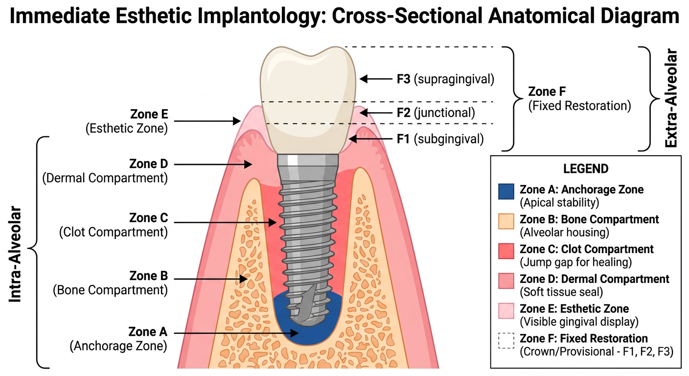
- Zone A - Anchorage Zone (apical implant engagement in bone)
- Zone B - Bone Compartment (surrounding alveolar bone walls)
- Zone C - Clot Compartment (jump gap between implant and socket wall)
- Zone D - Dermal Compartment (gingival soft tissue envelope)
- Zone E - Esthetic Zone (visible gingival display)
- Zone F - Fixed Restoration (crown, subdivided into F1/F2/F3)