
What / Where is Keratin in skin
Reading File
Finding Sources
Finding Sources
Reading File
Reading File
Reading File
Loading Image
Finding Sources
Reading File
Reading File
Loading Image
Now I have everything needed for a thorough, well-illustrated answer.
Keratin in Skin: What It Is and Where It Is
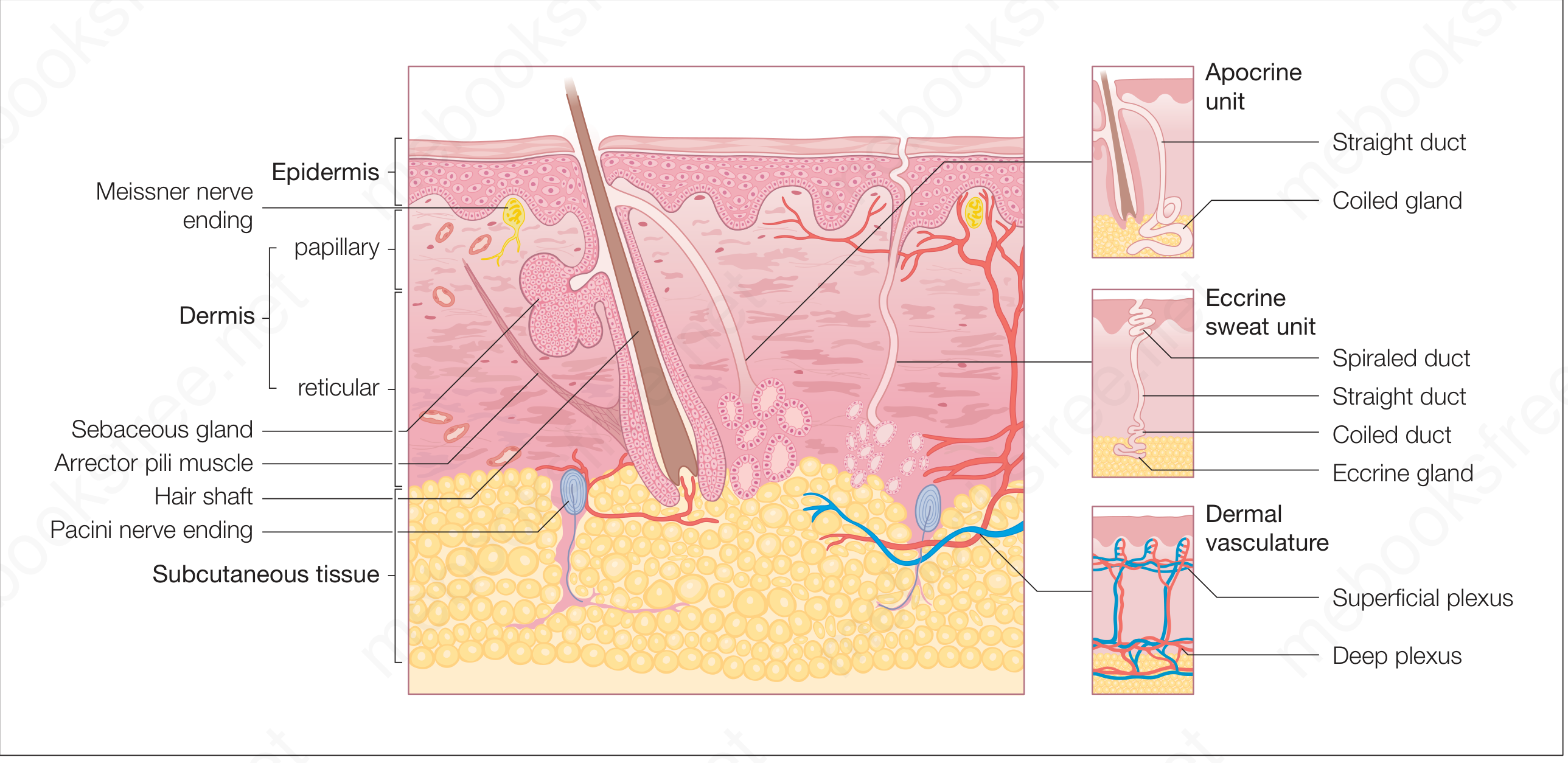
What Is Keratin?
Keratin is a fibrous structural protein that forms intermediate filaments (7-9 nm diameter) inside keratinocytes - the predominant cell type of the epidermis (85% of all epidermal cells). It exists in two subfamilies:
- Acidic keratins (Type I)
- Basic keratins (Type II)
One acidic and one basic keratin gene product always pair together to form a heteropolymer. There are multiple distinct keratin types, each expressed at different layers or structures. Keratins constitute nearly 85% of the total protein in a fully differentiated keratinocyte.
There are two broad categories:
- Soft keratin - found in the epidermis; cells eventually desquamate
- Hard keratin - found in hair cortex and nails; densely packed, does not desquamate
Where Exactly Is Keratin in the Skin?
Keratin is found throughout the epidermis, but its amount and form change progressively as cells move from the deepest to the most superficial layer. This process is called keratinization (cornification).
The Epidermal Layers (from deep to surface):
| Layer | Location | Keratin Status |
|---|---|---|
| Stratum basale (germinativum) | Deepest single-cell layer on basement membrane | Scattered keratin (cytokeratin) filaments begin; mainly K5 and K14 |
| Stratum spinosum (prickle layer) | 2-5 layers above basal | Keratin filaments (tonofilaments) radiate outward and anchor into desmosomes; K1 and K10 begin to appear |
| Stratum granulosum | 1-3 flat layers | Keratohyalin granules appear - contain filaggrin precursors that will aggregate keratin filaments; lamellar bodies release lipids to form the water barrier |
| Stratum lucidum | Only in thick skin (palms, soles) | Cells filling with keratin; nuclei disappear; contains eosinophilic eleidin |
| Stratum corneum | Outermost layer | Cells (corneocytes) are entirely filled with keratin filaments - no nucleus, no organelles; 15-20 layers of densely packed keratinized squames |
The Process (Keratinization)
The keratinocyte undergoes two phases on its way from basal cell to dead "horn cell":
- Synthetic phase - the cell accumulates keratin intermediate filaments arranged in an alpha-helical coiled pattern. These tonofilaments bundle up and terminate at desmosomes (cell-cell junctions).
- Degradative phase - organelles and nucleus disappear; all cytoplasmic content consolidates into a mixture of filaments and amorphous protein envelopes. This programmed cell death is called terminal differentiation.
Total epidermal turnover from basal cell to shed corneocyte takes approximately 47 days.
Keratin Beyond the Epidermis
Keratin is not only in the skin's outer surface. It is the structural protein of:
- Hair - hard keratin forms the hair cortex
- Nails - hard keratin, densely packed filaments, does not desquamate
- Merkel cells - contain a paranuclear whorl of keratin filaments (specifically K20), identifiable by immunostaining
Clinical Significance of Specific Keratins
| Keratin | Location | Disease when mutated |
|---|---|---|
| K5 + K14 | Basal layer | Epidermolysis bullosa simplex (fragile basal cells) |
| K1 + K10 | Suprabasal layers | Epidermolytic hyperkeratosis |
| K2 | Granular layer | Superficial epidermolytic ichthyosis |
| K20 | Merkel cells | Marker for Merkel cell carcinoma |
Deficiency of vitamin A leads to excessive keratinization (hyperkeratosis), as vitamin A normally regulates the keratinization process.
Sources: Andrews' Diseases of the Skin, p. 12-13; Histology: A Text and Atlas (Ross & Pawlina), pp. 1288-1290, 1379; Color Atlas of Human Anatomy Vol. 2, pp. 678-679; Junqueira's Basic Histology, 17e
This is a shared conversation. Sign in to Orris to start your own chat.