
Physiological changes in pregnancy and anesthesia
Reading File
This is a broad, complex medical topic. I'll run parallel searches across anesthesia and obstetric textbooks.
Searching sources...
Finding Sources
Finding Sources
Finding Sources
Finding Sources
Good - I can see anesthesiology and obstetrics books. Let me search those specifically.
Finding Sources
Finding Sources
Excellent! Found exactly what I need. Let me read the key sections in parallel - "Physiologic Changes During Pregnancy and Delivery" from Miller's, and "Anesthetic Implications of Pregnancy Physiology" from Barash.
Reading File
Reading File
Excellent content. Now let me read more of the Miller's block for respiratory, CNS, GI, and pharmacology sections.
Reading File
Reading File
Now let me get the respiratory and CNS/pharmacology sections:
Reading File
I now have very rich content from Miller's Anesthesia covering cardiovascular, respiratory (upper airway, lung volumes, O2 consumption), uterine blood flow, aortocaval compression, and anesthetic implications. I also have Barash's summary of anesthetic implications. Let me now compile the comprehensive answer.
Searching Images
physiological changes pregnancy cardiovascular respiratory diagram
Let me fetch the aortocaval compression figure from the textbook:
Loading Image
I now have comprehensive content from both Miller's Anesthesia and Barash's Clinical Anesthesia, plus a textbook figure. Here is the full, detailed answer:
Physiological Changes in Pregnancy and Their Anesthetic Implications
Sources: Miller's Anesthesia 10e; Barash, Cullen & Stoelting's Clinical Anesthesia 9e
1. Cardiovascular System
Anatomic Changes
The gravid uterus elevates the diaphragm, shifting the heart anteriorly and to the left. This produces left-axis deviation and apparent left ventricular hypertrophy on ECG - both normal findings. Auscultation reveals an accentuated S1, increased splitting, a benign systolic ejection murmur at the left sternal border (tricuspid annular dilation), and frequently an S3 in the third trimester.
Intravascular Volume
- Plasma volume increases 50-55% above baseline by term
- Driven by the renin-angiotensin-aldosterone axis (sodium retention + water retention) - likely triggered by rising progesterone
- Albumin falls by 25%, total protein by 10% → colloid osmotic pressure drops from 27 to 22 mm Hg
- This "physiologic dilutional anemia" is normal
- Volume returns to prepregnancy baseline at 6-9 weeks postpartum
Cardiac Output
| Stage | CO Change |
|---|---|
| End of 1st trimester | +35-40% |
| End of 2nd trimester | +40-50% |
| During 1st stage labor | +10-25% above prelabor |
| During 2nd stage labor | +40% above prelabor |
| Immediately postdelivery | +80-100% (autotransfusion) |
The postpartum surge is secondary to autotransfusion of uteroplacental blood as the uterus contracts, reduced vascular capacitance, and release of IVC compression. This is the highest cardiac load - critical for patients with fixed valvular stenosis or pulmonary hypertension.
Heart Rate and Stroke Volume
- HR increases 15-25% above baseline
- Stroke volume increases 25-30%
Systemic Vascular Resistance
- SVR decreases due to vasodilatory effects of progesterone, prostaglandins, and the low-resistance uteroplacental bed
- BP falls 5-20% by 20 weeks, then gradually recovers toward term
- Diastolic falls more than systolic → pulse pressure widens
- CVP and PCWP do not change (venous capacitance accommodates the volume increase)
Aortocaval Compression (Supine Hypotensive Syndrome)
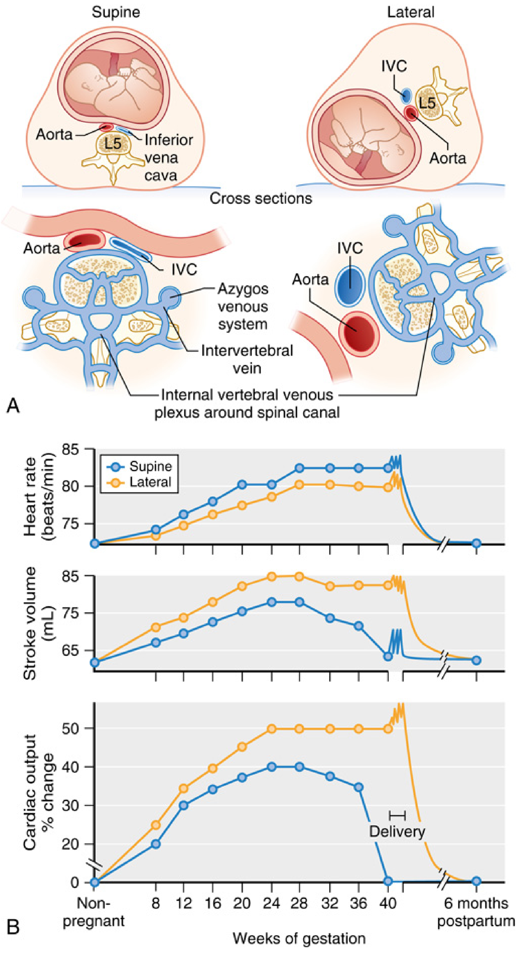
- IVC is compressed in nearly ALL term parturients in the supine position
- Symptomatic hypotension (>15 mmHg MAP drop + HR >20 bpm rise) occurs in only 8-10%
- IVC compression → blood returns via azygos/epidural/vertebral veins (these become engorged)
- Aortoiliac compression occurs in 15-20%
- Supine position reduces CO by 10-20% vs upright
- Anesthetic relevance: neuraxial/general anesthesia abolishes the compensatory sympathetic vasoconstriction, making hypotension far more severe
- Management: left uterine displacement (15-30° left tilt or lateral position); right hip elevation by 10-15 cm
2. Respiratory System
Upper Airway
- Capillary engorgement, mucosal edema, and friability begin in the first trimester throughout the oropharynx, larynx, and trachea
- Nasal congestion and epistaxis are common
- Critical anesthetic implication: airway edema worsens with Valsalva, fluid overload, and preeclampsia; a smaller endotracheal tube (6.0-6.5 mm) is recommended; awake fiberoptic intubation threshold should be low
- Mallampati score worsens progressively through labor
Lung Volumes
| Parameter | Change | Magnitude |
|---|---|---|
| Tidal volume (TV) | Increases | +45% |
| Respiratory rate | Slight increase | ~+15% |
| Minute ventilation (MV) | Increases | +45-50% |
| Functional residual capacity (FRC) | Decreases | -20-25% |
| Expiratory reserve volume (ERV) | Decreases | -25% |
| Residual volume (RV) | Decreases | -15% |
| Total lung capacity (TLC) | Slightly decreased | -5% |
| Inspiratory capacity | Increases | +5% |
FRC decrease is driven by diaphragm elevation. Closing capacity is near or below FRC in the supine or Trendelenburg position, causing airway closure during tidal breathing.
O2 Consumption and Gas Exchange
- O2 consumption increases 20-35% at term (higher in labor)
- PaO2 increases (respiratory alkalosis shifts oxyhemoglobin curve)
- PaCO2 decreases to ~30-32 mmHg (progesterone-driven hyperventilation)
- pH is slightly alkalotic (~7.44), compensated by renal bicarbonate excretion (HCO3 falls to ~20 mEq/L)
Key implication: The combination of increased O2 demand + reduced FRC means pregnant patients desaturate extremely rapidly during apnea. Pre-oxygenation with 100% O2 (3-5 minutes or 4 vital capacity breaths) is mandatory before induction. SpO2 can fall to <90% within 90 seconds of apnea at term.
3. Gastrointestinal System
- Lower esophageal sphincter (LES) tone decreases after 20 weeks of gestation
- The uterus displaces the stomach cephalad and rotates it, increasing intragastric pressure
- Gastric acid secretion increases (placental gastrin)
- Gastric emptying is essentially normal in uncomplicated pregnancy but can be markedly slowed in labor (pain, opioids, anxiety) and emergency situations
- All pregnant patients >20 weeks are considered at full-stomach risk
- Management: antacid prophylaxis (sodium citrate 30 mL PO), H2 antagonist or PPI, metoclopramide; rapid sequence induction (RSI) with cricoid pressure for general anesthesia
4. Central Nervous System and Pharmacology
MAC Reduction
- Minimum alveolar concentration (MAC) of volatile anesthetic agents decreases by approximately 25-40% during pregnancy
- Begins as early as 8-10 weeks
- Mechanism: progesterone has direct anesthetic and sedative properties; also endorphin elevation
- Implication: lower doses of volatile agents are needed to prevent awareness
Local Anesthetic Sensitivity
- Sensitivity to neuraxial and peripheral local anesthetics increases significantly
- Dose of local anesthetic should be reduced by 25-30% during any trimester
- Mechanisms: engorgement of epidural venous plexus (reduces epidural space volume), increased sensitivity of nerve membranes (progesterone effect), decreased protein binding
- Higher spread of spinal anesthesia is expected with the same intrathecal dose
Pharmacokinetics
- Increased volume of distribution (both hydrophilic and lipophilic drugs)
- Decreased protein binding (albumin and alpha-1-acid glycoprotein both fall) → increased free drug fraction
- Hepatic enzyme activity is variably affected
- Renal clearance increases (GFR up 50%)
5. Renal System
- GFR increases ~50% above baseline
- Renal plasma flow increases ~70%
- Serum creatinine and BUN fall (normal pregnancy creatinine <0.8 mg/dL; normal urea is lower than nonpregnant range)
- Glycosuria is common without hyperglycemia (reduced tubular reabsorption threshold)
6. Hematology and Coagulation
- RBC mass increases ~30%, but plasma volume increases ~50% → dilutional anemia (hemoglobin ~11-12 g/dL is normal at term)
- Leukocytosis up to 12,000-15,000/mm³ is normal
- Hypercoagulable state: factors I, VII, VIII, X, XII, and vWF all increase; fibrinogen increases dramatically (from ~300 to ~450-600 mg/dL)
- Protein S decreases; APC resistance increases
- This is physiologically appropriate for delivery but creates DVT risk
- Platelet count may slightly decrease (gestational thrombocytopenia)
7. Uterine Blood Flow
- Increases from ~100 mL/min (nonpregnant) to 700-900 mL/min at term (~10% of cardiac output)
- 80% goes to the intervillous space (placenta), 20% to myometrium
- Minimal autoregulation - vasculature is essentially maximally dilated
- Uterine blood flow = (Uterine arterial pressure - Uterine venous pressure) / Uterine vascular resistance
Factors that reduce uterine blood flow:
- Maternal hypotension (hypovolemia, spinal anesthesia, aortocaval compression)
- Vasopressors that increase uterine vascular resistance (historically alpha-agonists)
- Extreme hypocapnia (PaCO2 <20 mmHg) - direct uterine vasoconstriction + oxyhemoglobin curve shift
- Uterine hypertonus (oxytocin excess, abruptio)
- Valsalva/bearing down (increases uterine venous pressure)
Vasopressor choice:
Historically, ephedrine was preferred because pure alpha-agonists (methoxamine, metaraminol) reduced uterine blood flow in animal models. However, recent human trials show phenylephrine for spinal-induced hypotension is associated with less fetal acidosis and base deficit than ephedrine. Phenylephrine is now the first-line vasopressor for spinal hypotension in obstetric anesthesia.
Summary: Anesthetic Implications at a Glance
| System | Change | Anesthetic Implication |
|---|---|---|
| Airway | Edema, friability | Smaller ETT (6.0-6.5), RSI, lower threshold for awake FOB |
| Respiratory | ↓FRC, ↑O2 demand | Rapid desaturation; mandatory pre-oxygenation |
| Respiratory | ↑MV, ↓PaCO2 | Faster inhalation induction/emergence; avoid hyperventilation |
| CVS | ↑CO, ↓SVR | Careful hemodynamic monitoring |
| CVS - Supine | Aortocaval compression | Left uterine displacement always |
| CNS | ↓MAC (~25-40%) | Reduce volatile agent doses |
| Neuraxial | Engorged epidural veins, ↑nerve sensitivity | Reduce LA dose 25-30%; expect higher spread |
| GI | ↓LES tone, ↑gastric acid | Full-stomach precautions from 20 weeks; RSI mandatory |
| Pharmacology | ↓protein binding, ↑Vd | Altered drug kinetics; titrate carefully |
| Uterine flow | Low autoregulation | Avoid hypotension; phenylephrine preferred over ephedrine |
Miller's Anesthesia 10e, Chapter 58 (Physiologic Changes During Pregnancy and Delivery); Barash, Cullen & Stoelting's Clinical Anesthesia 9e, Chapter 41 (Anesthetic Implications of Pregnancy Physiology)
This is a shared conversation. Sign in to Orris to start your own chat.