
Epstein barr virus in gastric carcinoma risk factor
Epstein-Barr virus gastric carcinoma risk factors
PMID: 40234237
Epstein-Barr virus gastric carcinoma in situ hybridization EBV positive
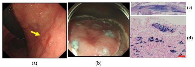
This composite image illustrates the clinical and histopathological findings of Epstein-Barr virus-associated gastric cancer (EBVaGC). Panel (a) shows an ordinary endoscopic view of the middle gastric body, identifying a superficial depressed-type early gastric cancer lesion (indicated by a yellow arrow). Panel (b) depicts the lesion during endoscopic submucosal dissection (ESD) using en bloc resection. Panel (c) is a low-power microscopic view of the H&E-stained ESD specimen, demonstrating submucosal invasion (0.8 mm) and a carcinoma with lymphoid stroma (CLS) pattern without lymphovascular involvement. Panel (d) shows a high-power microscopic view using EBER-1 in situ hybridization (ISH); the tumor cell nuclei exhibit strong positive (dark) staining for EBV-encoded RNA, while surrounding tumor-infiltrating lymphocytes (TILs) and non-cancerous gastric epithelium remain negative. This case demonstrates the diagnosis and therapeutic evaluation of EBVaGC, emphasizing the use of ISH for definitive classification in early-stage mucosal lesions.
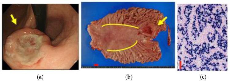
A multi-panel clinical and diagnostic figure demonstrating Epstein-Barr virus-associated gastric cancer (EBVaGC). Panel (a) is an endoscopic clinical photograph showing a gastric lesion near the cardia, characterized by a depressed morphology and a submucosal tumor (SMT)-like protrusion (marked by a yellow arrow). Panel (b) shows the macroscopic specimen from a total gastrectomy, displayed on a blue background with a 10 mm scale bar. The lesion (yellow arrow) is situated in the upper stomach near the gastric mucosal atrophic border, which is demarcated by yellow lines. Panel (c) is a histopathology image showing EBER-1 in situ hybridization (ISH). The image displays numerous tumor cell nuclei with positive dark purple staining, characteristic of carcinoma with lymphoid stroma (CLS) associated with EBV. The educational focus is on the endoscopic and macroscopic identification of EBVaGC, highlighting its typical location in the upper stomach and its distinctive histological signature.
Epstein-Barr Virus (EBV) in Gastric Carcinoma - Risk Factors & Pathogenesis
Epidemiology
Established Risk Factors for EBVaGC
1. EBV Infection Itself
- EBV infection alone: OR 4.98 (95% CI 3.17-7.85) for developing gastric cancer vs. non-infected controls
- H. pylori + EBV co-infection: OR 2.57 (95% CI 1.65-4.01) - a 2.57-fold elevated risk, suggesting synergistic interaction
2. H. pylori Co-infection
3. Male Sex
4. Prior Gastric Surgery (Remnant Stomach)
5. Proximal Stomach Location
Molecular Mechanisms of EBV-Driven Carcinogenesis
| Mechanism | Effect |
|---|---|
| EBV microRNAs | Suppress host tumor suppressor gene expression |
| CpG island hypermethylation | Epigenetic silencing of tumor suppressor genes (extreme DNA hypermethylation - the most methylated of all GC subtypes) |
| PIK3CA mutations | Recurrent activating mutations driving PI3K/AKT/mTOR pathway |
| JAK2 / CD274 / PDCD1LG2 amplification | Overexpression of PD-L1/PD-L2 - immune checkpoint upregulation enabling immune evasion |
| EBER (EBV-encoded small RNA) | Detected by in situ hybridization (ISH) - the diagnostic hallmark |
| LMP2A (Latent Membrane Protein 2A) | Mimics BCR signaling, promotes cell survival |
| TP53 mutations rare | Unlike other GC subtypes, suggesting a distinct pathogenic route |
- TCGA molecular classification cited in Yamada's Textbook of Gastroenterology; Sabiston Textbook of Surgery
TCGA Molecular Classification (Relevance to EBVaGC)
- EBV-positive - recurrent PIK3CA mutations, extreme hypermethylation, JAK2/PD-L1/PD-L2 amplification
- Microsatellite unstable (MSI)
- Genomically stable (enriched for diffuse type, RHOA mutations)
- Chromosomally unstable (CIN)
Morphological Features (Diagnostic Clues)
- Predilection: proximal stomach (cardia, fundus, body)
- Growth pattern: typically diffuse
- Histology: often carcinoma with lymphoid stroma (lymphoepithelioma-like) - dense TIL infiltrate
- Diagnosis confirmed by EBER-1 in situ hybridization (ISH) - strong nuclear positivity in tumor cells
Complete Risk Factor Summary (Box)
- EBV latent infection (OR ~5.0 for GC)
- H. pylori infection (OR ~1.4-6x for GC); co-infection synergistic (OR 2.57)
- Atrophic gastritis / intestinal metaplasia
- Prior gastric surgery
- Male sex (male predominance in EBVaGC)
- Younger age at diagnosis compared to other GC subtypes
- Epigenetic susceptibility (CpG hypermethylation background)
- PIK3CA pathway alterations
-
Smoked/salted/pickled foods, high nitrate intake
-
Smoking and alcohol
-
Low fruit/vegetable intake
-
Low socioeconomic status, lack of refrigeration
-
Sabiston Textbook of Surgery, Risk Factors Box 87.1