I now have all the information needed. Here is the complete IAP-format Pediatric Case Presentation for VSD:
Case History and Examination: Ventricular Septal Defect (VSD)
As per IAP Pediatric Case Presentation Format
CASE VIGNETTE
Patient: Master Arjun K., 4-month-old male infant
Informant: Mother (reliable historian)
Date of admission: [Date]
Registering Unit: Pediatric Cardiology OPD
PART I - HISTORY
1. CHIEF COMPLAINTS
(In chronological order of appearance)
- Difficulty in feeding since 6 weeks of age
- Excessive sweating during feeds - 6 weeks
- Fast breathing (tachypnea) at rest and with feeds - 6 weeks
- Poor weight gain since 6 weeks of age
- Recurrent respiratory tract infections - 2 episodes in past 2 months
- Murmur detected by local doctor - 3 months ago
2. HISTORY OF PRESENT ILLNESS
Master Arjun K., a 4-month-old male infant, born at term by normal vaginal delivery, presented with the above complaints. The infant was apparently well at birth. At around 6 weeks of age, the mother noticed that the baby was taking longer than usual to complete a feed (>30 minutes per feed), often becoming breathless and sweaty during feeding. The baby would tire easily and stop feeding before adequate intake, necessitating frequent, small-volume feeds. The mother noticed the baby breathing faster than normal, both at rest and especially during feeds. The infant has had two episodes of respiratory tract infection (cough, cold, and one episode requiring hospital admission for respiratory distress) in the past 2 months. A heart murmur was detected by the local doctor at 1 month of age, prompting referral to this center.
Symptoms elaborated:
| Symptom | Description |
|---|
| Difficulty feeding | Takes >30 min/feed, tires easily, stops before satiated |
| Sweating | Drenching sweats over forehead during feeds (increased sympathetic tone) |
| Tachypnea | Respiratory rate >60/min at rest; worse with feeds |
| Poor weight gain | Birth weight 3.2 kg; current weight 4.1 kg (expected ~5.5 kg at 4 months) |
| Recurrent RTIs | 2 episodes - one requiring hospitalization |
| Murmur | Detected at 1 month; referred for evaluation |
No history of: cyanosis at rest or cry, squatting spells, syncope, frank cardiac failure, anasarca, oliguria.
3. BIRTH HISTORY
- Antenatal: Regular antenatal checkups; no maternal diabetes, rubella, or teratogen exposure; no consanguinity; no family history of congenital heart disease; no fetal echocardiography done
- Natal: Born at 38 weeks gestation, NVD at district hospital; birth weight 3.2 kg; no birth asphyxia; APGAR score 8/9 (reported)
- Postnatal: Discharged on Day 2; no NICU admission; neonatal period uneventful apart from murmur noted at 1-month check
4. FEEDING HISTORY
- Exclusively breastfed since birth; struggling with feeds since 6 weeks
- No complementary feeds introduced (appropriate for age)
- Feeds of 15-20 mL taken at a time with diaphoresis and tachypnea
5. DEVELOPMENTAL HISTORY
- Social smile: Present (at 6 weeks - normal)
- Neck holding: Partial (in progress - consistent with 4 months)
- Gross motor: Appropriate for age
- No developmental delay (developmental milestones within normal limits for age)
6. IMMUNIZATION HISTORY
- BCG, OPV-0 at birth: Given
- DPT/OPV/IPV/HepB/Hib 6-week doses: Given
- 10-week doses: Pending (due this month)
- Immunization as per IAP schedule (up to date for age)
7. PAST MEDICAL HISTORY
- 2 episodes of URTI/LRTI - 1 requiring inpatient admission at 3 months for bronchiolitis-like presentation
- No surgical history
- No history of infective endocarditis
- No previous cardiac investigations
8. FAMILY HISTORY
- No consanguinity between parents
- No family history of congenital heart disease or sudden cardiac death
- No siblings with congenital anomalies
- Parents healthy
9. SOCIOECONOMIC HISTORY
- Parents: Father - daily wage laborer; Mother - homemaker
- Socioeconomic status: Lower middle class (Modified Kuppuswamy Scale)
- Housing: Semi-pucca house, adequate ventilation
- Diet: Breastfeeding only (appropriate)
PART II - PHYSICAL EXAMINATION
1. GENERAL EXAMINATION
| Parameter | Findings |
|---|
| Conscious | Yes, alert, irritable when disturbed |
| Activity | Mildly lethargic, consolable |
| Posture | Supine, no preferential positioning |
| Nutritional status | Malnourished - weight-for-age < -2 SD (underweight) |
| Pallor | Mild (secondary to cardiac failure/poor feeding) |
| Icterus | Absent |
| Cyanosis | Absent (central and peripheral) - important - acyanotic CHD |
| Clubbing | Absent (would develop in Eisenmenger syndrome) |
| Edema | Absent (pedal/facial) |
| Lymphadenopathy | Absent |
Vital Signs:
| Parameter | Finding | Normal for Age |
|---|
| Heart Rate | 148/min, regular | 100-160/min |
| Respiratory Rate | 62/min | 30-60/min - tachypneic |
| Temperature | 37.2°C (afebrile) | Normal |
| SpO2 | 97% on room air | >95% |
| Blood Pressure | Right arm: 82/60 mmHg | Normal |
| Weight | 4.1 kg | Expected ~5.5 kg (below -2SD) |
| Length | 58 cm | Expected ~62 cm (below -1SD) |
| Head Circumference | 40 cm | Normal |
Anthropometry:
- Weight-for-age: < -2 SD (underweight)
- Length-for-age: < -1 SD
- Weight-for-length: < -2 SD (wasted - suggests acute malnutrition secondary to CHF)
2. SYSTEMIC EXAMINATION
A. CARDIOVASCULAR SYSTEM
Inspection:
- Precordial bulge present bilaterally (cardiomegaly causing chest wall deformity in infancy)
- Increased precordial activity visible on inspection
- No visible pulsations in neck (no elevated JVP)
- No visible dilated veins on chest wall
Palpation:
| Finding | Description |
|---|
| Apex beat | Shifted laterally and inferiorly - located at 5th ICS, anterior axillary line (cardiomegaly) |
| Character of apex | Hyperdynamic, forceful (volume-overloaded LV) |
| Parasternal heave | Present - left parasternal heave indicating RV enlargement / pulmonary hypertension |
| Thrills | Systolic thrill palpable at LLSB (3rd-4th ICS, left sternal border) (indicates moderate-large VSD with significant pressure gradient) |
| Liver | Palpable 3 cm below right costal margin, soft, tender - hepatomegaly (congestive cardiac failure) |
Percussion:
- Cardiac dullness increased (suggests cardiomegaly)
- Liver dullness: 3 cm below RCM
Auscultation:
| Finding | Description |
|---|
| S1 | Normal intensity |
| S2 | Narrowly split or single (pulmonary component P2 may be loud - indicates pulmonary hypertension in large VSD) |
| Additional sounds | Mid-diastolic rumble at apex - indicates functional mitral stenosis from increased pulmonary venous return (indicates Qp:Qs > 2:1 - surgical shunt) |
| Murmur | Grade 3-4/6 harsh holosystolic (pansystolic) murmur - heard loudest at lower left sternal border (LLSB), 3rd-4th ICS; occupies entire systole; thrill palpable at same site |
| Radiation | No significant radiation (differentiates from MR which radiates to axilla) |
Axiom: "The louder the murmur, the smaller the VSD." In large VSDs with equalized pressures (Eisenmenger), the murmur may actually become soft or disappear. (MSD Manual)
Summary of murmur characteristics:
| Feature | VSD Murmur |
|---|
| Timing | Holosystolic / pansystolic |
| Quality | Harsh, blowing |
| Grade | 3-5/6 (moderate-large); may be 2/6 (small) |
| Location | LLSB (3rd-4th ICS) |
| Thrill | Present with moderate-large VSD |
| Radiation | Limited; may go to entire precordium |
| Associated | Apical mid-diastolic rumble with large shunt |
B. RESPIRATORY SYSTEM
| Finding | Description |
|---|
| Inspection | Tachypnea (RR 62/min); mild subcostal and intercostal retractions; no nasal flaring |
| Palpation | Increased tactile vocal fremitus at bases bilaterally |
| Percussion | Dull at both lung bases (bilateral pleural effusion / basal consolidation from pulmonary congestion) |
| Auscultation | Coarse crepitations at bilateral bases; mild wheeze; no bronchial breathing |
C. ABDOMEN
- Liver: 3 cm palpable below RCM, soft, slightly tender - hepatomegaly (right-sided heart failure/venous congestion)
- Spleen: Not palpable
- Kidneys: Not palpable
- No ascites
D. CENTRAL NERVOUS SYSTEM
- Conscious, alert
- Fontanelle: Anterior fontanelle open, flat, normotensive
- Tone, reflexes: Normal
- No focal neurological deficit
E. MUSCULOSKELETAL
- No dysmorphic features (check for Down syndrome - trisomy 21 associated with AVSD/VSD), Turner syndrome, DiGeorge (22q11 deletion)
- No skeletal anomalies
PART III - DIAGNOSIS (PROVISIONAL)
Provisional Diagnosis:
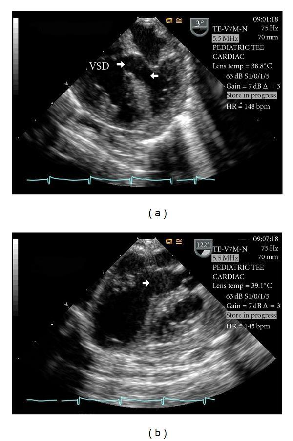
Moderate-to-large Perimembranous Ventricular Septal Defect (VSD) with Congestive Cardiac Failure and Failure to Thrive
Basis:
- 4-month-old male with feeding difficulty, diaphoresis, tachypnea, poor weight gain, recurrent RTIs
- Acyanotic; cardiomegaly on exam
- Harsh holosystolic murmur at LLSB + systolic thrill
- Mid-diastolic apical rumble (Qp:Qs likely >2:1)
- Hepatomegaly (CCF)
- Parasternal heave (RV/pulmonary pressure elevation)
- Features consistent with moderate-large left-to-right shunt
PART IV - INVESTIGATIONS PLANNED
| Investigation | Expected Finding in Large VSD |
|---|
| Chest X-ray (PA view) | Cardiomegaly (increased CTR); increased pulmonary vascular markings (plethora); prominent pulmonary artery segment |
| ECG (12-lead) | Small VSD: Normal; Medium: LVH ± LAE; Large VSD: Biventricular hypertrophy (BVH) ± LAE; pure RVH |
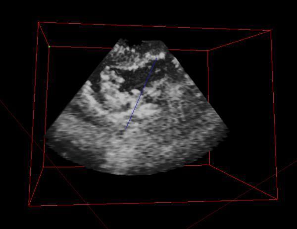
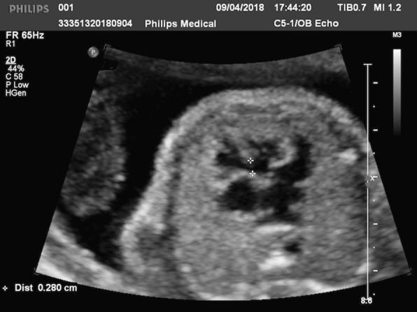
| 2D Echocardiography with Doppler | Confirmatory: Visualizes defect, measures size, location (perimembranous vs. muscular); estimates Qp:Qs; pulmonary artery pressure; LV/RV dimensions; rules out associated lesions |
| SpO2 | Normal (97-100%) in left-to-right shunt (acyanotic) |
| Hemogram | May show mild anemia (dilutional); polycythemia if Eisenmenger develops |
| Serum electrolytes, BUN, creatinine | Baseline before starting diuretics |
| Liver function tests | Assess hepatic congestion |
| Karyotype | If dysmorphic features (rule out trisomy 21) |
PART V - DIFFERENTIAL DIAGNOSIS
| Condition | Differentiating Feature |
|---|
| ASD (Atrial Septal Defect) | Wide fixed split S2; systolic ejection murmur at LUSB; no thrill usually |
| Mitral Regurgitation | Murmur radiates to axilla; apex displaced but softer S1 |
| Tricuspid Regurgitation | LLSB murmur increases on inspiration (Carvallo sign) |
| PDA (Patent Ductus Arteriosus) | Continuous "machinery" murmur at LUSB; wide pulse pressure |
| Innocent murmur (Still's) | Vibratory, musical; no thrill; no cardiomegaly; normal ECG |
| AVSD | Associated with Down syndrome; superior QRS axis on ECG |
PART VI - MANAGEMENT OUTLINE (BRIEF)
Medical (for CCF):
- Furosemide (diuretic) - reduces pulmonary congestion
- Spironolactone - potassium sparing
- Digoxin - improves myocardial contractility
- Captopril (ACE inhibitor) - reduces afterload, decreases L-to-R shunt
- High-calorie feeds / nasogastric tube feeding if inadequate oral intake
Surgical / Interventional:
- Indications for closure: Large VSD with CCF not responding to medical therapy; Qp:Qs > 2:1; failure to thrive; pulmonary artery pressure elevation
- Methods: Surgical patch closure (standard); catheter-based device closure (perimembranous/muscular VSDs)
- Timing: Within first year of life to prevent irreversible pulmonary vascular disease (Eisenmenger)
- Small VSDs with Qp:Qs < 1.5 and normal pulmonary pressure: observe for spontaneous closure (up to 80% close by 1 month; ~25% by 12 months)
APPENDIX: VSD Types (Anatomical Classification)
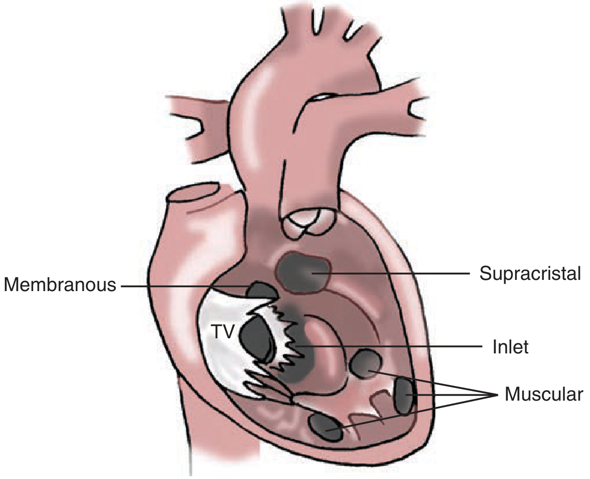
Figure: Types of VSD - Membranous (most common, ~80%), Supracristal (outlet), Inlet, and Muscular. (Schwartz's Principles of Surgery, 11th Ed.)
| Type | Location | Frequency | Notes |
|---|
| Perimembranous | Membranous septum, below aortic valve | ~80% | Most common requiring surgery |
| Muscular | Trabecular septum (anterior, mid, posterior, apical) | Common | May be multiple ("Swiss cheese") |
| Outlet/Supracristal | Conal septum, below pulmonary valve | Less common | Associated with aortic regurgitation |
| Inlet (AV Canal type) | Beneath tricuspid valve | Less common | Associated with Down syndrome |
Sources: Harriet Lane Handbook 23rd Ed. (Pediatric Cardiology section), Schwartz's Principles of Surgery 11th Ed., Goldman-Cecil Medicine, Tintinalli's Emergency Medicine, MSD Manual Pediatrics,
IAP Indian Guidelines for CHD Management,
Medscape VSD Clinical Presentation