
Give me all infromation for intrnal bledding plus patient in shock in short
Internal Bleeding + Shock (Hemorrhagic/Hypovolemic Shock) - Quick Summary
Key point on volume loss vs. signs
- ~10% blood loss: almost no change in BP/CO (compensated)
- ~15-20% loss without intact reflexes can be fatal; with intact sympathetic reflexes the body tolerates 30-40% loss before collapse
- 40-45% loss: CO and BP fall toward zero (Guyton, p. 2152-2159)
Classic clinical signs (internal bleeding + shock)
- Tachycardia, weak/rapid pulse
- Hypotension (late sign - appears only after significant loss because sympathetic vasoconstriction compensates first)
- Cold, clammy, pale skin (peripheral shutdown - blood shunted to brain/heart)
- Anxiety/confusion → decreasing consciousness as perfusion worsens
- Tachypnea
- Falling urine output
- Distension/pain/rigidity or a palpable mass if bleeding is intra-abdominal or pelvic; persistent shock despite fluid resuscitation with a falling hematocrit strongly suggests ongoing internal bleeding even without visible blood loss (Park's Preventive & Social Medicine; Pye's Surgical Handicraft, p. 1186).
Mechanism (compensation)
Management priorities
- ABC - airway, oxygen, two large-bore IVs
- Identify and stop the source of bleeding - this is the definitive treatment; fluids alone do not fix ongoing hemorrhage (surgery, endoscopy, embolization as indicated)
- Volume resuscitation - crystalloids initially, early blood products (packed RBCs, plasma) for significant hemorrhage; avoid over-resuscitation with crystalloid alone in trauma
- Identify occult sources - suspect internal bleeding when shock persists/hematocrit keeps falling despite resuscitation; use FAST/CT/serial exams to localize (pelvis, abdomen, chest, retroperitoneum, GI tract)
- Monitor - vitals, urine output, mental status, lactate/hematocrit trend
- Treat the underlying injury/disease definitively once identified
How to diagnose if its gi problem
Diagnosing GI Bleeding as the Source of Internal Bleeding + Shock
1. History & clinical clues
- Hematemesis (vomiting fresh blood) or coffee-ground emesis → active/recent upper GI bleed (proximal to ligament of Treitz)
- Melena (black, tarry, foul-smelling stool) → usually upper GI bleed, but can be from small bowel/right colon
- Hematochezia (bright red/maroon blood per rectum) → usually lower GI bleed, but ~10% of hematochezia is actually a brisk upper GI bleed (blood transits too fast to become melena) - Tintinalli's Emergency Medicine, p. 3115
- Ask about NSAID/anticoagulant use, alcohol/liver disease, known varices/ulcers, prior GI bleeds, weight loss, abdominal pain
2. Exam
- Vitals for shock (tachycardia, hypotension, orthostatic drop)
- Abdominal exam - tenderness, mass, ascites, stigmata of liver disease (spider angiomata, jaundice → varices)
- Digital rectal exam - confirms melena/hematochezia, checks for mass
3. Labs
- CBC (Hb/Hct - note initial Hct may look normal before hemodilution catches up), coagulation panel/INR (INR ≥1.5 predicts higher mortality in upper GI bleed on anticoagulants), BUN/creatinine (disproportionately elevated BUN:Cr ratio suggests upper GI source from blood digestion), liver function tests, type & crossmatch, lactate
4. Bedside/early diagnostic steps
- NG tube lavage/aspirate: bloody or coffee-ground aspirate confirms upper GI source; a clear bilious aspirate makes upper GI bleed less likely but doesn't fully exclude it (e.g., duodenal bleed distal to pylorus)
- Rule out other internal bleeding sources first (abdominal exam, FAST/CT for trauma, pelvic/retroperitoneal causes) if the picture isn't classic for GI
5. Definitive diagnosis
- Upper endoscopy (EGD) - gold standard for upper GI bleed; done within 24 hours once hemodynamically stabilized; identifies and can treat ulcers, varices, Mallory-Weiss tears, Dieulafoy lesions
- Colonoscopy - for suspected lower GI bleed once bowel prepped/stable
- CT angiography - useful when bleeding is brisk/ongoing and endoscopy is not immediately available or non-diagnostic
- Tagged RBC scan or angiography - for obscure/intermittent bleeding, especially if endoscopy is negative; angiography also positive for active arterial bleeding and allows embolization
- Capsule endoscopy/enteroscopy - for suspected small bowel (obscure) bleeding when EGD and colonoscopy are negative
Key differentiator vs. non-GI internal bleeding
How to diagnose if its gi bleeding and blood vomit
Diagnosing GI Bleeding When There's Blood Vomit (Hematemesis)
Step 1 - Confirm it's really GI bleeding
- Fresh red blood vomited = active, brisk bleeding
- Coffee-ground emesis (dark, granular) = blood that has been in contact with gastric acid for a while = slower or older bleeding
- Rule out mimics: swallowed blood from a nosebleed, dental extraction, or hemoptysis coughed up and then swallowed can look like hematemesis but isn't a GI source - Tintinalli's Emergency Medicine, p. 2957.
Step 2 - Use symptom pattern to narrow the cause
| Presentation | Likely cause |
|---|---|
| Painless hematemesis | Swallowed blood (non-GI), Mallory-Weiss tear |
| Hematemesis + epigastric/chest pain | Peptic ulcer, H. pylori gastritis, esophagitis/gastritis |
| Hematemesis + known liver disease/cirrhosis | Esophageal/gastric varices (portal hypertension) |
| Hematemesis + coagulopathy history | Bleeding diathesis worsening any lesion |
| (Tintinalli's, Table 134-3) |
Step 3 - Assess severity (vitals over hemoglobin)
- Heart rate and BP are the most reliable early indicators - postural tachycardia/hypotension, then resting tachycardia, then hypotension as bleeding worsens
- Hemoglobin is misleading early on - it doesn't drop immediately because you lose whole blood (plasma and red cells proportionally); it can take up to 72 hours to fully reflect the loss. A "normal" Hb right after a big bleed does NOT rule out a serious bleed - Harrison's, p. 1538.
- Elevated BUN (disproportionate to creatinine) and hyperactive bowel sounds also support an upper GI source (digested blood protein raises BUN).
Step 4 - Risk-stratify
Step 5 - Confirm and localize with endoscopy
- Upper endoscopy (EGD) is the definitive test - done within 24 hours (sooner if unstable) - identifies ulcers, varices, tears, gastritis, and allows immediate treatment (clipping, banding, injection).
- NG tube aspirate: blood or coffee-ground return confirms an upper GI source right away at the bedside, before endoscopy.
When to worry it's NOT simple GI bleeding
Brown blood
Brown Blood - What It Means
Brown blood in vomit ("coffee-ground emesis")
- Vomit that looks dark brown/black with a grainy, coffee-ground appearance = blood that has sat in the stomach long enough for gastric acid to convert it to hematin
- Suggests bleeding that is slower or has slowed/stopped recently (as opposed to bright red hematemesis, which means active, brisk bleeding)
- Still indicates an upper GI source (stomach, duodenum, esophagus) - same workup applies (NG lavage, endoscopy within 24h) - Sleisenger and Fordtran's GI and Liver Disease, p. 1348; Frameworks for Internal Medicine, p. 335.
- Caution: iron supplements or red wine can also cause a coffee-ground-like appearance without true bleeding - S. Das Manual on Clinical Surgery, p. 608.
Dark brown, foul-smelling vomit ("faeculent vomiting")
- If the vomit is dark brown and smells like feces, this is different - it points to bowel obstruction (advanced low small bowel obstruction) or late peritonitis, where bowel contents mix with altered blood, not simple GI bleeding - S. Das Manual on Clinical Surgery, p. 586, 6381. This is a surgical emergency (obstruction/peritonitis), distinct from a bleeding ulcer or varix.
Brown blood in stool
- Normal stool is brown because of stercobilin (a bilirubin breakdown product) - this is completely normal, not bleeding - Rosen's Emergency Medicine, p. 2589.
- If you mean dark, tarry, black-brown stool with a strong odor - that's melena, which does indicate upper or right-colon GI bleeding that has been in the gut ≥14 hours.
- Bright red blood on/mixed with normal brown formed stool usually means a lower source (anal, rectal, sigmoid, e.g., hemorrhoids) - Harrison's Internal Medicine, p. 1272.
Can endoscopy done in only conscious patient?
How endoscopy is typically done
- Conscious sedation (moderate sedation) - the most common approach. The patient is drowsy/relaxed but arousable and breathing on their own. This is fine for routine, stable patients - Bailey and Love's Short Practice of Surgery, p. 1103; Maingot's Abdominal Operations, p. 1579.
- Unsedated - some upper/lower endoscopies can even be done fully awake with no sedation at all.
When the patient is NOT conscious (or shouldn't be sedated lightly)
- General anesthesia with endotracheal intubation is used instead of conscious sedation when:
- The patient is unconscious, obtunded, or has depressed mental status (common in massive GI bleeding with shock)
- There's active, massive hematemesis or high risk of aspiration
- The patient has severe comorbidities and can't safely tolerate conscious sedation
- It's an emergency variceal bleed or unstable patient
- Pediatric foreign body/impaction cases
- Fischer's Mastery of Surgery, p. 835: "Severe comorbidities and inability to tolerate conscious sedation preclude safe endoscopy [with sedation alone], although general anesthesia can be used in select cases."
- Barash, Cullen, and Stoelting's Clinical Anesthesia, p. 1870: "Under general anesthesia, tracheal intubation is the gold standard to protect the airway" during upper GI endoscopy.
- For a patient with obtunded mental status and massive hematemesis, guidelines (Washington Manual) specifically recommend intubation before endoscopy to protect the airway from aspiration.
Bottom line for your scenario (internal bleeding + shock)
Classification of shock
Classification of Shock
1. Physiologic Classification (4 major types) - Harrison's Principles of Internal Medicine, Table 314-1
| Type | Primary problem | Examples |
|---|---|---|
| Hypovolemic | Reduced circulating blood volume | Hemorrhage (trauma, GI bleeding, ruptured ectopic pregnancy), GI fluid losses, burns, polyuria (DKA, diabetes insipidus) |
| Cardiogenic | Pump failure - heart can't generate enough output | MI, myocarditis, arrhythmia, severe valve disease (aortic/mitral insufficiency) |
| Obstructive | Physical obstruction blocking forward blood flow | Tension pneumothorax, cardiac tamponade, constrictive pericarditis, pulmonary embolism, aortic dissection |
| Distributive | Loss of vascular tone (vasodilation), SVR drops | Septic shock, anaphylactic shock, neurogenic shock, pancreatitis, severe burns, adrenal crisis/endocrine shock |
2. Severity Classification for Hemorrhagic/Hypovolemic Shock (ATLS - relevant to your internal bleeding scenario)
| Class | Blood volume lost | Approx. volume | Clinical picture |
|---|---|---|---|
| Class I | <15% | <750 mL | Usually asymptomatic, maybe mild tachycardia |
| Class II | 15-30% | 750-1,500 mL | Tachycardia, tachypnea, anxiety, clammy skin - fluids usually sufficient |
| Class III | 30-40% | 1,500-2,000 mL | Hypotension, tachycardia, tachypnea, pallor, altered mental status - usually needs transfusion |
| Class IV | >40% | >2,000 mL | Severe shock, weak/absent pulse, imminent risk of death - fluids first, transfusion mandatory |
Why this matters for your case (internal bleeding + shock)
Hemorrhagic gi bleeding in detail
"gastrointestinal hemorrhage" AND management
Hemorrhagic GI Bleeding - Detailed Overview
1. Classification (Anatomic)
| Type | Location | Landmark |
|---|---|---|
| Upper GI bleed (UGIB) | Esophagus, stomach, duodenum | Proximal to ligament of Treitz |
| Lower GI bleed (LGIB) | Jejunum, ileum, colon, rectum | Distal to ligament of Treitz |
| Obscure/small bowel bleed | Small intestine, not seen on standard EGD/colonoscopy | Requires capsule endoscopy/enteroscopy |
2. Causes
- Duodenal ulcer, gastric ulcer (peptic ulcer disease - most common overall cause)
- Erosive gastritis/esophagitis
- Esophageal/gastric varices - due to portal hypertension in cirrhosis; can cause massive, life-threatening hematemesis
- Mallory-Weiss tear (mucosal tear from forceful vomiting/retching)
- Dieulafoy lesion (abnormally large submucosal artery - covered by normal mucosa, notoriously hard to find and treat)
- Angiodysplasia/AVMs, malignancy, duodenal diverticula
- Diverticulosis - single most common cause of LGIB, ~30% of cases
- Angiodysplasia/vascular ectasia - especially in older adults
- Colitis (ischemic, infectious, inflammatory bowel disease)
- Hemorrhoids, rectal varices
- Post-polypectomy bleeding
- Colorectal malignancy
- Radiation proctitis
- Meckel's diverticulum (pediatric - heterotopic gastric mucosa causes ileal ulceration)
3. Clinical Presentation
- Hematemesis / coffee-ground emesis → UGIB
- Melena (black, tarry, foul stool) → blood present ≥14 hrs in gut, usually UGIB or proximal source
- Hematochezia (bright red/maroon PR blood) → usually LGIB, but ~10% is actually a brisk UGIB
- Occult bleeding → no visible blood; presents as iron-deficiency anemia or positive fecal occult blood test
4. Pathophysiology of the Shock Component
5. Risk Stratification
6. Diagnostic Workup
- Vitals + orthostatics (best initial assessment)
- Labs: CBC, coagulation profile/INR, BUN/creatinine (elevated BUN:Cr ratio suggests UGIB), liver function tests, type & crossmatch, lactate
- NG lavage - blood/coffee-ground aspirate confirms UGIB at bedside
- Digital rectal exam - confirms melena/hematochezia
- Endoscopy is the definitive diagnostic and therapeutic tool:
- Upper endoscopy (EGD) within 24 hours for suspected UGIB
- Colonoscopy (after prep) for suspected LGIB once stable
- Capsule endoscopy/enteroscopy for obscure bleeding
- CT angiography or tagged RBC scan / catheter angiography - for brisk/ongoing bleeding not localized by endoscopy, or when endoscopy isn't feasible (unstable patient); angiography also permits embolization
7. Management
- Airway protection - intubate first if obtunded/massive hematemesis (aspiration risk) before endoscopy
- Two large-bore IVs, crystalloid then blood products for ongoing/severe bleeding
- Restrictive transfusion threshold: Hb <7 g/dL (randomized trial evidence shows this reduces rebleeding and death vs. a 9 g/dL threshold) - Harrison's, p. 1538
- Reverse coagulopathy (INR ≥1.5 predicts higher mortality in UGIB on anticoagulants)
- IV proton pump inhibitor for suspected peptic ulcer/UGIB - reduces rebleeding, started empirically before endoscopy
- Octreotide/somatostatin analogue + prophylactic antibiotics if variceal bleeding suspected (cirrhotic patient)
8. Recent Evidence (last 2 years)
- Shung & Laine, Aliment Pharmacol Ther 2024 (PMID: 38517201) - review of current UGIB evidence, covers PPI use, endoscopic timing, and variceal management updates
- Ma et al., Korean J Intern Med 2024 (PMID: 38062723) - systematic review/meta-analysis on risk factors for anticoagulant-associated GI hemorrhage
- Xu et al., World J Pediatr 2025 (PMID: 41045337) - updated standardized approach to pediatric GI bleeding
Esophageal/ gastric varices
esophageal varices endoscopy image
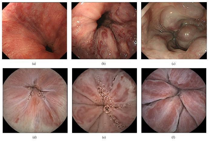
A multi-panel clinical diagnostic image comparing Esophagogastroduodenoscopy (EGD) and Capsule Endoscopy (CE) in the evaluation of esophageal varices (EVs). The top row (a-c) displays EGD views showing the progressive classification of varices by form: (a) Stage F1 presenting as straight, small-caliber longitudinal vessels; (b) Stage F2 showing moderately enlarged, beady or tortuous varices; and (c) Stage F3 showing markedly enlarged, nodular, or tumor-shaped varices that obstruct the esophageal lumen. The bottom row (d-f) provides corresponding circular-frame CE images of the same stages. The CE images demonstrate a wider field of view but varied resolution compared to conventional endoscopy. Key educational features include the visualization of red color signs (RC signs) such as red wale marks and cherry red spots, which are critical indicators of hemorrhage risk in portal hypertension. This comparison serves as a clinical reference for assessing the diagnostic accuracy and morphological presentation of esophageal varices across different endoscopic modalities.
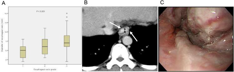
This composite image illustrates the correlation between diagnostic imaging and endoscopic findings for esophageal varices in a patient with liver cirrhosis. Panel A is a box plot showing a statistically significant positive correlation (p < 0.001) between endoscopic grading and the CT-measured diameter (mm) of esophageal varices. Panel B is an axial portal-phase CT scan demonstrating the distal esophagus with multiple nodular, hyper-enhancing, intraluminally protruding lesions (indicated by arrows), representing large esophageal varices measuring up to 10.7 mm. Panel C is an endoscopic view of the same patient, revealing multiple, tortuous, and congested large-caliber varices that cause significant distortion of the esophageal lumen. Reddish hues and surface congestion are visible, indicating high-risk features for hemorrhage. The content serves as a comparison between non-invasive radiological assessment (CT) and direct visualization (endoscopy) for managing portal hypertension complications.
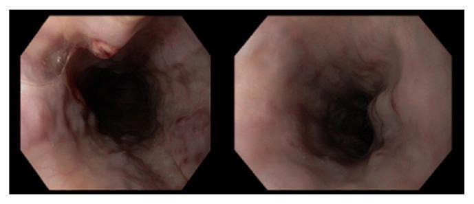
This diagnostic endoscopic image (esophagogastroduodenoscopy) provides a view of the esophagus, revealing extensive esophageal varices. The visual field shows a distended esophageal lumen with numerous large, tortuous, and dilated submucosal veins appearing as bluish-purple bulging columns along the longitudinal axis of the esophageal wall. The mucosal lining over these varices exhibits color variations, ranging from normal pink to focal areas of deep red. Critical visual markers of high bleeding risk are present, including 'red wale signs' (longitudinal red streaks resembling whip marks) and a 'nipple sign,' which indicates a site of recent or impending hemorrhage. These findings are characteristic of 'downhill' esophageal varices, often associated with superior vena cava (SVC) syndrome or portal hypertension. The image demonstrates severe vascular congestion and structural distortion of the esophageal mucosa, serving as a key educational example for identifying high-risk variceal stigmata during upper gastrointestinal endoscopy.
Esophageal and Gastric Varices
What they are
Classification
- GOV1 (gastro-esophageal varix type 1): extends 2-5 cm below the gastroesophageal junction along the lesser curve, in continuity with esophageal varices - most common
- GOV2: extends into the fundus
- IGV1 (isolated gastric varix type 1): in the fundus, no esophageal varices
- IGV2: ectopic, elsewhere in the stomach
Presentation
- Sudden, often massive hematemesis (bright red, large volume)
- Melena
- Signs of chronic liver disease: jaundice, ascites, splenomegaly, spider angiomata, caput medusae
- Can rapidly progress to hemorrhagic shock given the volume and pressure behind variceal bleeds
Acute Management (variceal hemorrhage is a true emergency)
- Resuscitate first: airway protection (intubate if massive hematemesis/altered mental status), IV access, blood products (restrictive transfusion, target Hb ~7-8 g/dL), correct coagulopathy
- Vasoactive drugs started immediately, even before endoscopy: octreotide (or somatostatin/terlipressin where available) causes splanchnic vasoconstriction and reduces portal pressure/bleeding - Current Surgical Therapy, p. 689
- Prophylactic antibiotics (e.g., ceftriaxone) - reduces infection risk and mortality in cirrhotic patients with GI bleeding
- Endoscopy within 12 hours once stabilized:
- Band ligation - preferred first-line therapy for esophageal varices, better than sclerotherapy for initial control - Sabiston Textbook of Surgery, p. 1791
- Sclerotherapy - alternative, less preferred now
- Cyanoacrylate (glue) injection - preferred for gastric varices, which don't respond as well to banding
- Balloon tamponade (Sengstaken-Blakemore tube) - temporary bridge if bleeding uncontrolled by endoscopy, before definitive therapy
- TIPS (transjugular intrahepatic portosystemic shunt) - salvage therapy for bleeding refractory to endoscopic + drug therapy; also superior to endoscopic therapy alone for preventing rebleeding long-term, though it carries a risk of hepatic encephalopathy - Yamada's Textbook of Gastroenterology, p. 2830
Prevention of rebleeding / primary prophylaxis
- Non-selective beta-blockers (propranolol, nadolol) reduce portal pressure and are used both for primary prophylaxis (known varices, not yet bled) and secondary prophylaxis (after a bleed), often combined with repeat band ligation sessions
- TIPS considered for recurrent bleeding despite combined drug + endoscopic therapy