
Heart
Reading File
The query "Heart" is very broad. Let me clarify what you're looking for before diving in.
Asking for Preferences
What aspect of the heart are you interested in?
Searching sources...
Finding Sources
Finding Sources
Finding Sources
Finding Sources
Finding Sources
Finding Sources
Gray's Anatomy for Students has a comprehensive heart anatomy section. Let me read it now.
Reading File
Reading File
Loading Image
Loading Image
Reading File
Loading Image
Reading File
Here is a comprehensive overview of Heart Anatomy & Structure, drawn from Gray's Anatomy for Students and the Color Atlas of Human Anatomy.
The Heart: Anatomy & Structure
Overview & Orientation
The heart has the general shape of a pyramid that has fallen on its side. The apex projects forward, downward, and to the left; the base faces posteriorly. The heart has four surfaces:
- Anterior (sternocostal) surface - formed mostly by the right ventricle
- Diaphragmatic (inferior) surface - formed by the left ventricle and a small part of the right ventricle
- Right pulmonary surface - formed by the right atrium
- Left pulmonary surface - broad, convex, formed by the left ventricle and part of the left atrium
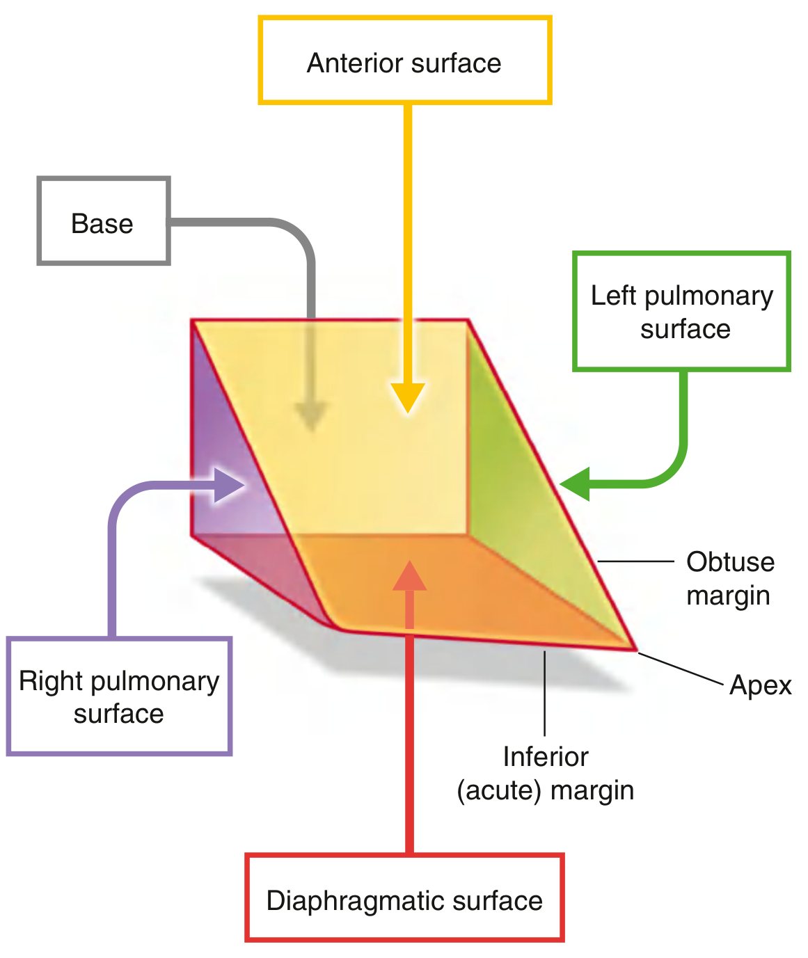
Cardiac orientation showing surfaces and margins - Gray's Anatomy for Students, Fig. 3.65
Anterior View with Key Structures
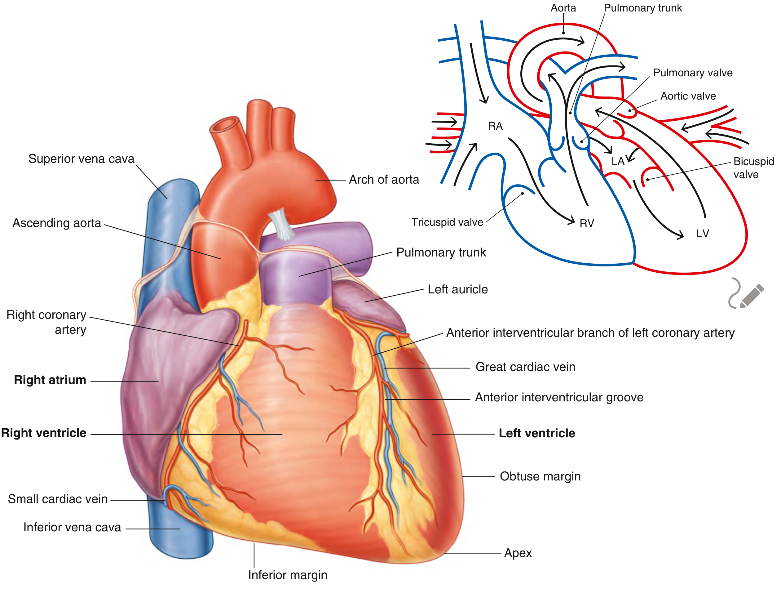
Anterior surface of the heart - Gray's Anatomy for Students, Fig. 3.67
Posterior (Base) View
The base is quadrilateral and directed posteriorly. It consists of:
- The left atrium (majority)
- A small portion of the right atrium
- Proximal parts of the great veins: superior/inferior venae cavae and pulmonary veins
The base is fixed posteriorly at vertebral levels TV to TVIII. The oesophagus lies immediately posterior.
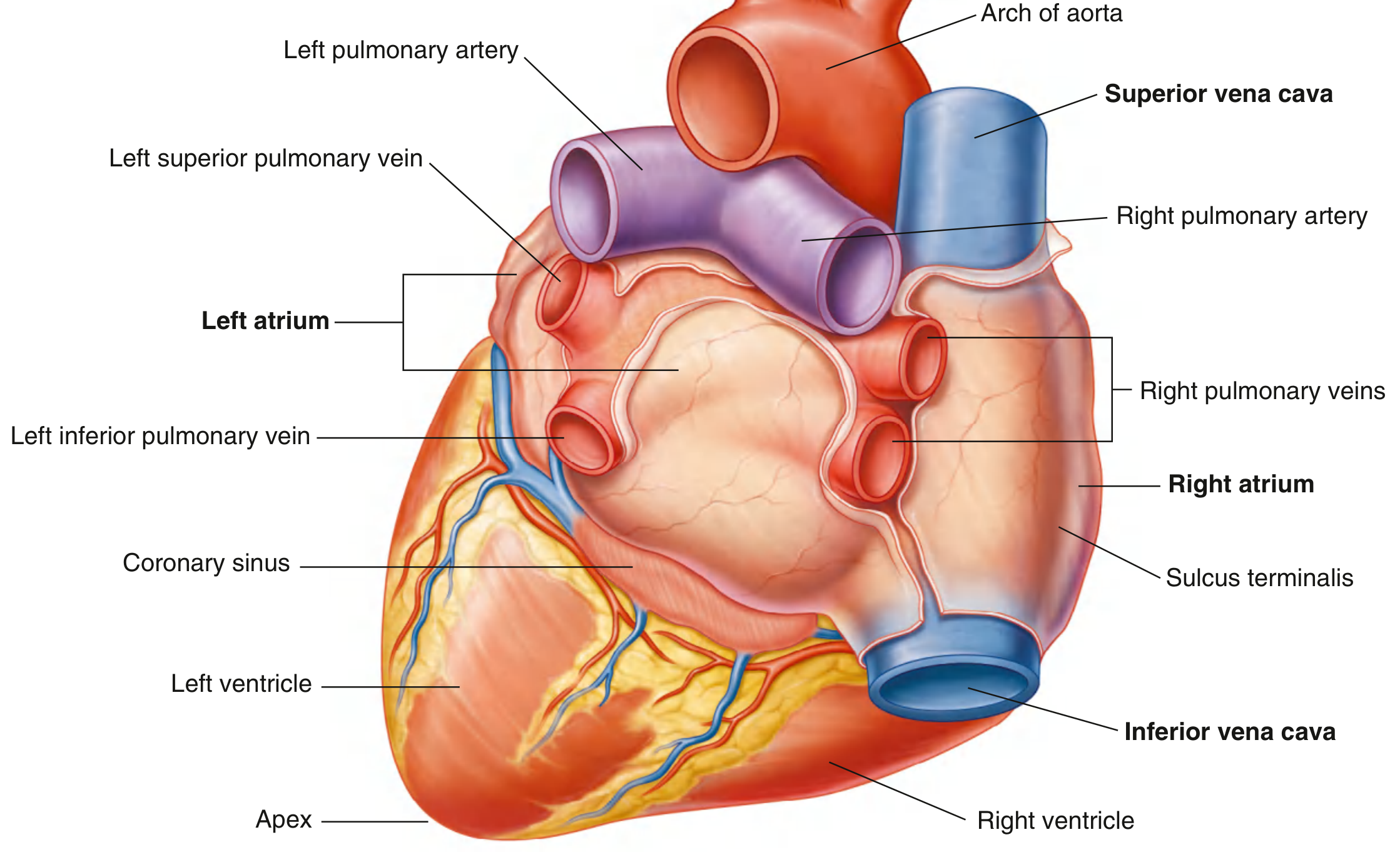
Base of the heart - Gray's Anatomy for Students, Fig. 3.66
Apex
The apex is formed by the inferolateral part of the left ventricle and lies in the left 5th intercostal space, 8-9 cm from the midsternal line.
The Four Chambers
Right Atrium
Receives deoxygenated blood from the systemic circulation via the superior and inferior venae cavae and the coronary sinus. The inner wall is divided by the crista terminalis into:
- A smooth posterior part (sinus venarum)
- A ridged anterior part containing the pectinate muscles
The fossa ovalis (remnant of the foramen ovale) is visible on the interatrial septum.
Right Ventricle
Receives blood from the right atrium through the tricuspid valve (3 cusps: anterior, posterior, septal). The outflow tract leads to the pulmonary valve and pulmonary trunk. The inner wall has:
- Trabeculae carneae (muscular ridges)
- Papillary muscles (anterior, posterior, septal) - anchor the chordae tendineae to the tricuspid valve cusps
- The septomarginal trabecula (moderator band), which carries the right bundle branch to the anterior papillary muscle
Left Atrium
Receives oxygenated blood from the lungs via four pulmonary veins (two right, two left). The smooth-walled body makes up most of the base of the heart. The left auricle has pectinate muscles.
Left Ventricle
The main pumping chamber for the systemic circulation. Has thicker walls (about 2-3x the thickness of the right ventricle). Receives blood via the mitral (bicuspid) valve (2 cusps: anterior, posterior) and ejects through the aortic valve. The outflow tract is called the aortic vestibule.
The Four Valves
| Valve | Location | Cusps | Opens into |
|---|---|---|---|
| Tricuspid | Right AV orifice | 3 (anterior, posterior, septal) | Right ventricle |
| Pulmonary | Pulmonary outflow | 3 semilunar | Pulmonary trunk |
| Mitral (Bicuspid) | Left AV orifice | 2 (anterior, posterior) | Left ventricle |
| Aortic | Aortic vestibule | 3 semilunar | Ascending aorta |
The aortic valve has three sinuses (right, left, posterior). The right and left coronary arteries originate from the right and left aortic sinuses respectively. As blood recoils after ventricular contraction and fills the aortic sinuses, it is automatically directed into the coronary arteries.
Cardiac Skeleton
The cardiac skeleton is a framework of dense fibrous tissue that:
- Provides attachment for the valve leaflets and leaflet roots
- Electrically isolates the atria from the ventricles (except at the AV bundle)
- Provides attachment for the myocardium
- Maintains the shape and size of the valve orifices
Coronary Circulation
Right coronary artery (RCA): Arises from the right aortic sinus, runs in the atrioventricular groove. Gives off the posterior interventricular artery (in ~70% of people, "right dominant") and supplies the SA and AV nodes in most individuals.
Left coronary artery (LCA): Arises from the left aortic sinus, quickly divides into:
- Anterior interventricular artery (LAD) - supplies the anterior interventricular septum and anterior left ventricle
- Circumflex artery - runs in the left AV groove, supplies the left atrium and posterior left ventricle
Venous drainage primarily via the coronary sinus (which drains into the right atrium), with smaller contributions from anterior cardiac veins and thebesian veins.
Cardiac Conduction System
The conduction system initiates and coordinates contraction in a unidirectional pathway:
- Sinoatrial (SA) node - the pacemaker, located at the superior end of the crista terminalis at the SVC-right atrium junction. Intrinsic rate ~60-100 bpm.
- Atrioventricular (AV) node - located near the opening of the coronary sinus, close to the septal cusp of the tricuspid valve. Delays the impulse (~0.1 sec) to allow ventricular filling.
- Atrioventricular (AV) bundle (Bundle of His) - the only electrical connection between atria and ventricles, runs along the lower border of the membranous interventricular septum.
- Right and Left bundle branches - run on their respective sides of the interventricular septum to reach the ventricles.
- Purkinje fibers (subendocardial plexus) - terminal network that distributes the impulse to ventricular myocardium from apex upward to the outflow tracts.
This arrangement means contraction begins at the apex and moves toward the outflow tracts, efficiently ejecting blood upward.
Cardiac Innervation
- Sympathetic (T1-T5 thoracic segments): increases heart rate and force of contraction (positive chronotropy and inotropy). Postganglionic fibers reach the heart via cardiac nerves.
- Parasympathetic (vagus nerve, CN X): decreases heart rate. Preganglionic fibers synapse in ganglia within the cardiac plexus.
The cardiac plexus lies at the base of the heart near the aortic arch and bifurcation of the trachea.
Radiological Borders (Chest X-Ray)
On a standard PA chest X-ray:
- Right border: Superior vena cava (top) → Right atrium (bottom)
- Left border: Aortic arch → Pulmonary trunk → Left auricle → Left ventricle (bottom)
Source: Gray's Anatomy for Students, Chapter 3 (Thorax) - Gray's Anatomy for Students
This is a shared conversation. Sign in to Orris to start your own chat.