
Explain decrebrate rigidity short
Decerebrate rigidity is a motor pattern caused by a brainstem lesion (at or below the level of the red nucleus, i.e., at the midbrain-pontine junction). It is characterized by extension of all four limbs, with the arms adducted, extended, and internally rotated, forearms pronated, wrists and fingers flexed - and the legs extended with plantarflexion.
Searching sources...
Reading File
Finding Sources
Reading File
Reading File
Reading File
Let me also grab the posture image for a visual reference:
Loading Image
Decerebrate Rigidity
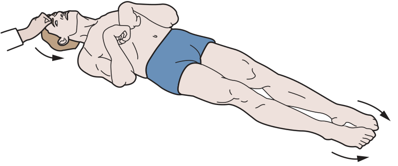
(Figure 12-12A from Ganong's Review of Medical Physiology)
Definition
Decerebrate rigidity is a stereotyped motor posture resulting from a brainstem transection (functionally or structurally) at the level of the lower midbrain / upper pons - i.e., below the red nucleus but above the pontine reticular formation.
Posture (Classic Features)
| Region | Position |
|---|---|
| Head & neck | Extended (opisthotonic) |
| Upper limbs | Extended, adducted, internally rotated; forearms pronated; wrists & fingers flexed |
| Lower limbs | Extended, with plantarflexion (toes pointed inward) |
It involves the antigravity muscles - neck, trunk, and limb extensors.
Mechanism / Pathophysiology
The key is an imbalance of descending motor pathways:
- Lesion site: Between midbrain and upper pons. This interrupts inhibitory input from the cerebral cortex, red nucleus, and basal ganglia to the medullary reticular nuclei.
- Result: The medullary reticulospinal inhibitory system becomes nonfunctional.
- The pontine reticulospinal (excitatory) tract and lateral vestibulospinal tract (from Deiters nucleus) are left unopposed - both powerfully activate extensor muscles.
- This causes hyperactivity of extensor (antigravity) muscles in all four limbs.
In simple terms: the excitatory brainstem pathways "run wild" without cortical inhibition, producing rigid extension.
Mechanism of Rigidity (Spasticity)
The rigidity is spastic in nature, driven via the gamma loop:
- Excitatory reticulospinal input activates gamma (γ) motor neurons
- These fire intrafusal fibers → stretch of spindle → Ia afferents → activate alpha (α) motor neurons
- Cutting dorsal roots immediately abolishes the rigidity (proving gamma-loop dependence)
In decerebellate animals (anterior cerebellum removed), rigidity is worsened and becomes alpha motor neuron-driven (gamma-loop independent).
- Ganong's Review of Medical Physiology, 26e
- Guyton and Hall Textbook of Medical Physiology
- Costanzo Physiology, 7e
Clinical Causes
- Uncal herniation (large hemispheric tumor or hemorrhage compressing the midbrain)
- Severe metabolic disorders (e.g., hypoxic-ischemic encephalopathy)
- Upper brainstem lesions (infarct, hemorrhage)
- Toxic (e.g., phenytoin toxicity at lethal levels)
It is associated with Hunt-Hess grade IV-V subarachnoid hemorrhage, indicating a poor prognosis.
Decerebrate vs. Decorticate - Quick Comparison
| Feature | Decerebrate | Decorticate |
|---|---|---|
| Lesion level | Lower midbrain / upper pons | Above midbrain (cerebral cortex) |
| Upper limbs | Extended, pronated | Flexed at elbow |
| Lower limbs | Extended, plantarflexed | Extended |
| Prognosis | Worse | Less severe |
The key mnemonic: De-COR-ticate = CORtex lesion = arms CURL in (flex); De-CERE-brate = CEREbellum level = all EXTEND.
Why the pontine reticulospinal did not lost its effect
Great question. The short answer: the decerebrate lesion is below the pontine reticular formation, so the pons itself is still intact and functional - it simply loses its cortical inhibition.
Reasoning...
Finding Sources
Finding Sources
Finding Sources
Reading File
Finding Sources
Reading File
This is the exact answer. Here is the full explanation:
Why Does the Pontine Reticulospinal System Keep Working in Decerebrate Rigidity?
The Core Reason: It Has Its Own Intrinsic Excitability
The pontine reticular nuclei have a high degree of natural (intrinsic) excitability - they do not depend on the cortex to stay active. They generate tonic excitatory output to extensor (antigravity) muscles on their own.
On top of that, they receive strong excitatory input from two sources that remain intact after decerebration:
- Vestibular nuclei - still functional below the lesion
- Deep cerebellar nuclei (fastigial nucleus) - still functional
So the pontine system keeps firing because its own fuel supply (vestibular + cerebellar input + intrinsic drive) is intact. (Guyton & Hall)
The Real Problem: The Medullary Inhibitory System Loses Its Input
The key is understanding the balance between two opposing systems:
| System | Location | Effect on extensors | Driven by |
|---|---|---|---|
| Pontine reticulospinal | Pons | Excitatory (facilitates) | Intrinsic + vestibular + cerebellum |
| Medullary reticulospinal | Medulla | Inhibitory (suppresses) | Cortex + red nucleus + rubrospinal tract |
In normal conditions, the medullary reticular system counterbalances the pontine system. The medullary nuclei get strong input from:
- The corticospinal tract (cortex)
- The rubrospinal tract (red nucleus)
- Other descending motor pathways
The decerebrate lesion (lower midbrain/upper pons) cuts off all of this input to the medullary reticular nuclei. Without it, the medullary inhibitory system goes silent. - Guyton & Hall, Costanzo Physiology
The Net Result
Normal:     Pontine excitation  ←balanced by→  Medullary inhibition
Decerebrate: Pontine excitation  ←   NOTHING   →  (medullary system silent)
                    ↓
          Unopposed extensor drive → RIGIDITY
To use Guyton's exact words: "when the pontine reticular excitatory system is unopposed by the medullary reticular system, it causes powerful excitation of antigravity muscles throughout the body."
The pontine system did not "gain" anything - it simply lost its opponent. The medullary inhibitory system collapsed because it was entirely dependent on cortical/rubrospinal drive, which the decerebrate lesion severed.
So give the reason for everything here short so I can remember for my exams
Here is the full logic chain, short and reason-linked:
Decerebrate Rigidity - The "Why" Behind Everything
1. Why does the lesion cause rigidity at all?
Lesion is at lower midbrain/upper pons → cuts off cortex + red nucleus + basal ganglia from reaching the medullary reticular nuclei → medullary inhibitory system goes silent → pontine excitatory system runs unopposed → extensor muscles fire continuously = rigidity
2. Why only extensors (antigravity muscles)?
The pontine reticulospinal + vestibulospinal tracts specifically target extensor/antigravity muscles (neck, trunk, limb extensors). They are built to hold the body upright. The flexors have no equivalent unopposed driver.
3. Why does the pontine system not get cut off too?
Because the lesion is above the pons - the pons is still physically intact. The pontine nuclei also have intrinsic excitability + receive input from vestibular nuclei and cerebellum (both below the lesion = still intact). So it never needed the cortex.
4. Why does the medullary system fail?
Because it has no intrinsic drive - it is 100% dependent on input from the cortex, red nucleus, and rubrospinal tract. The lesion severs all of these. No input → no inhibition.
5. Why is the rigidity spastic (not flaccid)?
It is driven via the gamma loop: pontine system activates γ-motor neurons → spindle stretch → Ia afferents → α-motor neurons fire. Cutting dorsal roots abolishes it, proving it is reflex-mediated spasticity, not direct stiffness.
6. Why are upper limbs extended (unlike decorticate)?
In decorticate (cortex lesion only), the red nucleus is still intact → rubrospinal tract still fires → flexes upper limbs. In decerebrate, the lesion is below the red nucleus → rubrospinal tract is also cut → no flexion signal → upper limbs extend too.
7. Why do lesions above the midbrain NOT cause decerebrate rigidity?
Because the pontine system and medullary system are both intact below such a lesion, and the medullary system still receives enough input from the red nucleus (midbrain) to maintain balance.
The One-Line Logic to Remember Everything:
Decerebrate = lesion below red nucleus → medullary inhibitor loses all input → pontine excitator (self-powered by vestibular + cerebellum) runs free → all four limbs extend.
Where is pontine and medullary system located
Both are part of the reticular formation - the diffuse network of neurons running through the brainstem core.
Searching sources...
Finding Sources
Reading File
Loading Image
Here is the exact anatomy, short and clear:
Location of the Two Reticular Systems
Both sit in the reticular formation - the diffuse core of the brainstem. They are in different brainstem levels and project down through different spinal cord columns.
Pontine Reticular Nuclei (Excitatory)
- Located: Lateral part of the pons, extending up into the mesencephalon
- Tract: Pontine reticulospinal tract → runs in the anterior (ventral) column of spinal cord
- Targets: Medial anterior horn motor neurons → axial + extensor muscles
- Effect: Excites antigravity muscles
Medullary Reticular Nuclei (Inhibitory)
- Located: Throughout the entire medulla, ventromedially near the midline
- Tract: Medullary reticulospinal tract → runs in the lateral column of spinal cord
- Targets: Same anterior horn motor neurons
- Effect: Inhibits antigravity muscles
Spinal Cord Cross-Section - Where Their Tracts Travel
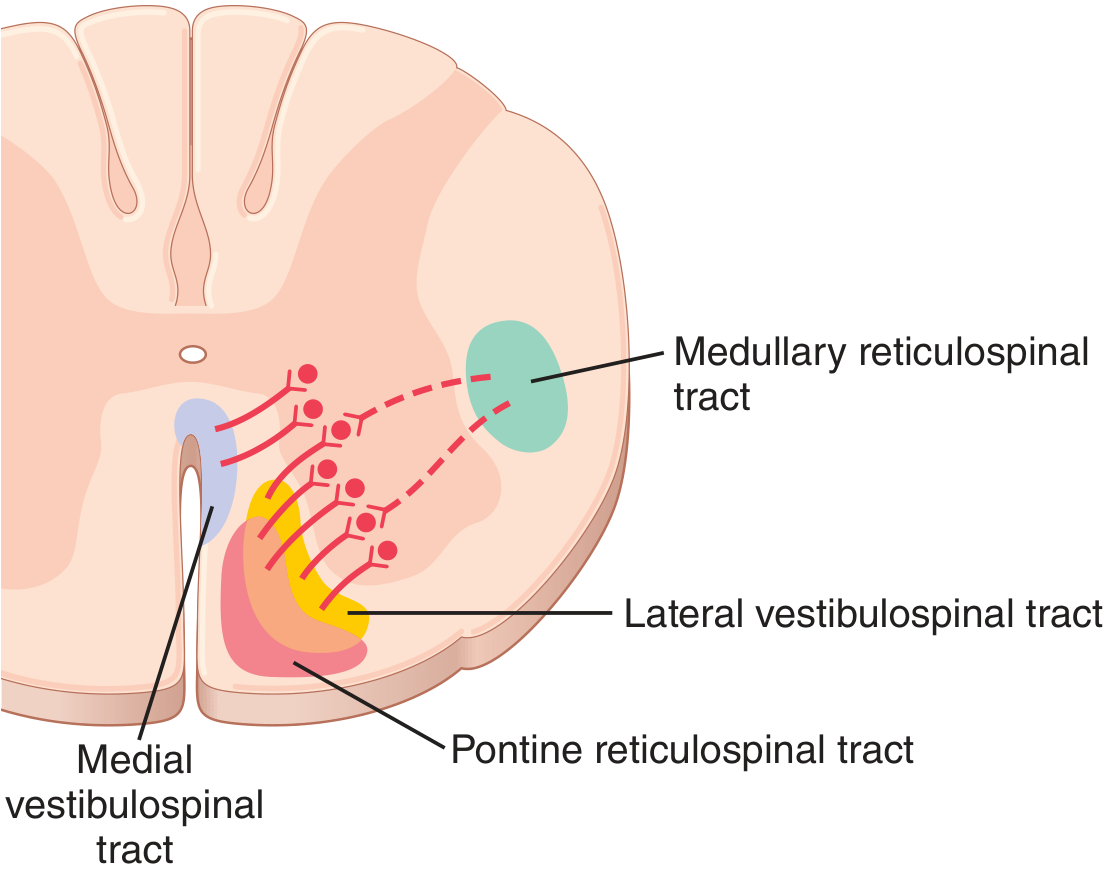
- Solid lines = excitatory (pontine + vestibulospinal)
- Dashed lines = inhibitory (medullary reticulospinal)
(Guyton & Hall)
Quick Memory Table
| Pontine | Medullary | |
|---|---|---|
| Location | Lateral pons + mesencephalon | Entire medulla, ventromedial |
| Spinal cord column | Anterior | Lateral |
| Effect | Excite extensors | Inhibit extensors |
| Driven by | Intrinsic + vestibular + cerebellum | Cortex + red nucleus |
| In decerebrate | Still active | Silent |
Memory tip: Pons = Push (excite). Medulla = Modulate/inhibit (needs cortex to work).
So why not decrebrate not preset above lower part of midbrain Short
Because the red nucleus is in the lower midbrain.
Why Lesions ABOVE the Lower Midbrain Don't Cause Decerebrate Rigidity
If the lesion is above the lower midbrain (e.g., cortex only = decorticate):
- Red nucleus is still intact
- Red nucleus → rubrospinal tract → still feeds the medullary inhibitory system
- Medullary inhibitor still works → still opposes the pontine excitator
- Balance is partially maintained → no full extensor rigidity
Upper limbs flex (rubrospinal activates flexors) = decorticate posture, not decerebrate.
The Simple Rule:
| Lesion level | Red nucleus intact? | Medullary inhibitor works? | Result |
|---|---|---|---|
| Above lower midbrain (cortex) | Yes | Yes (fed by red nucleus) | Decorticate |
| At/below lower midbrain | No (cut off) | No | Decerebrate |
One line to remember:
Decerebrate needs the red nucleus to be disconnected. As long as the red nucleus is intact and feeding the medullary inhibitor, you get decorticate - not decerebrate.
Clinical features of cerebellar lesion Exams style
Finding Sources
Finding Sources
Reading File
Reading File
Reading File
Here is the full exam-style answer:
Clinical Features of Cerebellar Lesion
Mnemonic: DANISH (Dysdiadochokinesia, Ataxia, Nystagmus, Intention tremor, Slurred speech, Hypotonia)
1. ATAXIA (Incoordination) - the hallmark
- Gait ataxia: Wide-based, unsteady, "drunken" gait; falls/deviates toward the side of the lesion
- Appendicular ataxia: Clumsy, incoordinated limb movements
- Truncal ataxia: Titubation (rhythmic tremor of trunk/head) - seen in midline lesions
- Romberg test: Negative (ataxia does NOT worsen with eyes closed - distinguishes from sensory ataxia)
2. DYSMETRIA
- Past-pointing (overshoot/undershoot) on finger-nose-finger test and heel-shin test
- "Loss of check" - arm overshoots when resistance suddenly released
3. INTENTION TREMOR
- Tremor that appears/worsens on movement, absent at rest
- Worsens as limb approaches target
- Distinguishes from Parkinsonian tremor (which is a resting tremor)
4. DYSDIADOCHOKINESIA
- Inability to perform rapid alternating movements (e.g., pronation/supination)
- Due to loss of synergy and rhythm (dysrhythmia)
5. NYSTAGMUS
- Usually gaze-evoked (gaze-paretic): fast phase toward the side of gaze
- Changes direction with direction of gaze (unlike peripheral vestibular nystagmus which is fixed direction)
- Vertical nystagmus may also occur
- Ocular dysmetria: saccades overshoot/undershoot target
6. SPEECH - Dysarthria
- Scanning (staccato/explosive) speech: irregular rate and volume
- Words come out slurred and broken up in syllables
- e.g., "BA-NA-NA" said as "BA... NA... NA"
7. HYPOTONIA
- Decreased muscle tone on the ipsilateral side
- Reduced resistance to passive movement
- Pendular knee jerk (leg swings freely without normal dampening)
8. VESTIBULOCEREBELLAR SIGNS (flocculonodular lobe)
- Impaired VOR suppression (nystagmus when fixating on moving object)
- Nausea, vomiting, vertigo
Key Exam Points
| Feature | Detail |
|---|---|
| Side of lesion | Signs are ipsilateral (cerebellum doesn't decussate before acting) |
| Romberg | Negative - ataxia present eyes open AND closed |
| Intention tremor | Present on movement, absent at rest |
| Midline lesion | Truncal ataxia, gait > limb involvement |
| Lateral lesion | Limb ataxia (appendicular) > gait |
| No weakness | Pure cerebellar = incoordination WITHOUT paralysis |
| No sensory loss | Pure cerebellar = no sensory deficit |
One-Line Rule for Exams:
Cerebellum = IPSILATERAL incoordination, intention tremor, nystagmus, dysarthria, hypotonia - with NO weakness and NO sensory loss.
(Neuroanatomy through Clinical Cases, 3e)
Short few lines Decorticate rigidity
Decorticate Rigidity
Lesion: Above the midbrain (cerebral cortex damage) — red nucleus is intact
Posture:
- Upper limbs: Flexed at elbow, wrist, fingers (rubrospinal tract still active → excites flexors)
- Lower limbs: Extended with plantarflexion (same as decerebrate — pontine system still unopposed below)
- Head: Extended
Why upper limbs flex: Red nucleus → rubrospinal tract → activates upper limb flexors. This pathway is spared because the lesion is above the midbrain.
Why lower limbs extend: Rubrospinal tract has minimal influence on lower limbs, so pontine + vestibulospinal extensor drive still dominates below.
Memory trick:
De-COR-ticate = CORtex lesion = arms CURL in Think: patient is curling arms toward the core of the body
| Decorticate | Decerebrate | |
|---|---|---|
| Lesion | Above midbrain | Below midbrain |
| Red nucleus | Intact | Disconnected |
| Arms | Flexed | Extended |
| Legs | Extended | Extended |
| Prognosis | Less severe | Worse |
But there is medullary inhibitors system active through why still extension of Lowe limb
Great catch. Here is the precise reason:
Why Lower Limbs Still Extend in Decorticate (Despite Medullary Inhibitor Being Active)?
The medullary inhibitor IS active in decorticate - but it is only partially restored.
It gets input from:
- Red nucleus ✅ (intact)
- Rubrospinal tract ✅ (intact)
- Cortex ❌ (damaged - that's the lesion)
- Corticospinal tract ❌ (damaged)
So the medullary inhibitor is running on partial fuel (red nucleus only), not full power.
The Critical Point: Rubrospinal tract is strong for upper limbs, weak for lower limbs
The rubrospinal tract predominantly controls upper limb flexors. Its influence on lower limbs is minimal.
So:
- Upper limbs: Red nucleus input enough to activate flexors → arms flex
- Lower limbs: Rubrospinal influence too weak → pontine excitatory + vestibulospinal system still dominates → legs extend
One Line:
Medullary inhibitor is partially restored (by red nucleus), but only enough to flip upper limbs to flexion - lower limbs remain extended because rubrospinal tract barely reaches them.
This is a shared conversation. Sign in to Orris to start your own chat.