
An ulcer on glans penis
Reading File
Now let me search the medical library in parallel for the key causes and features of penile glans ulcers.
Finding Sources
Finding Sources
Finding Sources
Good results. Now let me read the key sections in parallel - especially the S Das clinical surgery section (which covers differential diagnosis of penile ulcers well) and the Harrison's syphilis section, plus search for other STI ulcers.
Reading File
Reading File
Finding Sources
Let me read more of the S Das section and also search for chancroid and herpes specifically.
Reading File
Finding Sources
Finding Sources
I now have excellent material. Let me read a bit more from S Das and Bailey & Love for the complete differential and features, and fetch the key Harrison's syphilis image.
Reading File
Reading File
Loading Image
I now have comprehensive material for a thorough answer. Let me compile this into a well-organized response.
Ulcer on Glans Penis - Differential Diagnosis & Clinical Approach
An ulcer on the glans penis is a common presentation with causes ranging from sexually transmitted infections to malignancy. The key clinical distinction is painful vs. painless.
Differential Diagnosis
1. Hunterian Chancre (Primary Syphilis) - PAINLESS
The classic and most important cause to recognize.
Features:
- Caused by Treponema pallidum
- Incubation: ~3-4 weeks after exposure
- Single (occasionally multiple) painless papule that erodes into a well-defined ulcer
- Indurated base - cartilaginous, "button-like" consistency on palpation - this is the hallmark
- Well-defined, clean edges; serosanguinous discharge
- Associated non-tender inguinal lymphadenopathy (bilateral)
- Heals spontaneously in 3-6 weeks, but systemic disease progresses
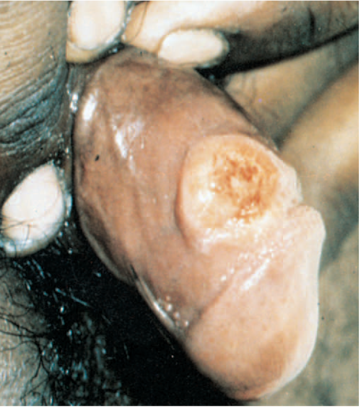
2. Chancroid (Soft Sore) - PAINFUL
- Caused by Haemophilus ducreyi (Gram-negative facultative anaerobe)
- Incubation: 3-10 days (short - distinguishes it from syphilis)
- Multiple, painful ulcers with ill-defined, oedematous/ragged margins
- Soft base; purulent discharge
- Associated tender inguinal lymphadenopathy (bubo), which may suppurate
- "Kissing ulcers" by autoinoculation (frenulum to glans) are characteristic
- Endemic in Africa, Southeast Asia, India, Latin America
- Treatment: ceftriaxone or azithromycin
3. Genital Herpes (HSV-2, less commonly HSV-1) - PAINFUL
- Multiple small grouped vesicles that rupture to form painful shallow ulcers
- Burning/tingling prodrome
- Recurrent episodes are common
- Heals without scarring in 1-3 weeks
- Associated tender inguinal lymphadenopathy
- Treatment: acyclovir/valacyclovir
4. Lymphogranuloma Venereum (LGV)
- Caused by Chlamydomia trachomatis (serovars L1-L3)
- Primary lesion: small (2-3 mm), fleeting herpetiform vesicle or erosion on the glans, prepuce, or coronal sulcus - often unnoticed by the patient
- Incubation: 3-20 days
- Main feature is painful inguinal/femoral lymphadenopathy (buboes) - the "groove sign"
5. Granuloma Inguinale (Donovanosis) - PAINLESS
- Caused by Klebsiella granulomatis
- Incubation: 1-4 weeks; starts as painless vesicle or indurated papule
- Erodes to form a beefy-red, painless ulcer with raised, clean edges; bleeds easily on contact
- Progressive, destructive tissue destruction if untreated
- Intracellular "Donovan bodies" (bipolar-staining in macrophages) on smear
- No lymphadenopathy (distinguishes it from LGV and chancroid)
6. Carcinoma of the Penis - PAINLESS, INDURATED
- Disease of older age; rare in circumcised men
- Appears as a non-tender ulcer (with raised, everted, indurated edges and necrotic floor) or warty growth at the coronal sulcus or glans
- Often hidden under a tight prepuce (phimosis)
- Inguinal lymph node enlargement (~50% due to secondary infection rather than metastasis)
- Pre-malignant lesions to recognize:
- Erythroplasia of Queyrat - dark red flat indurated patch on glans/inner prepuce (essentially Bowen's disease of the penis)
- Leukoplakia - white plaque
- Paget's disease - chronic red eczema-like patch
7. Balanitis / Balanoposthitis
- Inflammation of glans (balanitis) ± prepuce
- Can lead to superficial erosions/ulcers
- Candidal balanitis is common in diabetics
- Creamy, malodorous subpreputial discharge
Quick Comparison Table
| Feature | Syphilitic Chancre | Chancroid | Herpes | LGV | Donovanosis | Carcinoma |
|---|---|---|---|---|---|---|
| Cause | T. pallidum | H. ducreyi | HSV-1/2 | C. trachomatis | K. granulomatis | Squamous cell |
| Incubation | ~3-4 weeks | 3-10 days | 2-7 days | 3-20 days | 1-4 weeks | - |
| Pain | Painless | Painful | Painful | Minimal | Painless | Painless |
| Number | Usually single | Multiple | Multiple | Single/small | Progressive | Single |
| Edge | Well-defined, indurated | Ragged, soft | Irregular | - | Raised, clean | Everted, indurated |
| Base | Clean, indurated | Purulent, soft | Shallow | - | Beefy red | Necrotic |
| Lymph nodes | Non-tender bilateral | Tender, suppurates | Tender | Massive buboes | None | Enlarged |
Clinical Approach
History: Time of onset relative to sexual exposure is key - 4 days = chancroid; 4 weeks = syphilitic chancre. Ask about pain, number of partners, previous episodes.
Investigations:
- Dark-field microscopy of ulcer exudate - for T. pallidum (syphilis)
- VDRL / RPR (screening); TPHA / FTA-ABS (confirmatory) - for syphilis
- Swab for culture or PCR - for H. ducreyi (chancroid)
- Tzanck smear / HSV PCR - for herpes
- Tissue smear (Giemsa) for Donovan bodies - donovanosis
- Biopsy - if malignancy suspected or ulcer not healing
- HIV testing - mandatory in all genital ulcer disease (GUD); ulcers facilitate HIV transmission bidirectionally
Remember: Multiple STIs frequently coexist. Always test for HIV and syphilis in any patient presenting with genital ulcer disease.Circumcision provides significant protection against penile carcinoma (Muslims and Jews nearly immune due to early religious circumcision) and also reduces susceptibility to ulcerative STIs and HIV transmission.
Sources: S Das - A Manual on Clinical Surgery 13th Ed; Bailey & Love's Short Practice of Surgery 28th Ed; Harrison's Principles of Internal Medicine 22E; Andrews' Diseases of the Skin; Fitzpatrick's Dermatology 5e

IMG_20260716_040911.jpg
Near frenulum
analyse_media
Based on the image analysis, this is more consistent with a frenular fissure/tear than a classical STI ulcer. Here is the clinical assessment:
Clinical Assessment
What the Image Shows
There is a linear fissure (split) along the midline of the frenulum on the ventral aspect of the glans penis. Key features:
- Linear, narrow split (~5-8 mm long, <2 mm wide)
- Clean, sharp edges - not ragged or everted
- Shallow base - raw, pink mucosal tissue; not deeply ulcerated or necrotic
- No induration - the tissue appears pliable, not button-hard
- No visible discharge - no pus, no serous fluid
- Surrounding erythema - mild-moderate inflammation of the frenular area
- No lymphadenopathy visible
This pattern does not match a classical STI ulcer. It is most consistent with:
Most Likely Diagnosis: Frenular Tear / Fissure
Causes:
- Mechanical trauma (frenulum breve / short frenulum) - friction during intercourse or masturbation causes splitting at the point of maximum tension. This is the most common cause of frenular fissures.
- Balanitis-related fissuring - inflammation (fungal, bacterial, or irritant) makes the tissue inelastic and prone to linear splits.
- Recurrent fissuring - if this has happened before, a short or scarred frenulum is very likely.
Can an STI Be Ruled Out?
Not completely from a photo alone. Features that argue against a primary STI ulcer:
- Linear, fissure-like morphology (STI ulcers are typically oval/round)
- No indurated base (syphilitic chancre feels like a button)
- No purulent discharge (chancroid)
- No grouped vesicles (herpes)
- No beefy red, raised edges (donovanosis)
However, HSV (herpes) can occasionally present with linear fissures in this location, especially on first episode or with frenular involvement. It cannot be excluded clinically.
What to Do
Immediate:
- Avoid sexual activity until healed
- Keep area clean and dry; avoid harsh soaps
- Do not apply OTC creams without medical advice
See a doctor for:
- Swab of the lesion for HSV PCR (to rule out herpes)
- VDRL/RPR + dark field microscopy if sexual exposure history warrants it
- Assessment for frenulum breve - if the frenulum is congenitally short, this will recur. Treatment options include frenuloplasty (surgical lengthening) or circumcision.
Red flags requiring urgent review:
- Ulcer fails to heal in 2 weeks
- Develops induration, lymph node swelling, or purulent discharge
- Systemic symptoms (fever, rash, malaise)
Important: This is a clinical image assessment for educational purposes. A physical examination by a doctor or sexual health clinic is necessary for definitive diagnosis and management. Do not self-treat a genital lesion.
This is a shared conversation. Sign in to Orris to start your own chat.