


A 18 yrs old male pt presents with such lesions on dorsum of hand with pain swelling and pus discharge tlc count was elevated no itching was there he has some likely lesions on groin and inner thighs also no family members involved no itching treatment given was tab clavam 625 mg bd for seven days alongwith fusidic acid topical mahamanjishtadi kwath and permithrin 5 % he has improved as shown in the next picture what should be the next treatment plan and what is probable diagnosis
nodular scabies treatment management
nodular scabies dorsum hand
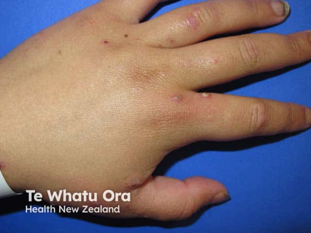
A high-resolution clinical photograph captures the dorsum of the hand demonstrating scattered pustular papules and small erosions on an erythematous base. The image was acquired using clinical photography in visible light with a neutral background to ensure color fidelity; no contrast agents or staining were used. The primary anatomical focus is the skin of the dorsal hand, including the metacarpal dorsum and interdigital spaces. Pustules are nonfollicular, approximately 2–4 mm in diameter, with surrounding mild erythema and shallow crusts, consistent with secondary bacterial infection superimposed on a scabies eruption. The appearance is compatible with infected scabies (scabies with impetiginization) and may show superficial crusting or pustulation at burrow sites. Clinically significant features include pruritus, linear burrows that may be subtle, and a crusted/pustular infestation pattern. Diagnostic significance lies in identifying secondary infection that necessitates antibiotic therapy (e.g., anti-staphylococcal coverage) in addition to scabicidal treatment. This image is valuable for dermatology education, clinical case repositories, and differential diagnosis training (impetigo, eczema herpeticum, papular urticaria). Potential use cases include teledermatology consultations, image-based teaching modules, and research on mite-related skin lesions and superinfection dynamics.
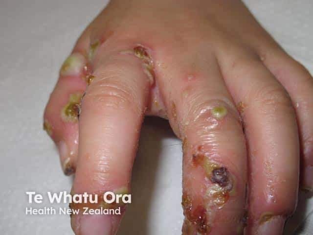
Clinical photograph of the dorsum and interdigital surfaces of the hand demonstrating extensive crusted skin lesions with yellow-brown crusts, eroded margins, and surrounding erythema. The appearance is characteristic of crusted (Norwegian) scabies with superimposed bacterial infection; in this case the crusts and purulent debris are more accentuated by a secondary pyococcal infection. Numerous discrete plaques are distributed across several digits, including the proximal and middle phalanges, with fissuring in interdigital spaces. In addition to crusts, there are vesicles or pustules and areas of tissue breakdown, suggesting active inflammation and possible secondary necrosis. The overall picture emphasizes a high infectious burden and contagiousness, requiring urgent dermatologic and infectious disease management. The clinical relevance includes recognizing crusted scabies as a risk factor for disseminated bacterial colonization and sepsis if untreated. Management implications entail combination therapy with specific scabicides (permethrin cream, ivermectin) and broad-spectrum antibiotics active against Pseudomonas aeruginosa or equivalent pyogenic organisms; strict contact precautions are advised in clinical settings. The image provides educational value for diagnosing crusted scabies, secondary impetigo, and differential diagnoses such as eczema with crusting or bullous impetigo, and supports teaching on infection control, wound care, and outpatient vs inpatient management.
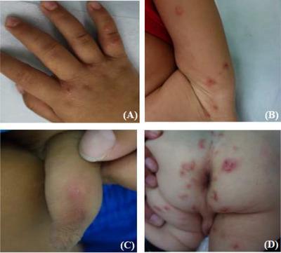
This composite clinical photograph illustrates the diverse dermatological manifestations of scabies in a pediatric patient across four anatomical regions. Panel A shows the dorsal hand and interdigital spaces featuring erythematous papules, papulovesicles, and distinct blisters. Panel B displays the axillary region with scattered red papules and inflammatory nodules. Panel C depicts the genital area (penis) presenting with characteristic pruritic red nodules, where subtle linear, whitish 'tunnels' or burrows—pathognomonic for Sarcoptes scabiei infestation—are visible on the nodular surfaces. Panel D captures the buttocks and perianal area showing extensive polymorphic lesions, including widespread erythema, inflammatory papules, and nodules interspersed with secondary lesions such as excoriations (scratches), shallow ulcers, and hemorrhagic scabs resulting from chronic pruritus. The collection serves as an educational reference for identifying both primary sarcoptic lesions and secondary infections or irritations common in childhood scabies cases.
Probable Diagnosis: Scabies with Secondary Bacterial Infection (Impetiginized Scabies) - Now Resolving to Post-Scabies Nodules
Image Analysis
- Diffuse lichenification and hyperpigmentation of the dorsal hand
- Multiple tense pustules and purulent vesicles (clustered, cloudy)
- Raw ulceration/erosion on the index finger with serosanguinous discharge
- Honey-colored and hemorrhagic crusting (classic impetiginization)
- Scattered excoriated papules and nodules
- Background xerosis with exaggerated skin markings
- Significant resolution of active infection
- Discrete, flat-topped, hypopigmented/smooth papules (2-8 mm) on the dorsal hand
- Grouping over the MCP and PIP joints
- Some knuckle area hyperkeratosis persisting
- Lesions suggesting possible Koebner distribution (linear arrangement)
- Overall picture consistent with post-inflammatory hypopigmentation + residual nodules of scabies
Diagnosis: Scabies with Secondary Pyoderma (Impetiginized Scabies)
- 18-year-old male
- Dorsum of hand + groin/inner thighs involvement (classic predilection sites - circle of Hebra)
- Painful swelling and pus discharge = secondary bacterial infection (Staph aureus/Streptococcus)
- Elevated TLC (neutrophilic leucocytosis from secondary infection)
- No itching - this is actually consistent: sensitization to scabies mite typically produces intense itch but secondary bacterial infection may dominate the picture; some primary presentations (especially early or with superinfection) can have variable itch. Additionally, in some atypical/post-sensitization scabies, itch can be minimal
- No family members involved (common but not mandatory; close physical contact needed for transmission)
- Responded to clavam (antibiotic) + fusidic acid (topical antibiotic) + permethrin 5% (scabicide) - this combination response strongly confirms the diagnosis
Next Treatment Plan
1. Confirm Eradication of Scabies
- Repeat permethrin 5% cream - full-body single application (neck to toes), left on for 8-10 hours, repeated after 7 days as some mites/eggs may survive a single cycle. The patient improved but nodules may have been present from the start.
- Alternatively or additionally: Oral ivermectin 200 mcg/kg (approximately 12 mg for ~60 kg adult) given as two doses 7-14 days apart - this is now standard of care especially when there is persistent/treatment-resistant disease.
2. Treat Post-Scabies Nodules
- Intralesional triamcinolone acetonide (10 mg/mL, 0.1-0.2 mL per nodule) - first-line for persistent nodules
- Potent topical corticosteroids under occlusion (e.g., clobetasol 0.05% cream under a bandage overnight for 2-4 weeks)
- Topical pimecrolimus or tacrolimus (calcineurin inhibitors) - useful for steroid-resistant nodular scabies (Fitzpatrick's, 2005 citation: "Treatment of steroid-resistant nodular scabies with topical pimecrolimus")
- Coal tar preparations - as an adjunct
- Mahamanjishtadi kwath (already prescribed, reasonable Ayurvedic adjunct for its anti-inflammatory/detoxifying properties)
3. Address Post-Inflammatory Hyperpigmentation/Hypopigmentation
- Mild topical retinoids (e.g., tretinoin 0.025%) or azelaic acid 20% once nodules flatten
- Broad-spectrum sunscreen to prevent UV-worsening of hyperpigmentation
- Reassure patient: PIH in dark skin types resolves over months
4. General Measures (Mandatory)
| Measure | Detail |
|---|---|
| Treat all close contacts simultaneously | Even if asymptomatic - sexual partner, household members with direct skin contact |
| Launder all clothing/bed linen | Hot wash (60°C) or seal in plastic bag for 72 hours |
| Avoid topical steroids alone | Will not treat the mite and may allow proliferation |
| Counsel on re-infestation prevention | Close physical/sexual contact is the main transmission route |
5. Monitor for
- Persistent fever or rising TLC despite antibiotic course - consider deeper infection / MRSA
- Post-streptococcal glomerulonephritis if GAS was the causative secondary pathogen (check urine in 2-3 weeks)
- Itch recurrence after treatment - distinguishes true treatment failure from post-scabies itch (which is immunological and can persist 4-6 weeks after cure)
Summary Table
| Phase | Drug / Intervention | Duration |
|---|---|---|
| Scabies eradication | Ivermectin 200 mcg/kg orally x 2 doses (7-14 days apart) + permethrin 5% repeat | 2 weeks |
| Post-scabies nodules | Intralesional triamcinolone 10 mg/mL OR clobetasol 0.05% under occlusion | 4-6 weeks |
| If steroid-resistant nodules | Topical tacrolimus 0.1% or pimecrolimus 1% BD | 4-8 weeks |
| PIH | Sunscreen + azelaic acid 20% or tretinoin 0.025% | 3-6 months |
| Contacts | Simultaneous treatment of all close contacts | Same day |
| Environment | Launder/bag all clothing + linen | Day of treatment |
- Andrews' Diseases of the Skin: Clinical Dermatology, Mites section, p. 517-519
- Fitzpatrick's Dermatology, Vol 1-2, Persistent nodular arthropod bite reactions and nodular scabies, p. 2145
- Dermatology 2-Volume Set 5e, Clinical Features section (nodular scabies images)