
diabetic foot ulcer surgery clinical examination
diabetic foot ulcer clinical examination Wagner classification
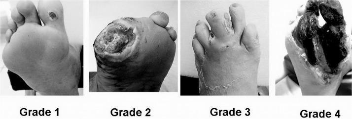
A comparative clinical photograph set illustrating the Wagner Classification System for diabetic foot ulcers across four stages (Grade 1 to Grade 4). Grade 1 (Superficial Ulcer) shows a small, localized skin lesion on the plantar surface near the base of the toes with no deep tissue involvement. Grade 2 (Deep Ulcer) depicts a larger, deeper wound penetrating to the tendon or joint capsule without abscess or osteomyelitis. Grade 3 (Osteitis/Infection) demonstrates a deep ulcer associated with significant inflammation, purulent discharge, and clinical signs of underlying bone infection or deep tissue abscess. Grade 4 (Forefoot Gangrene) shows extensive necrosis and blackening of the tissue localized to the forefoot or toes, indicating severe vascular compromise and tissue death. This series serves as an educational tool for medical professionals to differentiate between stages of diabetic foot pathology, guiding clinical management and prognosis assessment.
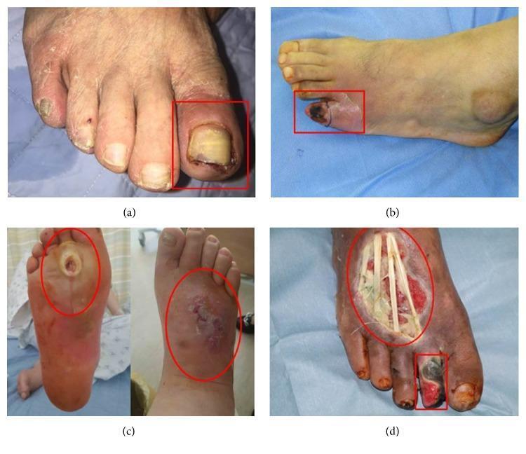
A series of four clinical photographs (a-d) demonstrating the progression of diabetic foot ulcers categorized by the Wagner classification system. (a) Wagner Grade 1: A superficial ulcer on the left hallux (big toe) featuring erythema, periungual inflammation, and a thickened, discolored yellowish toenail, indicative of an ischemic etiology. (b) Wagner Grade 2: A deep ulcer on the fifth digit (pinky toe) showing more extensive tissue involvement and localized cyanosis. (c) Wagner Grade 3: A neuropathic perforating ulcer extending from the plantar surface to the dorsum of the foot, associated with deep infection, abscess, and clinical signs of osteomyelitis. (d) Wagner Grade 4: Advanced diabetic foot pathology exhibiting local gangrene of the second toe and a large, deep dorsal foot ulcer. The necrotic tissue is blackened, and the wound bed is exposed, revealing underlying anatomical structures such as tendons. This set illustrates the clinical manifestations of ischemic, neuropathic, and mixed-type diabetic foot disease for educational use in wound care and endocrinology.
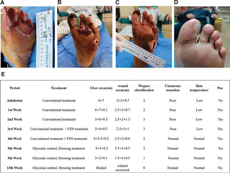
This composite figure presents a clinical progression timeline of a complex diabetic foot ulcer (DFU) on the left foot, categorized under the Wagner classification system. (A) Initial presentation shows a large, 6x7 cm ulcer on the plantar forefoot with surrounding maceration and a lateral abscess. (B) Following initial debridement, the wound exhibits deep tissue involvement (Wagner Grade 3), with necrotic tissue extending to the tendons and musculature of the first through third toes, accompanied by a deep 2x2x0.7 cm abscess cavity. (C) Mid-treatment status demonstrates significant clinical improvement; the wound bed appears ruddy with healthy granulation tissue, reduced exudate, and diminishing necrotic debris. (D) Final follow-up at three months shows complete secondary intention healing with full epithelialization and stable scar tissue formation. (E) An accompanying summary table provides longitudinal data on treatment phases (including Fu's subcutaneous needling), ulcer/wound dimensions, Wagner grade transition (from Grade 3 to 0), and clinical markers such as cutaneous sensation, skin temperature, and presence of pus.
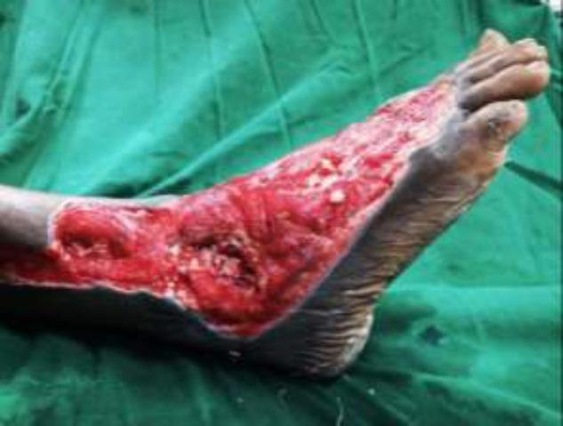
Clinical photograph of a right foot demonstrating a severe, extensive Wagner Grade III or IV diabetic foot ulcer. The wound spans the lateral and dorsal aspects of the foot, extending from the distal lower leg across the ankle joint to the midfoot region. The ulcer bed is characterized by beefy red granulation tissue and deep cavitation, with visible exposure of underlying musculoskeletal structures including the ankle joint. There are focal areas of yellow/white slough and fibrin, indicative of potential infection or chronic inflammatory processes. The wound margins are irregular but well-demarcated against the surrounding hyperpigmented, intact skin. This image serves as a clinical example of complex diabetic wound progression requiring advanced surgical intervention, such as debridement and microvascular flap reconstruction. Key educational concepts include diabetic foot classification, wound bed assessment, and the management of deep tissue infections with joint involvement.
diabetic foot neuropathy ischemia monofilament test ankle brachial index
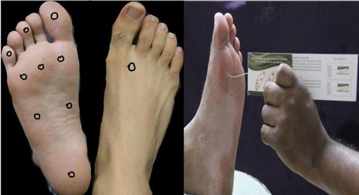
This composite educational image illustrates the Semmes-Weinstein 10 g monofilament test, a standard clinical procedure for assessing loss of protective sensation (LOPS) in patients with diabetes mellitus. The left panel demonstrates the specific testing sites marked with circles: nine sites on the plantar surface—including the distal great, third, and fifth toes; the first, third, and fifth metatarsal heads; the medial and lateral midfoot; and the heel—and one site on the dorsal surface between the first and second metatarsals. The right panel displays the clinical application of the test. A clinician’s hand is shown holding the monofilament perpendicularly to the skin of the foot's dorsal surface. The monofilament is bent into a C-shape, indicating the correct application of a 10 g buckling force. This visual serves as a procedural guide for medical professionals to screen for peripheral neuropathy and identify patients at risk for diabetic foot ulceration and secondary complications like gangrene.
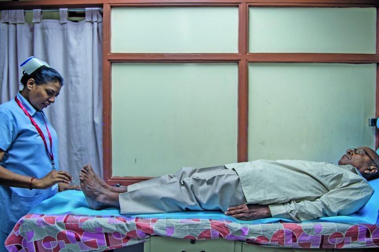
A clinical photograph capturing a diabetic foot examination in a healthcare setting. The image shows a medical professional, identified as a nurse in a blue scrub suit and cap, performing a sensory assessment on an elderly male patient. The patient is lying supine on an examination table. The nurse is utilizing a specialized instrument, consistent with a Semmes-Weinstein monofilament, to test for peripheral neuropathy by applying pressure to specific points on the patient's toes and plantar surface. This procedure is a critical diagnostic step in identifying loss of protective sensation (LOPS) in patients with diabetes mellitus, aimed at preventing ulcers and complications related to diabetic foot disease. The setting includes clinical furniture, patterned linens, and privacy curtains, illustrating a standard outpatient diabetic screening environment.
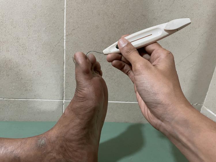
Clinical photograph of a sensory assessment being performed on a human foot using a 5.07 Semmes-Weinstein 10 g monofilament. The image shows a clinician's hand holding a white plastic monofilament device, applying the thin black filament to the dorsal surface of the distal hallux (big toe). The filament is buckled into a characteristic 'C' shape, indicating that exactly 10 g of pressure is being applied to test for Loss of Protective Sensation (LOPS). The foot exhibits several clinical signs associated with diabetic peripheral neuropathy, including skin dryness, hyperpigmentation, and a scaly, hyperkeratotic lesion near the lateral malleolus. The toenails appear thickened and dystrophic. This procedure is a critical screening tool for identifying patients at high risk for diabetic foot ulceration and neuropathic complications.
Diabetic Foot Ulcer (DFU): Clinical Examination and Surgical Management
1. History Taking
- Duration and onset of the ulcer (often unknown due to sensory neuropathy - patient may not recall trauma)
- Prior episodes of ulceration, prior amputations, or Charcot neuroarthropathy
- Symptoms of peripheral neuropathy: paresthesias, numbness, burning, loss of sensation
- Symptoms of claudication or rest pain (peripheral arterial disease)
- Local wound care being used, footwear type (shoes should be inspected for wear patterns)
- Systemic symptoms: fever, chills (ascending infection requiring urgent admission)
- Comorbidities: impaired vision, renal disease, HbA1c control
- Social history: smoking, immobility, nutritional status
Campbell's Operative Orthopaedics 15th Ed 2026 - Ch. 91
2. Physical Examination - Structured Approach
General Inspection
- Examine both feet without socks at every visit
- Document: wound size, depth, location, edges (raised vs. macerated), wound bed appearance (granulation, slough, eschar)
- Diabetic neuropathic ulcers: typically pink center with granulation tissue and raised edges, most commonly under the 1st or 5th metatarsal head
- Look for structural deformities: claw toes, hammer toes, Charcot deformity, hallux valgus - these create abnormal pressure points
Neurological Assessment
- 10-g Semmes-Weinstein monofilament test: the cornerstone for loss of protective sensation (LOPS). Test at 9 plantar sites (distal great toe, 3rd and 5th toes; 1st, 3rd, 5th metatarsal heads; medial and lateral midfoot; heel) and 1 dorsal site (between 1st and 2nd metatarsals). The monofilament should buckle into a "C" shape at each site.
- At least one additional modality: pinprick, temperature, vibration (128 Hz tuning fork), or proprioception
- Assess motor function: plantar/dorsiflexion strength, intrinsic muscle wasting (leads to clawing)
- Look for autonomic signs: dry skin, fissures, absence of sweating, anhidrosis
Vascular Assessment
- Peripheral pulses: dorsalis pedis and posterior tibial. Note: calcified/non-compressible vessels are common in diabetics, so absent pulse doesn't always equal ischemia.
- Ankle-Brachial Index (ABI):
- Normal: 0.9-1.2
-
1.3 = non-compressible vessels (medial calcinosis - falsely elevated)
- <0.5 = unlikely to heal without vascular intervention
- Ankle systolic pressure >60-90 mmHg needed for healing
- A side-to-side difference >0.15 is significant
- Toe-Brachial Index (TBI): preferred when vessels are calcified (digital vessels are often spared)
- TBI >0.7 = normal; >0.4 or absolute toe pressure >40 mmHg indicates adequate perfusion for wound healing
- Absolute toe pressure >70 mmHg is ideal
- Capillary refill time, skin temperature, colour (pallor on elevation, dependent rubor = Buerger's sign)
Wound Examination
- Debride overriding callus first - callus typically obscures the true wound extent
- Measure wound size (length x width x depth in cm) at every visit - a 50% reduction in 4 weeks predicts healing
- Probe-to-bone test: probe wound with a sterile blunt probe or back of cotton swab - bone contact is highly suggestive of osteomyelitis (PPV ~90%)
- Assess for undermining, sinus tracts, fistulae
- Wound bed: granulation tissue, slough, necrosis, eschar
- Exudate: amount, character - maceration may indicate infection or fluid overload
- Peri-wound skin: erythema (measure extent), warmth, induration, blistering
- Spreading erythema + purulent drainage + fascial plane penetration = active infection requiring antibiotics and surgical evaluation urgently
Infection Assessment
- Local signs: erythema, warmth, swelling, purulence, crepitus (gas gangrene)
- Probe-to-bone: positive result strongly suggests osteomyelitis
- WIFI Classification: guides amputation risk - score Wound (0-3), Infection (0-3), Foot Ischemia (0-3)
- Systemic signs: fever, tachycardia, leukocytosis (ascending/limb-threatening infection)
3. Ulcer Classification
Wagner Classification (most widely used)
| Grade | Description |
|---|---|
| 0 | No ulcer; pre-ulcerative or healed lesion |
| 1 | Superficial ulcer - skin depth only, no bone/tendon/joint |
| 2 | Deep ulcer penetrating to tendon, bone, or joint capsule |
| 3 | Deep ulcer with osteomyelitis, abscess, or joint sepsis |
| 4 | Localized gangrene - forefoot/toes |
| 5 | Extensive gangrene - whole foot |
Current Surgical Therapy 14e - Wound grading section
4. Investigations
| Investigation | Purpose |
|---|---|
| X-ray foot (3 views) | Osteomyelitis (cortical destruction), Charcot changes, prior amputations, heterotopic ossification, gas in tissues |
| ESR, CRP, WBC | Infection/osteomyelitis screening |
| HbA1c | Glycaemic control - elevated HbA1c increases risk of surgical failure |
| MRI foot | Gold standard for osteomyelitis; soft tissue extent |
| Bone scan / WBC scan | If MRI unavailable |
| Bone biopsy | Histology + culture for definitive osteomyelitis diagnosis (recommended by guidelines) |
| Vascular duplex / CTA / MRA | If revascularization being considered |
| ABI / TBI | Bedside vascular screening |
| Deep wound swab / tissue culture | Guide antibiotic therapy (surface swabs unreliable) |
| Nutritional markers | Albumin, pre-albumin, transferrin (critical before amputation) |
5. Surgical Management
Principles
- Do not aggressively debride until adequate perfusion is confirmed (unless severe, life-threatening infection)
- Multidisciplinary limb preservation team is standard of care: vascular surgery, orthopaedics/podiatry, endocrinology, infectious disease, plastic surgery, nutrition, orthotics
- Address the triad: Debridement + Vascular supply + Offloading
Surgical Options
A. Debridement
- Remove all infected, necrotic, and non-viable tissue
- Sharp/surgical debridement is first-line for most DFUs
- Negative pressure wound therapy (VAC): accelerates healing in midfoot/hindfoot wounds; less benefit in small forefoot wounds or severe PAD
- Biological options: platelet-rich plasma, growth factors, acellular dermal matrices
B. Revascularization
- Mandatory if ABI <0.5 or inadequate toe pressures
- Options: percutaneous angioplasty (preferred first-line) or surgical bypass
- Proximal revascularization may enable distal infection surgery to succeed
- Perform revascularization before definitive soft tissue surgery when feasible
C. Infection Control
- Mild infection: oral antibiotics targeting gram-positive organisms (Staphylococci)
- Moderate-severe infection: IV antibiotics covering polymicrobial flora (gram-positive, gram-negative, anaerobes)
- Surgical drainage of abscess + debridement of necrotic tissue for severe infection
- Osteomyelitis: surgical debridement + 6 weeks antibiotics; bone resection if needed for tension-free closure
D. Amputation
- Toes/ray resection: single toe or ray (metatarsal + toe) for localized gangrene
- Transmetatarsal amputation (TMA): forefoot gangrene; requires ABI assessment pre-op
- Syme amputation: through-ankle; requires intact heel pad
- Below-knee amputation (BKA): extensive foot disease; 25% more energy expenditure vs. normal walking
- Above-knee amputation (AKA): used when BKA unlikely to heal
- Nearly 30% of initial amputations progress to more proximal level; half of hindfoot ulcers progress to BKA or higher
Surgical Technique Tips (Campbell's)
- Avoid excessive pressure on skin edges with forceps
- Use thick skin flaps
- Avoid tourniquet use in ischaemic limbs
- Control haemostasis meticulously
- For midfoot/forefoot ulcers with tight Achilles: assess dorsiflexion, consider Achilles tendon lengthening
- Consider plastic/reconstructive surgeon early for free flaps when extensive coverage needed
6. Special Considerations
Charcot Neuroarthropathy
- Bony destruction from repeated microtrauma without pain perception
- Clinically: hot, swollen, erythematous foot without pain - can mimic infection
- X-ray: fragmentation, subluxation, "rocker bottom" deformity
- Management: offloading (total contact cast), surgical stabilization in select cases
Osteomyelitis
- Probe-to-bone positive = high suspicion; MRI confirms
- Treatment: surgical resection + antibiotics; bone biopsy for culture
- Elevated HbA1c significantly increases transmetatarsal amputation failure rates
Gangrene (Bailey & Love)
- Dry gangrene: desiccated, clear demarcation line - wait for demarcation then amputate
- Wet gangrene: partially perfused, no clear line - urgent debridement
- Gas gangrene (Clostridium): rapidly spreading, crepitus - emergency surgery + penicillin
- Drainage of pus + liberal debridement + antibiotics + revascularization; primary amputation for life-threatening systemic sepsis
Key Summary Box (Bailey & Love's)
- Most important risk factor for osteomyelitis = presence of a foot ulcer
- Ulcer swabs are unreliable for identifying osteomyelitis pathogens
- Bone biopsy should be considered in complex infections
- In severe disease: surgical debridement of collections/necrosis, then culture-targeted antibiotics
- Full vascular assessment is mandatory before any foot surgery in diabetics
- Wound healing after surgery is unreliable if vascular compromise or neuropathy present
- Campbell's Operative Orthopaedics 15th Ed 2026, Ch. 91 (Diabetic Foot) and Ch. 17 (Amputation and Diabetic Patient)
- Current Surgical Therapy 14e (Evaluation of the Diabetic Foot; WIFI Classification)
- Fischer's Mastery of Surgery 8th Ed (Diagnosis, Clinical Presentation, Major Amputations)
- Bailey and Love's Short Practice of Surgery 28th Ed (Diabetic Foot Infection, Diabetic Gangrene)
what are the examination that can be done
Examinations for Diabetic Foot Ulcer
A. NEUROLOGICAL EXAMINATION
1. Semmes-Weinstein 10g Monofilament Test
- Purpose: Detect loss of protective sensation (LOPS)
- Method: Apply monofilament perpendicular to skin until it buckles into a "C" shape (= 10g force)
- Sites tested (10 points):
- Plantar: distal great toe, 3rd toe, 5th toe
- 1st, 3rd, 5th metatarsal heads
- Medial midfoot, lateral midfoot, heel
- Dorsal: between 1st and 2nd metatarsals
- Interpretation: Inability to feel = LOPS = high ulceration risk
2. Vibration Sense
- Tool: 128 Hz tuning fork
- Applied to bony prominences (big toe interphalangeal joint, medial malleolus)
- Loss of vibration = large fibre neuropathy
3. Pinprick / Sharp-Blunt Discrimination
- Tests pain sensation (small fibre neuropathy)
- Use a disposable pin or Neurotip
4. Temperature Sensation
- Use warm and cold objects or a TipTherm device
- Loss = early small fibre neuropathy
5. Proprioception
- Passive movement of big toe - ask patient if moving up or down
- Loss = severe sensory neuropathy
6. Ankle and Knee Reflexes
- Absent ankle jerk = early sign of peripheral neuropathy
7. Motor Assessment
- Plantar/dorsiflexion strength
- Intrinsic muscle wasting (causes clawing, hammer toes)
- Assess for foot drop (peroneal nerve involvement)
8. Autonomic Signs (Indirect)
- Dry skin, fissures, absent sweating (anhidrosis)
- Distended veins on dorsum of foot (sympathetic denervation)
B. VASCULAR EXAMINATION
1. Peripheral Pulse Palpation
- Femoral, popliteal, dorsalis pedis, posterior tibial
- Rate on 0-3+ scale
- Note: calcified vessels may be palpable but non-compressible (medial calcinosis)
2. Ankle-Brachial Index (ABI)
- Non-invasive, bedside test
- Doppler probe over dorsalis pedis / posterior tibial + sphygmomanometer
- Formula: Ankle systolic BP ÷ Brachial systolic BP
| ABI Value | Interpretation |
|---|---|
| 0.9 - 1.2 | Normal |
| 0.7 - 0.9 | Mild PAD |
| 0.5 - 0.7 | Moderate PAD |
| < 0.5 | Severe - unlikely to heal without revascularization |
| > 1.3 | Non-compressible (calcified) - result invalid |
- A difference >0.15 between limbs is significant
3. Toe-Brachial Index (TBI)
- Used when ABI is invalid (calcified vessels are common in diabetes)
- Digital vessels are often spared from calcification
- Normal TBI: >0.7
- Healing likely if: TBI >0.4 or absolute toe pressure >40 mmHg
- Ideal for healing: toe pressure >70 mmHg
4. Buerger's Test (Elevation-Dependency Test)
- Elevate legs to 45° for 1-2 minutes - pallor = arterial insufficiency
- Then hang legs dependent - reactive hyperaemia (rubor) within 2 minutes = PAD
- The lower the angle at which pallor appears, the worse the ischaemia
5. Capillary Refill Time
- Press nail bed for 5 seconds, release
- Normal: <2 seconds
-
3 seconds = impaired perfusion
6. Skin Temperature & Colour Assessment
- Coolness, pallor, cyanosis, dependent rubor
- Compare both limbs symmetrically
C. WOUND EXAMINATION
Step 1 - Callus Debridement First
- Overriding callus must be debrided before wound assessment
- Callus hides true wound size and prevents accurate grading
Step 2 - Wound Measurement
- Record length × width × depth in cm at every visit
- A ≥50% reduction in 4 weeks predicts eventual healing
Step 3 - Wound Bed Assessment
- Granulation tissue: healthy healing (pink/red)
- Slough: yellow/white fibrinous debris
- Eschar: hard black dead tissue
- Necrosis: extent and depth
Step 4 - Wound Edge Assessment
- Raised/rolled edges (chronic)
- Undermining or sinus tracts (probe carefully)
- Macerated vs. dry edges
Step 5 - Probe-to-Bone Test
- Use sterile blunt probe or back of cotton swab
- If probe contacts bone = highly suggestive of osteomyelitis (PPV ~90%)
- Assess for sinus tracts and depth of penetration
Step 6 - Exudate Assessment
- Serous, serosanguinous, purulent, haemorrhagic
- Macerated wound = infection or fluid overload
Step 7 - Peri-wound Skin
- Erythema: measure extent with a skin marker
- Warmth, induration, blistering, crepitus
- Spreading erythema + purulence + fascial plane involvement = surgical emergency
D. MUSCULOSKELETAL / STRUCTURAL EXAMINATION
- Foot deformities: claw toes, hammer toes, hallux valgus, rocker-bottom foot
- Fixed flexion deformities: interphalangeal joints of hallux, PIP joints
- Charcot foot: hot swollen erythematous foot without pain (mimics cellulitis)
- Ankle dorsiflexion: tight Achilles → forefoot pressure → ulceration; assess with knee extended and flexed
- Gait assessment: abnormal pressure distribution
- Footwear inspection: areas of abnormal wear reveal pressure points
E. SYSTEMIC EXAMINATION
- Temperature, pulse, BP (signs of systemic sepsis)
- Lymphadenopathy: inguinal nodes (ascending infection)
- Eyes: diabetic retinopathy (marker of microvascular disease severity)
- Kidneys: assess renal function (affects drug dosing and wound healing)
- Cardiovascular system: heart failure, hypertension (wound healing impaired)
- Nutritional status: body habitus, muscle wasting (even obese patients can be malnourished)
F. SPECIAL / ADJUNCT EXAMINATIONS
| Test | What It Detects |
|---|---|
| Handheld Doppler | Audible waveform quality (triphasic = normal, biphasic = mild PAD, monophasic = severe PAD) |
| Transcutaneous Oxygen (TcPO2) | Tissue oxygen delivery; >30 mmHg needed for healing |
| Skin Perfusion Pressure | Alternative to TBI in calcified vessels |
| Wagner Classification | Grades ulcer 0-5 for surgical planning |
| WIFI Score | Wound + Ischemia + Foot Infection → predicts 1-year amputation risk |
Summary: Examination Sequence at a Glance
1. INSPECT both feet without socks
2. Neurological: monofilament → vibration → pinprick → temperature → reflexes
3. Vascular: pulses → ABI → TBI (if ABI invalid) → Buerger's test
4. Wound: debride callus → measure → probe-to-bone → grade (Wagner)
5. Musculoskeletal: deformities → Achilles tightness → gait
6. Systemic: vitals → lymph nodes → eyes → nutrition
Sources: Campbell's Operative Orthopaedics 15th Ed 2026; Current Surgical Therapy 14e; Fischer's Mastery of Surgery 8th Ed; Bailey and Love's 28th Ed
diabetic foot ulcer surgery notes
diabetic foot surgery debridement amputation transmetatarsal surgical technique
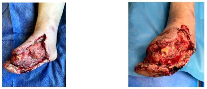
Clinical photographs of a diabetic foot with a large, complex surgical wound following excisional debridement and transmetatarsal amputation. The images show a deep, irregular wound bed spanning the dorsal and medial aspects of the foot. The tissue composition is heterogeneous, featuring areas of beefy red granulation tissue interspersed with patches of yellowish slough and fibrin. Some deep structures, potentially including exposed bone or fascia, are visible. The wound margins are irregular, exhibiting erythema and edema indicative of localized inflammation. Areas of clotted blood and dark, devitalized tissue are present, reflecting the immediate post-debridement state before Vacuum-Assisted Closure (VAC) therapy. The periwound skin shows signs of chronic peripheral arterial disease and venous insufficiency, including discoloration, pallor, and mottled skin distal to the ankle, suggesting compromised regional circulation. This visual illustrates the surgical management of critical limb ischemia and diabetic foot ulcers in an educational context.
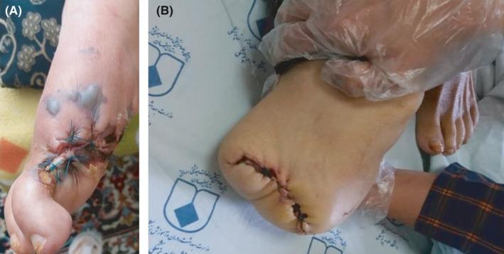
Two-panel clinical photograph demonstrating progressive infectious necrosis and subsequent surgical management in a diabetic patient. Panel A shows a dorsal view of a right foot featuring a grade 3, stage D lesion localized to the hallux (big toe). The area displays extensive tissue necrosis, dark eschar, and surgical sutures indicating a prior intervention. Multiple fluid-filled bullae and purplish-gray ecchymotic patches are visible on the dorsal midfoot, suggesting spreading infection. Panel B displays the same limb following a transmetatarsal or forefoot amputation. The surgical stump is closed with primary sutures, though the incision line appears dark and potentially necrotic. The remaining skin of the foot is pale. A gloved hand is shown stabilizing the amputated limb against a clinical background. These images illustrate the rapid progression of a fungal infection (Sarocladium spp.) requiring escalating surgical debridement and amputation in an immunocompromised, diabetic host.
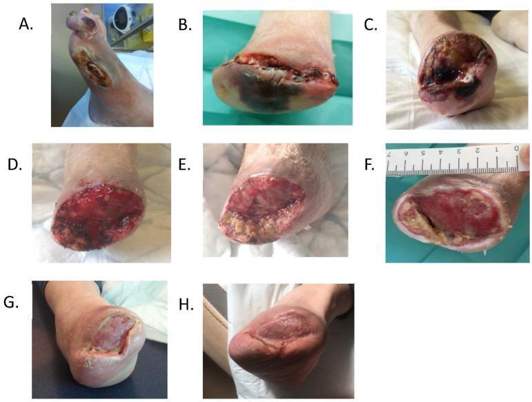
A longitudinal series of eight clinical photographs (A–H) documenting the management and healing progression of a diabetic foot ulcer following various levels of amputation. Image (A) shows initial hallux and first ray necrosis with infection. Subsequent images (B–C) depict the surgical site following transmetatarsal and Lisfranc amputations, characterized by deep, irregular wound beds with fibrin, slough, and dark necrotic tissue. Images (D–E) illustrate the transition to a healthy, beefy-red granulation tissue bed following wound environment control and debridement. Image (F) shows the prepared wound bed prior to skin grafting, with a metric ruler for scale. Final images (G–H) track the outcome of the skin graft over three months, demonstrating progressive contraction and complete epithelialization. The series highlights clinical markers of healing, including the reduction of necrotic slough, the emergence of vascularized granulation tissue, and the final transition to mature, pigmented skin coverage.
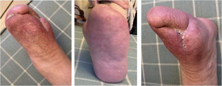
Clinical photograph showing three views (dorsal, plantar, and medial/lateral) of a left foot 18 months after percutaneous Deep Vein Arterialization (pDVA) for chronic limb-threatening ischemia (CLTI). The foot demonstrates successful salvage and complete epithelialization following a transmetatarsal amputation and previous toe debridement. Visually, the skin across all views exhibits a healthy, pinkish-red hue indicative of adequate distal perfusion. The plantar view shows smooth, intact skin without active ulceration. The dorsal and side views reveal a well-healed, linear surgical scar at the amputation site with well-approximated edges and no signs of inflammation, infection, or necrotic tissue. The educational focus of the image is to demonstrate the long-term clinical outcome and wound healing success of pDVA in a diabetic patient with severe calcific occlusive disease, showing the preservation of the foot and resolution of ischemic rest pain.
DIABETIC FOOT ULCER (DFU) - SURGERY NOTES
1. PATHOPHYSIOLOGY (Surgical Relevance)
    NEUROPATHY
        |
    ISCHAEMIA  +  INFECTION  =  TISSUE LOSS  →  AMPUTATION
        |
    BIOMECHANICAL PRESSURE
- Sensorimotor neuropathy: Loss of protective sensation (LOPS), intrinsic muscle wasting → claw toes → abnormal plantar pressure → ulceration
- Autonomic neuropathy: Dry skin, fissures, distended dorsal veins, arteriovenous shunting
- Peripheral arterial disease (PAD): 4x higher in diabetics; impairs wound healing and determines amputation level
- Infection: Most important determinant of outcome; spreads rapidly in subfascial planes due to neuropathy masking pain
2. PRE-OPERATIVE ASSESSMENT
A. History Red Flags
- Prior amputations (predictive of future amputation level)
- Known Charcot neuroarthropathy
- Claudication or rest pain (severe PAD)
- Systemic symptoms (fever, chills = limb-threatening infection)
- HbA1c and glycaemic control
- Nutritional status (do not assume obese = well-nourished)
B. Pre-operative Investigations
| Test | Significance |
|---|---|
| HbA1c | >7% = higher complication risk; >pre-op glucose >200 mg/dL = increased nonunion, infection, wound failure |
| FBC, ESR, CRP | Infection/osteomyelitis baseline |
| Renal function | Affects drug dosing, wound healing |
| ABI / TBI | Determines vascular adequacy for healing (ABI <0.5 = revascularize first) |
| Doppler duplex / CTA / MRA | If revascularization planned |
| X-ray foot (3 views) | Osteomyelitis, Charcot changes, gas in soft tissue, structural deformity |
| MRI foot | Gold standard for osteomyelitis extent; soft tissue planes |
| Bone scan / WBC scan | If MRI contraindicated (metal implants, pacemaker) |
| Deep tissue / bone culture | Before antibiotics; surface swabs are unreliable |
| Nutritional markers | Albumin, pre-albumin, transferrin |
C. Pre-operative Optimization
- Glycaemic control: Target HbA1c <7%; involve endocrinology
- Vascular: Revascularize before elective surgery if ABI <0.5 or toe pressure <40 mmHg
- Nutrition: Dietary supplementation if malnourished before proceeding
- Antibiotics: Based on final culture sensitivities (not empiric) - prescribe at time of definitive debridement
- Tobacco cessation
- Antiplatelet therapy if PAD present (low-dose aspirin)
3. SURGICAL DECISION FRAMEWORK
| Domain | Grade 0 | Grade 1 | Grade 2 | Grade 3 |
|---|---|---|---|---|
| Wound | No ulcer/pre-ulcer | Shallow, no bone | Exposed bone/tendon, heel ulcer | Extensive ulcer/gangrene |
| Ischaemia | Toe pressure ≥60 mmHg | Mild | Moderate | Severe |
| Foot Infection | None | Local <2 cm | >2 cm / abscess / osteomyelitis | SIRS |
- Infected ulcers = 40-55% chance of some form of amputation
- Hindfoot ulcers = 50% progress to BKA or higher
4. SURGICAL OPTIONS - OVERVIEW
SURGICAL LADDER (Limb-Sparing → Amputation)
├── Debridement (sharp, surgical)
├── Abscess drainage
├── Revascularization (endovascular / bypass)
├── Osteomyelitis resection
├── Structural correction (Achilles lengthening, metatarsal head resection)
├── Toe amputation
├── Ray amputation (1st / 5th / central / multiple)
├── Transmetatarsal amputation (TMA)
├── Lisfranc / Chopart amputation
├── Syme amputation
├── Below-knee amputation (BKA)
└── Above-knee amputation (AKA)
5. DEBRIDEMENT
Indications
- All infected or necrotic DFUs
- Non-healing wound despite 4-6 weeks maximal wound care
- Abscess / deep space infection
- Exposed bone with osteomyelitis
Types
| Type | Description |
|---|---|
| Sharp/surgical | Scalpel/curette; first-line; removes all necrotic/infected tissue |
| Mechanical | Wet-to-dry dressings (less specific; painful) |
| Autolytic | Moisture-retentive dressings (slow, for non-infected wounds) |
| Enzymatic | Collagenase (Santyl), MediHoney - for fibrinous tissue |
| Negative Pressure (VAC) | Vacuum-Assisted Closure - see below |
Key Principles
- Debride callus first before wound assessment - callus hides true wound extent
- In infected wounds: send deep tissue samples for microbiology at time of debridement
- Dirty/infected wounds = leave open initially, re-inspect at 48-96 hours
- Serial debridement until clean wound base achieved
- Staged closure once source control confirmed
- In wounds with exposed tendon or bone: use Integra dermal regeneration template then split-thickness skin graft after 3-4 weeks
Pedal Sepsis (Surgical Emergency)
- Signs: spreading cellulitis, purulence, fever, tachycardia, hypotension, altered mental status, gas on imaging
- Management: immediate surgical debridement + drainage regardless of vascular status
- Do not wait for angiography; source control is the priority
6. NEGATIVE PRESSURE WOUND THERAPY (VAC)
- Best suited: midfoot and hindfoot wounds, larger open wounds post-debridement
- Mechanism: removes exudate, reduces oedema, promotes granulation, draws wound edges together
- Less benefit: small forefoot wounds, wounds with severe PAD
- Evidence: heals diabetic foot wounds proximal to transmetatarsal level faster than moist gauze
- Use as bridge to definitive closure or skin grafting
7. REVASCULARIZATION
Indications
- ABI <0.5, toe pressure <40 mmHg
- Non-healing wound despite 4-6 weeks maximal care (even with borderline ABI 60-80 mmHg in diabetics)
- Pre-amputation (to improve healing potential)
Options
| Method | Details |
|---|---|
| Endovascular (PVI) | First-line; angioplasty ± stenting; lower perioperative risk; lower 4-year patency than bypass |
| Open surgical bypass | Single-segment saphenous vein graft preferred; translocated (not reversed) fashion for tibial size-match; arm vein if no saphenous |
| Hyperbaric oxygen | Last resort when revascularization not possible; indications: osteomyelitis, gangrene, open wound with inadequate perfusion |
Timing
- Infected wound: debride first (source control), then angiography
- Subacute/chronic infection: angiography first, then debridement (avoids ischaemia-mediated necrosis)
8. SURGICAL TECHNIQUES - AMPUTATIONS
A. Toe Amputation (Single Toe)
- Fashion long plantar + short dorsal skin flaps
- Dorsal incision at intended bone level, medial to lateral; plantar flap slightly longer than dorsoplantar diameter
- Dissect flaps proximally to level of bone section
- Divide flexor and extensor tendons (let retract)
- Isolate, ligate, and divide digital nerves and vessels
- Section bone at selected level (minimum 1 cm from base of phalanx); smooth end with rasp
- Close flaps with interrupted non-absorbable sutures
- If wound infected: leave open, inspect at 48-96 hours
B. Metatarsophalangeal Joint Disarticulation (Technique 17.4)
- Incision to MTP joint level, extend distally and circumferentially plantarward
- Acute flexion of toe → incise dorsal capsule first → divide flexor tendons and neurovascular bundles
- For hallux: remove sesamoids in insensate foot if metatarsal head is retained
- For 1st/5th digit: longer plantar flap if skin allows
- Cauterize neurovascular bundles
- Close with interrupted non-absorbable sutures
C. Ray Amputation
- 1st / 5th ray (border ray) - Technique 17.5
- Central ray (2nd, 3rd, 4th) - Technique 17.6
- Multiple ray - if >2 rays needed, consider TMA instead
- Base incision medially (1st) or dorsolaterally (5th), extending from medial/lateral eminence proximally and plantarward to mid-metatarsal level
- In diabetic with osteomyelitis: take incision to bone immediately (tissue planes are obscured, thin flaps will not survive)
- Raise dorsal and plantar full-thickness flaps to metatarsal
- Disarticulate digit at MTP joint first, then resect metatarsal shaft
- For 1st metatarsal: section from proximal-plantar-medial to distal-dorsal-lateral; preserve penetrating branch of dorsalis pedis if disarticulating at medial cuneiform
- For 5th ray: preserve base of 5th metatarsal to maintain peroneus brevis attachment; if entire 5th metatarsal removed, re-attach brevis or tenodese to peroneus longus
- Close in single layer non-absorbable suture; leave open if wound not surgically clean
- For 2nd ray resection: consider Lapidus procedure to prevent valgus deformity
- For 3rd + 4th ray removal: osteotomy of 5th metatarsal base facilitates closure
- In deep central space abscess with necrotic intrinsic muscles: debride necrotic tissue + excise affected rays
- In compromised vascularity or incomplete debridement: retention sutures only (loose closure) + allow secondary intention healing; negative pressure wound therapy facilitates healing
D. Transmetatarsal Amputation (TMA)
- Deep infection, chronic ulceration, or gangrene involving multiple digits and/or dorsal skin of forefoot
- Multiple ray involvement
- Poor vascularity (check pedal arch patency)
- Deep infection tracking proximal to forefoot
- Patient unable/unwilling to accept revision surgery if needed
- No prosthesis required - shoe filler + steel shank + rocker sole
- Preserves limb length; maintains reasonable gait
- Achilles tendon lengthening is almost always required simultaneously (prevent plantarflexion contracture from shortened lever arm; also reduces terminal pressure during walking)
- Do Achilles lengthening first to prevent contamination and facilitate dorsiflexion correction
- Patency of pedal arch (dorsal-plantar connection) is key to healing
- Fashion long plantar + short dorsal full-thickness flaps
- Dorsal incision: from anteromedial foot, curve slightly distal, end at midpoint of lateral foot
- Plantar incision: carry distally beyond metatarsal heads, curve back to lateral midpoint; slightly longer medially
- Plantar flap includes: subcutaneous fat + layer of plantar muscles
- Remove toes at MTP joints
- Section metatarsals bevelled dorsal-distal to plantar-proximal at junction of middle and distal thirds
- CRITICAL: Preserve essentially all plantar skin proximal to MTP joints
- Close - or if dirty: leave open + staged closure
E. Syme Amputation (Through-Ankle)
- Requires: Intact heel pad (essential for weight bearing)
- Preserves more limb length than BKA
- Walking possible with modified shoe or simple prosthesis
F. Below-Knee Amputation (BKA / Transtibial)
- Expends 25% more energy in walking compared to intact limb
- Indicated when forefoot/midfoot salvage not possible
- Nearly 30% of DFU amputations progress to more proximal level
G. Above-Knee Amputation (AKA / Transfemoral)
- When BKA unlikely to heal or further proximal disease
- Highest functional impairment; prosthesis fitting mandatory
9. OSTEOMYELITIS - SURGICAL MANAGEMENT
- Probe-to-bone positive = assume osteomyelitis; treat surgically
- Surgical resection of infected bone is standard (not antibiotics alone for most cases)
- Send tissue samples for microbiology at time of surgery
- Antibiotic duration post-resection:
- Simple toe amputation: 2 weeks oral antibiotics
- Metatarsal resection (ray/partial ray/more proximal): 4-6 weeks even with disease-free margins (residual infection risk)
- Bone biopsy required for histology and culture in complex/extensive infection
10. WOUND CLOSURE OPTIONS
| Closure Type | Indication |
|---|---|
| Primary closure | Clean, tension-free wound |
| Delayed primary closure | Infected wound - close at 48-96 hours after open inspection |
| Secondary intention | Compromised vascularity, incomplete debridement |
| Negative pressure (VAC) | Open wounds; bridge to closure |
| Integra dermal template | Exposed tendon or bone; insufficient skin for grafting |
| Split-thickness skin graft (STSG) | After 3-4 weeks on Integra (when silicone layer removed) |
| Local flaps | Advancement, rotation, transposition, pedicle flaps for limited coverage |
| Free flap (vascularized) | Extensive coverage needs; consult plastics early |
11. ADVANCED / ADJUNCT SURGICAL PROCEDURES
| Procedure | Purpose |
|---|---|
| Achilles tendon lengthening | Reduces forefoot/midfoot pressure; essential with TMA; threshold low for midfoot/forefoot ulcers |
| Metatarsal head resection | Corrects deformity; reduces recurrence; transfers pressure points |
| Gastrocnemius recession | Alternative to Achilles lengthening for equinus deformity |
| Lapidus procedure | After 2nd ray resection to prevent valgus deformity |
| Exostectomy | Remove bony prominences (Charcot foot) causing pressure |
| Charcot stabilization | Internal/external fixation for unstable rocker-bottom deformity |
| Liquid silicone injection | Plantar pressure reduction (adjunct) |
12. POST-OPERATIVE CARE
Wound Care
- Wound dressing choice based on wound characteristics:
- Healthy wound: collagen dressing
- Fibrinous tissue: Santyl (collagenase) or MediHoney
- Deep healthy wound: VAC dressing
- Locally infected: Iodosorb or Bactroban
- Macerated: alginate impregnated with silver / absorptive foam
- Peri-wound maceration: Calmoseptine to wound edges
Offloading (post-op and chronic management)
- Total contact cast (TCC): gold standard; extends above ankle; reduces plantar pressure up to 60%; non-removable; requires weekly cast changes; needs uninfected wound
- Fixed ankle walking boot / removable walker boot: for wounds needing frequent dressing changes
- Wedge shoe / heel-out shoe / DH walking boot: alternative offloading options
- Custom orthotics and therapeutic footwear: once healed, to prevent recurrence
Oedema Management
- Oedema severely impairs wound healing
- Multilayer compression: mainstay; prevents fluid exudation from wound bed
- Address underlying cause: venous hypertension, lymphoedema, fluid overload
Glycaemic Monitoring
- HbA1c every 3 months
- Target HbA1c <7%
- Tight glucose control can reverse neuropathy with prolonged good control
Systemic Medical Management
- ACE inhibitor: for hypertension control
- Statin: for hyperlipidaemia
- Antiplatelet (aspirin): if PAD present
- Tobacco cessation: address at every visit
13. COMPLICATIONS OF DFU SURGERY
| Complication | Notes |
|---|---|
| Wound dehiscence / failure | Most common; higher risk with HbA1c >7%, glucose >200, malnutrition |
| Proximal amputation revision | 30% of initial amputations progress to higher level |
| Osteomyelitis recurrence | Residual infection leads to further amputation |
| Plantarflexion contracture | After TMA if Achilles not lengthened |
| Transfer ulceration | After ray resection; pressure transfers to adjacent areas |
| Phantom limb pain | After amputation |
| Haematoma | Avoid tourniquet use; meticulous haemostasis |
| Skin edge necrosis | Avoid forceps pressure on skin; use thick flaps |
| Stump infection | Common in diabetics; culture-guided antibiotics |
14. MULTIDISCIPLINARY TEAM (MDT) - STANDARD OF CARE
| Specialist | Role |
|---|---|
| Vascular surgeon | Revascularization, amputation level |
| Orthopaedic / Podiatric surgeon | Debridement, amputations, deformity correction |
| Plastic surgeon | Flap coverage, skin grafting |
| Endocrinologist | Glycaemic optimization |
| Infectious disease | Antibiotic selection, osteomyelitis management |
| Nutritionist / Dietitian | Nutritional supplementation pre/post-op |
| Orthotist / Prosthetist | Offloading, footwear, post-amputation prosthetics |
| Wound care nurse | Dressing management, monitoring |
15. KEY SURGICAL RULES (Summary)
- Never aggressively debride without adequate perfusion - unless severe, life-threatening infection is present
- Revascularize before elective surgery if ABI <0.5 or toe pressure <40 mmHg
- Achilles tendon lengthening with any TMA - almost always required
- Preserve plantar skin - it cannot be replaced; key for all forefoot amputations
- Leave infected wounds open - staged closure at 48-96 hours
- Avoid tourniquet in ischaemic limbs
- Avoid thin flaps in diabetics - especially with osteomyelitis; take incision to bone
- Thick flaps, no forceps pressure on skin edges
- Culture before antibiotics - culture at time of debridement, not empirically from surface swab
- Glycaemic control is non-negotiable - glucose >200 mg/dL pre-op markedly increases complications
- Campbell's Operative Orthopaedics 15th Ed 2026, Ch. 17 (Amputation) and Ch. 91 (Diabetic Foot)
- Current Surgical Therapy 14e (Multidisciplinary Management of DFU; WIfI Classification)
- Fischer's Mastery of Surgery 8th Ed (Diabetic Foot Ulcer: Diagnosis, Management, Summary)
- Bailey and Love's Short Practice of Surgery 28th Ed (Diabetic Gangrene; Diabetic Foot Infection)
- Sabiston Textbook of Surgery 21st Ed (Negative Pressure Wound Therapy in DFU)