
Temporomendibular joint short notes
Reading File
Now let me search the medical library for TMJ content in parallel.
Finding Sources
Finding Sources
Excellent - found rich sources. Let me now read the most relevant blocks in parallel.
Reading File
Reading File
Reading File
Now let me fetch the detailed TMJ anatomy and clinical disorder sections.
Reading File
Reading File
Reading File
I have rich, comprehensive content now. Let me also fetch the anatomical diagram image and quickly check the imaging anatomy block.
Loading Image
Finding Sources
Reading File
Reading File
I now have comprehensive content from multiple authoritative textbooks. Let me compile the full short notes.
Temporomandibular Joint (TMJ) - Short Notes
1. Definition and Type
The temporomandibular joint (TMJ), also called the craniomandibular joint, is a synovial joint formed by the articulation of the mandibular condyle with the mandibular (glenoid) fossa of the temporal bone, just anterior to the external auditory canal.
2. Unique Features (Distinguishes TMJ from Other Synovial Joints)
| Feature | Detail |
|---|---|
| Intra-articular disc | Divides joint space into upper and lower cavities |
| Articular surfaces | Covered by fibrous tissue (not hyaline cartilage) - reflects intramembranous development |
| Secondary condylar cartilage | Present beneath fibrous surface until ~16 years of age |
| Tooth dependency | Movements are influenced by the occlusal relationships of the teeth |
Scott-Brown's Otorhinolaryngology Head & Neck Surgery
3. Anatomy
Bony Components
- Mandibular condyle - articular surfaces are anterior and superior
- Neck of condyle - thin bony projection; common site for mandibular fracture
- Pterygoid fovea - small depression on anterior neck; attachment of lateral pterygoid muscle
- Mandibular (glenoid) fossa + articular eminence of temporal bone
Movements
- Upper compartment: Gliding/translatory movements
- Lower compartment: Hinge movements
- Normal mouth opening: 35-55 mm (inter-incisal distance); <25 mm in a young adult = abnormal; >55 mm = hypermobility
4. Articular Disc (Meniscus)
- Dense fibrous consistency, avascular centrally (blood vessels only at periphery)
- Upper surface: concavo-convex (anteroposteriorly); lower surface: concave
- Shape provides a self-centering mechanism maintaining alignment with the condyle during movement
- Functions: spreads joint forces, stabilizes the condyle
Zones of the disc:
- Central (intermediate) zone: thinnest, crimped collagen fibres
- Anterior: fibrous bands connect to articular eminence (above) and condyle (below)
- Medial/lateral margins: attached to capsule and poles of condyle
- Posterior: attached via retrodiscal tissue (bilaminar zone):
- Superior lamina - loose, vascular, elastic fibres; attached to squamotympanic fissure
- Inferior lamina - relatively avascular, less extensible; attached to posterior condyle
The retrodiscal tissue increases 4-5x in volume via venous engorgement as the jaw opens (condyle moves anteriorly). Elastic recoil of the superior lamella helps return the disc to its resting position.
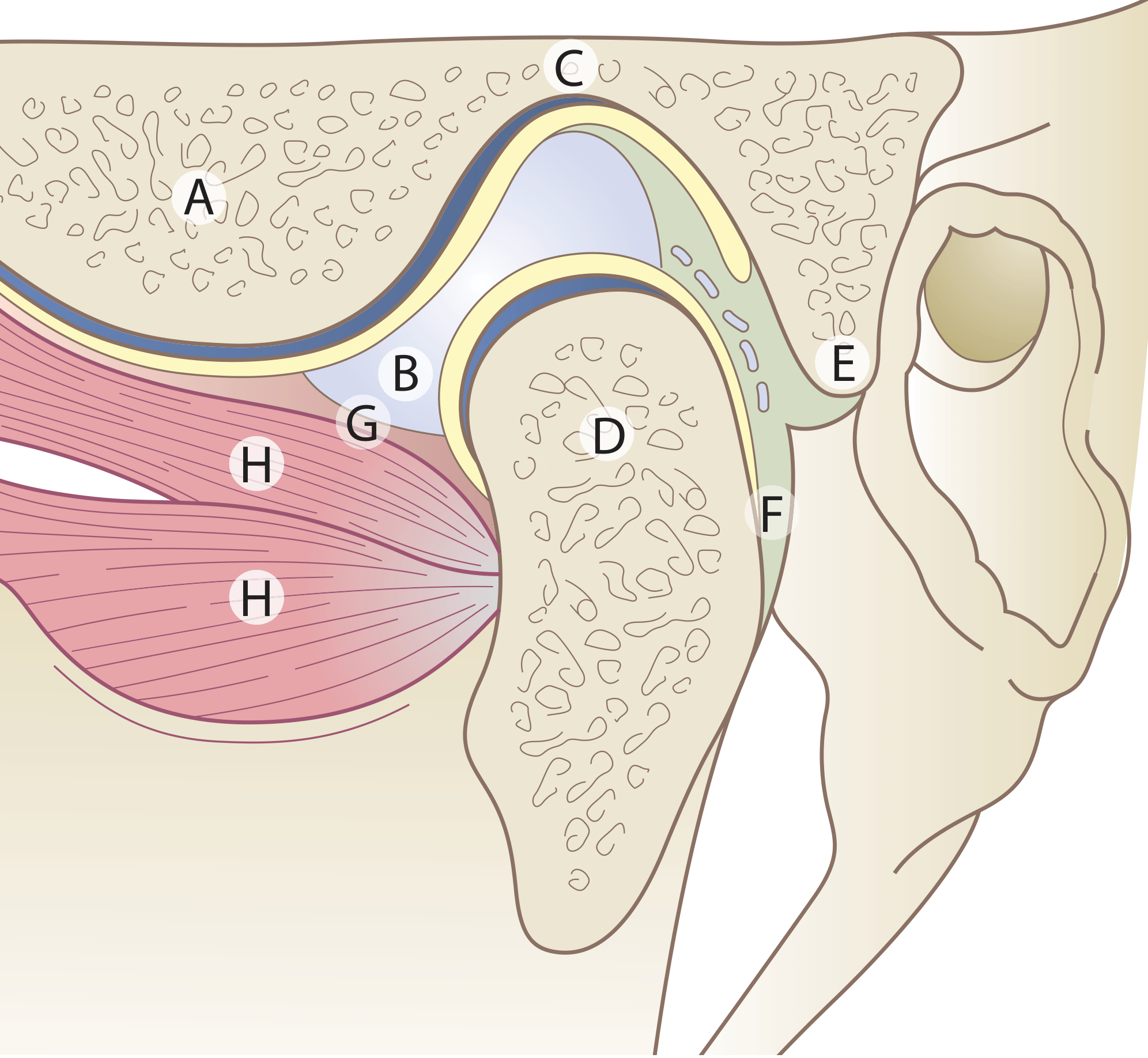
Figure: TMJ showing articular disc and lateral pterygoid attachments. A = articular eminence; B = articular disc; C = mandibular fossa; D = condyle; E = upper lamina (fibro-elastic); F = lower lamina (non-elastic); G = capsule; H = lateral pterygoid muscle.
5. Joint Capsule
- Attached above to mandibular fossa (anteriorly to just in front of the articular eminence crest; posteriorly to squamotympanic and petrotympanic fissures)
- Attached below to the neck of the condyle
- Thin, slack - does not limit mandibular movements
- Inner surface lined by synovial membrane (does not cover articular surfaces)
- Synovial fluid contains proteoglycans; pressure is subatmospheric at rest, rises greatly during mastication
6. Ligaments
| Ligament | Attachments | Function |
|---|---|---|
| Lateral (temporomandibular) ligament | Outer surface of articular eminence → neck of condyle (posteroinferiorly) | Main support; limits posterior and inferior condylar displacement |
| Sphenomandibular ligament | Spine of sphenoid → lingula of mandible | Remnant of perichondrium of Meckel's cartilage (1st branchial arch); becomes tense as condyle passes in front of lateral ligament |
| Stylomandibular ligament | Styloid process → posterior angle of mandible | Thickening of deep cervical fascia; limits excessive protrusion |
| Retinacular ligament (recently described) | Articular eminence → fascia over masseter | May maintain blood circulation during mastication |
7. Muscles of Mastication (Acting on TMJ)
| Muscle | Action |
|---|---|
| Masseter | Elevation (closing) |
| Temporalis | Elevation + retraction |
| Medial pterygoid | Elevation |
| Lateral pterygoid | Depression, protrusion, lateral excursion; upper head attaches to articular disc |
8. Nerve Supply
Branches of the mandibular nerve (V3):
- Auriculotemporal nerve
- Masseteric nerve
- Deep temporal branches
- Proprioceptive endings are critical for reflex control of mastication
9. Blood Supply
- Superficial temporal artery
- Maxillary artery (anterior tympanic and deep auricular branches)
- Also: deep temporal and transverse facial arteries
10. Clinical Disorders
A. TMJ Disorders (TMD) - Overview
- Multifactorial origin; common symptoms: jaw/facial pain, clicking, limited mouth opening
- Affects a considerable proportion of the population
B. Internal Derangement
1. Anterior Disc Displacement With Reduction (ADDR)
- Disc displaced anteromedially when jaw is closed
- On opening, condyle slips back under disc = reciprocal click (opening + closing click)
- Normal range of motion usually maintained
- Asymptomatic cases are common, often need no treatment
- If painful: soft diet, NSAIDs, splint therapy, physiotherapy
- Refractory cases: arthrocentesis, arthroscopy, or open surgery
2. Anterior Disc Displacement Without Reduction (Closed Lock)
- Disc remains anteriorly displaced, acts as a physical barrier to full condylar translation
- Presents with limited mouth opening (typically <35 mm), deviation to affected side
- No click (disc never reduces)
- Management: manual manipulation, arthrocentesis, arthroscopy
Cummings Otolaryngology Head and Neck Surgery
C. Degenerative Joint Disease (Osteoarthritis)
- Damage to articular surfaces → increased friction → disc degeneration
- Progressive disc displacement, eventual disc perforation
- Degenerative changes → crepitus (bone-on-bone contact)
- Synovial fluid shows increased proinflammatory cytokines
D. Inflammatory Arthritis (RA, JIA)
- Both juvenile and adult RA can affect TMJ
- Micrognathia may develop in children (arrested mandibular growth)
- Examination: palpate just anterior to external auditory canal; measure inter-incisal distance (normal 3-6 cm)
Firestein & Kelley's Textbook of Rheumatology
E. TMJ Syndrome (Myofascial Pain Dysfunction, MPD)
- Related to bruxism (tooth clenching/grinding)
- Thought to be a form of myofascial pain, similar to fibromyalgia
- Pain is muscular in origin, not intracapsular - not a surgical candidate
F. Joint Noises
| Sound | Cause | Treatment |
|---|---|---|
| Click | Disc displacement with reduction | Reassurance (no treatment if asymptomatic) |
| Crepitus | Scarring, degeneration | Reassurance if no symptoms; rule out arthritis |
G. Ankylosis
- Absolute indication for TMJ surgery
- Fibrous or bony fusion of joint; severe restriction of mouth opening
11. Surgical Management
Absolute indications for surgery: neoplasms, growth abnormalities, ankylosis
Relative indications: pain and dysfunction refractory to nonsurgical therapy + imaging evidence of pathology
| Procedure | Details | Success Rate |
|---|---|---|
| Arthrocentesis | Two 18-gauge needles into superior joint space; lysis + lavage with Ringer lactate; least invasive; office-based under IV sedation | 70-90% |
| Arthroscopy | Surgical telescope into superior joint space; can visualize pathology; done under GA | Similar to arthrocentesis |
| Open joint surgery | For severe cases; condylotomy, discectomy, disc repair, total joint replacement | Reserved for refractory cases |
Post-arthrocentesis care: non-chew diet, range-of-motion exercises, analgesics.
Cummings Otolaryngology Head and Neck Surgery
12. Imaging
| Modality | Use |
|---|---|
| OPG (Orthopantomogram) | Screening; bony condylar changes |
| CT scan | Bony anatomy, condylar erosion, fractures |
| MRI | Gold standard for disc position, disc morphology, soft tissue pathology |
| Arthrography | Historically used; largely replaced by MRI |
13. Key Points Summary
- TMJ is a bicondylar, synovial joint with an intra-articular disc dividing it into two compartments
- Articular surfaces are fibrocartilage (not hyaline), unlike other synovial joints
- The lateral pterygoid is the only muscle that opens the jaw (depresses mandible); all others close it
- Anterior disc displacement is the most common internal derangement; clicking = with reduction; closed lock = without reduction
- Normal inter-incisal opening = 35-55 mm; <25 mm in a young adult needs urgent referral
- Surgical intervention is reserved for cases refractory to conservative treatment; arthrocentesis is first-line surgical option
Sources: Scott-Brown's Otorhinolaryngology Head & Neck Surgery | Cummings Otolaryngology Head and Neck Surgery | Firestein & Kelley's Textbook of Rheumatology
Opening of diaphram short notes
Reading File
Your credits are exhausted. You can purchase a top-up or subscribe to a plan to continue using Orris. If you're already subscribed, your daily credits will reset tomorrow.
This is a shared conversation. Sign in to Orris to start your own chat.