
/teachme brachial plexus block
Reading File
Now let me search the library for brachial plexus block content in parallel:
Finding Sources
Finding Sources
Excellent sources found. Let me now read the key sections from Miller's Anesthesia and Morgan & Mikhail in parallel:
Reading File
Reading File
Good content found. Let me now read more of Miller's section on the specific approaches and Morgan & Mikhail's detailed technique sections:
Reading File
Reading File
Excellent content. Now let me read the specific approaches (supraclavicular, infraclavicular, axillary) from Miller's and the Morgan sections on technique:
Reading File
Reading File
Great content. Let me get the infraclavicular and axillary sections plus complications from Miller's, and the axillary/complications sections from Morgan:
Reading File
Reading File
I now have rich content from both Miller's Anesthesia (10e) and Morgan & Mikhail's (7e). Let me fetch the key anatomy diagram and one ultrasound image to include in the answer:
Loading Image
Loading Image
I now have all the material needed for a comprehensive teach-me session. Let me compile the full answer:
Brachial Plexus Block - Complete Teaching Guide
Sources: Miller's Anesthesia 10e, Morgan & Mikhail's Clinical Anesthesiology 7e
1. Anatomy of the Brachial Plexus
The brachial plexus arises from ventral rami of C5-T1. It is organized into a memorable sequence:
Roots → Trunks → Divisions → Cords → Branches ("Randy Travis Drinks Cold Beer")
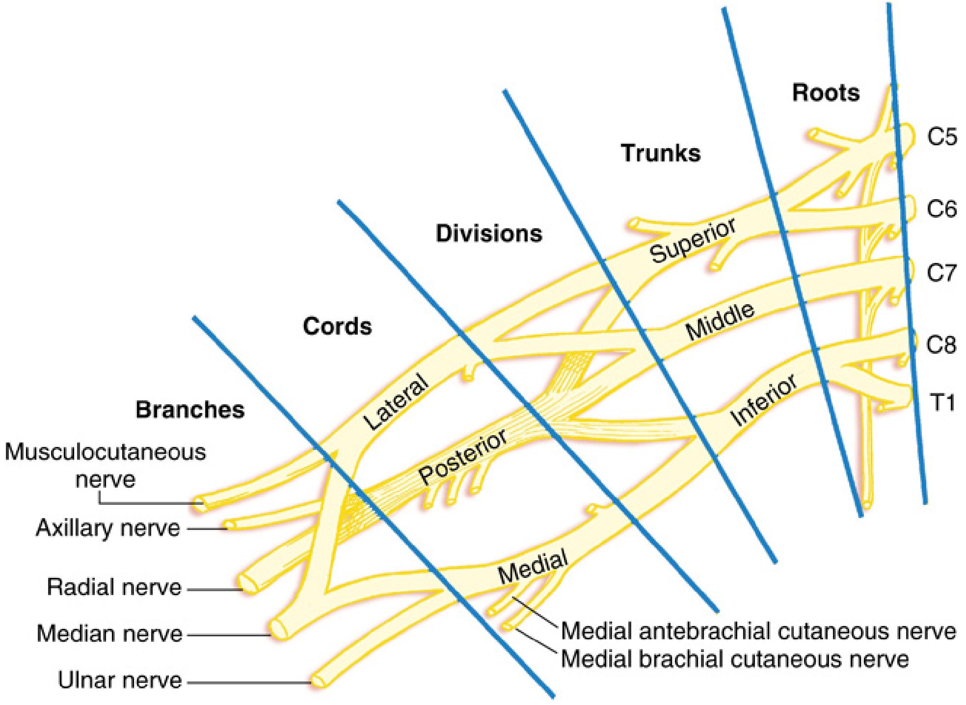
| Level | Components | Location |
|---|---|---|
| Roots | C5, C6, C7, C8, T1 | Between anterior & middle scalene muscles |
| Trunks | Superior (C5+C6), Middle (C7), Inferior (C8+T1) | Interscalene space → cephaloposterior to subclavian artery |
| Divisions | Anterior + posterior (from each trunk, 6 total) | Posterior to midportion of clavicle |
| Cords | Lateral, Posterior, Medial | Named relative to 2nd part of axillary artery |
| Branches | Terminal nerves | Lateral border of pectoralis minor |
Terminal nerve origins:
- Lateral cord → musculocutaneous nerve + lateral head of median nerve
- Medial cord → ulnar nerve + medial head of median nerve + medial antebrachial/brachial cutaneous nerves
- Posterior cord → axillary nerve + radial nerve
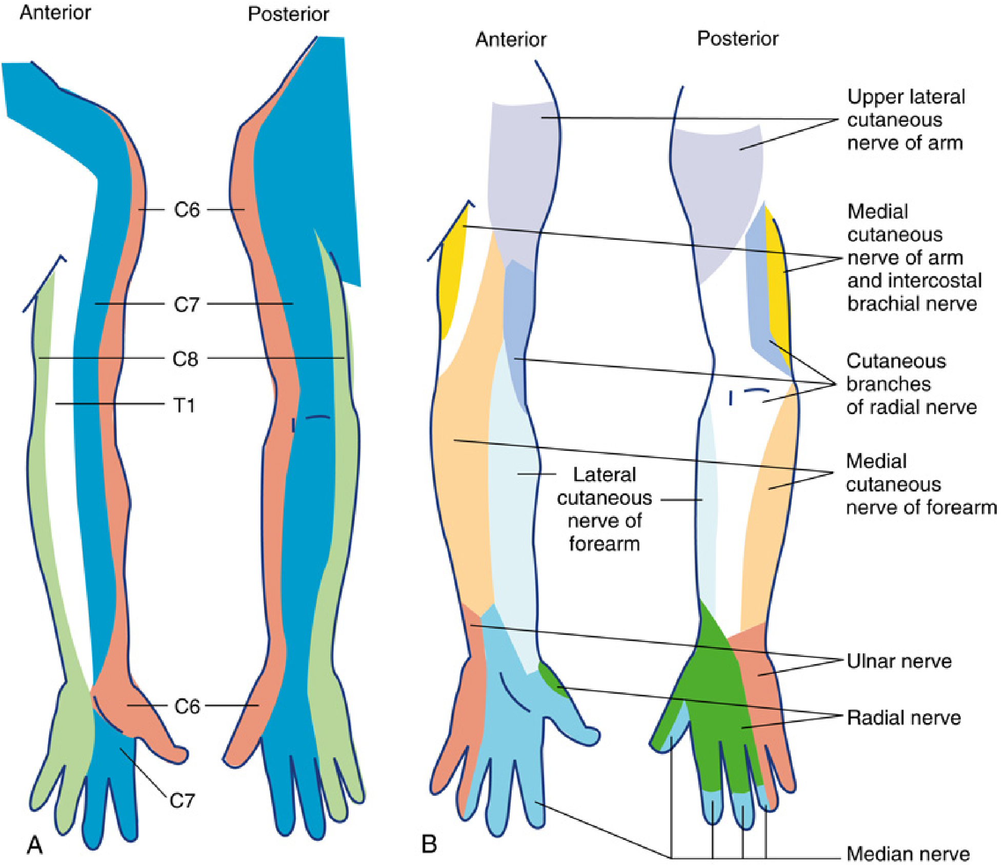
Cutaneous distribution:
2. The Four Main Approaches
The approach chosen depends on the surgical site. As you go more distal on the plexus, shoulder coverage is lost but ulnar coverage improves.
| Approach | Level Blocked | Best Indications | Notable Coverage Gap |
|---|---|---|---|
| Interscalene | Roots/upper & middle trunks | Shoulder, proximal humerus | C8-T1 (ulnar nerve) often spared |
| Supraclavicular | Distal trunk / proximal division | Elbow, forearm, hand | Suprascapular nerve (unreliable) |
| Infraclavicular | Cords | Elbow, forearm, hand (complete) | None - most complete coverage |
| Axillary | Terminal nerves | Hand, wrist, forearm | Musculocutaneous + intercostobrachial nerves spared |
3. Interscalene Block (ISB)
Indication
Shoulder surgery and proximal humerus procedures in patients without significant pulmonary disease.
Anatomy target
Superior and middle trunks, lying between the anterior scalene (AS) and middle scalene (MS) muscles - the classic "stoplight" sign on ultrasound (3 hypoechoic circles).
US-Guided Technique
- Patient supine, head turned away from operative side
- High-frequency linear probe placed at supraclavicular fossa, then traced cranially until trunks are visible between scalene muscles
- Needle advanced in-plane or out-of-plane (out-of-plane avoids the long thoracic and dorsal scapular nerves within the middle scalene belly)
- Volume: 10-15 mL; as low as 5 mL associated with decreased diaphragmatic paresis
- For surgical anesthesia: 20-30 mL commonly used
Complications (memorize these - high-yield)
| Complication | Frequency | Notes |
|---|---|---|
| Phrenic nerve block (hemidiaphragmatic paralysis) | ~100% | Reduces ipsilateral lung function ~25%; absolute contraindication with contralateral phrenic palsy or severe COPD |
| Horner's syndrome | Common | Stellate ganglion spread |
| Recurrent laryngeal nerve palsy | Common | Transient hoarseness |
| Vertebral artery injection | Rare but serious | |
| Subarachnoid/epidural injection | Rare | Needle directed medially |
Key rule: Bilateral interscalene blocks are always contraindicated. Phrenic nerve blockade is virtually inevitable with ISB.
4. Supraclavicular Block
Indication
Operations on the elbow, forearm, and hand. Historically called the "spinal of the arm" - rapid onset, dense reliable blockade because the plexus is most compact here (distal trunk / proximal division level).
Ultrasound Technique
- Patient supine, head turned 30° to contralateral side
- Linear high-frequency transducer in supraclavicular fossa, angled slightly toward thorax
- Identify subclavian artery in short-axis; plexus appears as a "cluster of grapes" cephalolateral to the artery, above the first rib
- Needle advanced in-plane from lateral to medial; avoid directing medially (pneumothorax risk)
- LA deposited around the plexus cluster; carefully trace cranially and caudally to ensure lower trunk is included (prevents ulnar sparing)
Complications
- Pneumothorax - most feared (risk reduced but not eliminated with ultrasound)
- Phrenic nerve palsy - ~50% of patients; apply same precautions as ISB
- Horner's syndrome
- Subclavian artery puncture
Note: Supraclavicular perineural catheters provide inferior analgesia compared with infraclavicular catheters and are more prone to displacement with movement.
5. Infraclavicular Block
Indication
Complete brachial plexus anesthesia for elbow, forearm, and hand. Best site for perineural catheter placement (stable, minimal displacement).
Anatomy target
The three cords (medial, lateral, posterior) in relation to the 2nd part of the axillary artery, deep to pectoralis major and minor.
Ultrasound Technique (Lateral Sagittal / Paracoracoid approach)
- Arm abducted to 90° (externallyrotates humeral head, retracts clavicle, straightens neurovascular bundle)
- Probe placed below and medial to coracoid process in sagittal orientation
- Axillary artery visible in short-axis, deep to pec major and minor
- Target: posterior to the axillary artery - 30-40 mL single injection produces complete block without needing to visualize all three cords
- Costoclavicular approach alternative: cords clustered lateral to 1st part of axillary artery - faster onset but higher pneumothorax risk
Complications
- Deeper block = decreased needle tip visibility (steep needle angles required)
- Pneumothorax (lower risk than supraclavicular)
- No phrenic nerve block - advantage over interscalene and supraclavicular
6. Axillary Block
Indication
Procedures on the hand, wrist, and forearm. Safest approach (no risk of pneumothorax or phrenic nerve palsy).
Critical anatomy - nerves ALREADY diverged at this level:
- Musculocutaneous nerve has already branched off (found between coracobrachialis heads or between coracobrachialis and biceps - must be separately blocked)
- Intercostobrachial nerve (T2) is always spared - causes tourniquet discomfort
- Musculocutaneous nerve = lateral antebrachial cutaneous nerve distally; must be blocked for lateral forearm coverage
Ultrasound Technique
- Patient supine, arm abducted 90° or hand placed behind head
- High-frequency linear probe in axilla - identify axillary artery and vein in cross-section
- Median (anteromedial), ulnar (posteromedial), and radial (posterolateral) nerves surround the artery
- Multiple injection technique - each nerve must be targeted individually (fascial separations exist between them)
- 5-10 mL around each nerve; musculocutaneous nerve blocked separately in coracobrachialis muscle
Key Limitations
- Axilla is a poor site for catheters - inferior analgesia vs. infraclavicular, increased infection and dislodgement risk
- Upper arm and shoulder not covered
- Requires more needle passes than other approaches
7. Lung-Sparing (Phrenic-Sparing) Alternatives for Shoulder
When shoulder surgery is needed in patients with pulmonary disease, newer targeted approaches include:
- Superior trunk block - targets the superior trunk (C5+C6) just proximal to the suprascapular nerve branch; noninferior pain scores vs. ISB with less hemidiaphragmatic paralysis
- Anterior suprascapular nerve block - LA deposited deep to omohyoid, shallow to prevertebral fascia; 5-15 mL; volumes >15 mL risk phrenic spread
8. Local Anesthetics Commonly Used
| Agent | Concentration | Onset | Duration |
|---|---|---|---|
| Ropivacaine | 0.5-0.75% | Moderate | Long (12-18h) |
| Bupivacaine | 0.25-0.5% | Slow-moderate | Long (12-18h) |
| Lidocaine | 1.5-2% | Fast | Intermediate (4-8h) |
| Mepivacaine | 1.5% | Moderate | Intermediate (4-8h) |
Epinephrine 1:200,000-1:400,000 may be added to prolong duration and serve as an intravascular injection marker.
9. General Complications (All Approaches)
| Complication | Prevention |
|---|---|
| Intravascular injection / LAST | Aspiration before injection; inject in increments; use epinephrine marker |
| Nerve injury | Avoid intraneural injection; stop if high resistance or pain on injection |
| Infection | Strict asepsis; monitor catheters |
| Hematoma | Respect anticoagulation guidelines (ASRA) |
Persistent neuropathic symptoms are more common after brachial plexus blocks than lower extremity blocks. Mechanisms include local ischemia, vasoconstrictors, neurotoxicity, or direct trauma.
10. Quick Reference: "Which Block for Which Surgery?"
| Surgical Site | Preferred Approach |
|---|---|
| Shoulder / rotator cuff | Interscalene (or superior trunk block if lung-sparing needed) |
| Proximal humerus / shoulder | Interscalene |
| Elbow (distal humerus / forearm) | Supraclavicular or Infraclavicular |
| Forearm / wrist / hand | Infraclavicular (best completeness + catheter) or Axillary |
| Hand only | Axillary (simple, safe) |
| Tourniquet analgesia | Intercostobrachial (T2) block supplement for any approach |
Key High-Yield Points to Remember
- The brachial plexus lies in a fascial sheath derived from prevertebral fascia - this is why a single injection at the right level can block the entire plexus
- Phrenic nerve palsy is inevitable with ISB and occurs in ~50% with supraclavicular block; it is absent with infraclavicular and axillary blocks
- The inferior trunk (C8-T1 / ulnar distribution) is the most commonly missed segment - check coverage explicitly
- Supraclavicular = most compact point of plexus = fastest onset, densest block
- Infraclavicular = best for catheters; axillary = safest (no pleural or phrenic risk)
- The intercostobrachial nerve (T2) is never blocked by any brachial plexus approach - it must be separately blocked for tourniquet tolerance
- Miller's Anesthesia 10e, Chapter 42 - Regional Anesthesia of the Upper Extremity
- Morgan & Mikhail's Clinical Anesthesiology 7e, Chapter 46 - Peripheral Nerve Blocks
/doubtkiller adductor canal block
Reading File
Finding Sources
Finding Sources
Reading File
Reading File
Reading File
Reading File
Reading File
Loading Image
I now have everything needed. Here is the focused /doubtkiller answer on adductor canal block:
Adductor Canal Block (ACB) - Doubt Killer
Sources: Miller's Anesthesia 10e, Morgan & Mikhail's Clinical Anesthesiology 7e, Barash Clinical Anesthesia 9e
The Core Concept in One Line
The ACB is a motor-sparing, purely sensory block that targets the saphenous nerve (and often the nerve to vastus medialis) inside the adductor canal of the mid-thigh, providing knee and medial leg analgesia without the quadriceps weakness caused by a femoral nerve block.
1. Anatomy - What is the Adductor Canal?
The adductor canal (Hunter's canal) is a triangular fascial tunnel in the anteromedial mid-thigh, bounded by:
| Wall | Structure |
|---|---|
| Roof / Anteromedial | Sartorius muscle |
| Anterolateral | Vastus medialis |
| Posterior / Floor | Adductor longus + magnus |
The vastoadductor membrane forms the roof of the deep canal between the vastus medialis and adductor muscles - this is the key landmark on ultrasound (seen as a double contour).
Contents of the adductor canal:
- Superficial femoral artery (SFA) - main landmark
- Superficial femoral vein (SFV)
- Saphenous nerve - lies anterior/anterolateral to the SFA
- Nerve to vastus medialis - lies outside the canal in its own fascial sheath (clinically important - see below)
- Posterior division of the obturator nerve (inconsistently)
2. The Big Clinical Question: ACB vs. Femoral Nerve Block (FNB)
This is the most tested comparison - know it cold.
| Feature | Femoral Nerve Block | Adductor Canal Block |
|---|---|---|
| Level blocked | Femoral nerve (mixed motor + sensory) | Saphenous nerve + nerve to VM (sensory dominant) |
| Quadriceps weakness | Up to 80% of patients | Minimal - motor sparing |
| Fall risk | High | Much lower |
| Analgesic quality for TKA | Good | Similar to FNB |
| Post-op ambulation (TKA) | Limited by weakness | Better day-1 ambulation |
| Coverage | Broader (anterior thigh + knee) | Knee + medial leg only |
Key fact (Morgan & Mikhail): Patients with continuous adductor canal catheters ambulate further on day 1 following TKA than patients with either femoral block (limited by weakness) or no block (limited by pain).
3. The "True ACB" Controversy - High-Yield Exam Trap
There is active debate about the correct injection site. Here's the crux:
- "True" adductor canal block - identified by sartorius medial border converging with adductor longus medial border; deeper in the canal
- Problem: The nerve to the vastus medialis (major contributor to knee pain after TKA) often lies outside the true adductor canal in its own fascial sheath - a block too far distal misses it
- Recommended technique (Jaeger et al.): Periarterial injection lateral to the femoral artery, deep to the sartorius, deep to the vastoadductor membrane, at the midpoint between the ASIS and patella - this position captures both the saphenous nerve AND the nerve to vastus medialis
- "High" ACB = injection under the sartorius lateral to the SFA, at the level where vastus medialis and saphenous nerves co-exist in the same fascial plane; essentially a transarterial perifemoral injection
4. Ultrasound-Guided Technique
Positioning: Supine, thigh in slight external rotation, knee extended - exposes inner thigh.
Probe: High-frequency linear transducer, transverse orientation at anteromedial mid-thigh, starting at the junction of middle and distal thirds.
What you see:
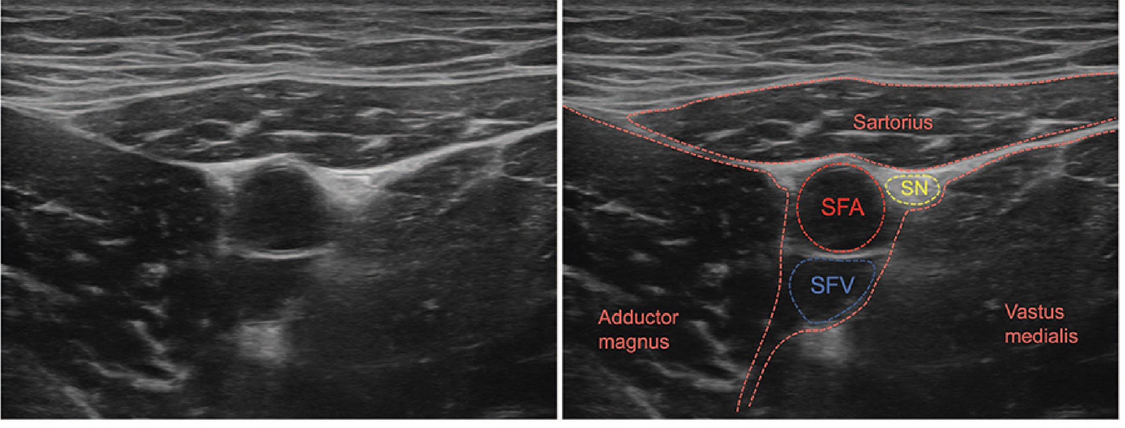
- SFA = large pulsatile round structure (compressible with pressure = vein; non-compressible = artery)
- SFV = adjacent, compressible
- Saphenous nerve (SN) = small hypoechoic oval, anterolateral to SFA
- Sartorius = superficial, roof of canal
- Vastoadductor membrane = echogenic double contour defining canal floor
Needle technique:
- In-plane approach, needle 2-3 cm lateral to probe, advanced anteromedially
- Target: deep to sartorius, anterior and lateral to the artery
- Inject after negative aspiration (highly vascular area)
- Volume: 10-15 mL (Miller's) / 15-20 mL (Morgan & Mikhail)
Warning: Higher volumes risk spread to the femoral triangle proximally → quadriceps weakness (defeats the purpose of the motor-sparing block).
5. Indications - Where ACB Fits
| Surgery | Role of ACB |
|---|---|
| Total knee arthroplasty (TKA) | Primary analgesic block (preferred over FNB for motor-sparing) |
| ACL reconstruction | Preferred block (often combined with sciatic or IPACK) |
| Knee arthroscopy | First-line |
| Bunionectomy | Combined with sciatic nerve block |
| Medial ankle / leg surgery | Saphenous nerve coverage |
| Patellar ORIF / tendon repair | FNB preferred (knee immobilizer used anyway) |
ACB does NOT cover:
- Posterior knee (add IPACK block for this)
- Lateral leg (peroneal territory)
- Tourniquet discomfort (needs femoral or sciatic coverage)
- Hip or thigh surgery
6. IPACK Block - The Perfect ACB Complement
For TKA, ACB alone misses the posterior knee capsule (innervated by tibial and common peroneal branches). The IPACK block (Interspace between the Popliteal Artery and the posterior Capsule of the Knee) fills this gap without motor block.
ACB + IPACK = complete knee analgesia without quadriceps weakness - this is the modern standard for TKA.
7. Complications
| Complication | Notes |
|---|---|
| Vascular injury / pseudoaneurysm | Rare; frequent aspiration mandatory (SFA is immediately adjacent) |
| Myonecrosis | Intramuscular LA spread → severe myonecrosis reported; stay in fascial plane |
| Unexpected thigh weakness | Prompts investigation for intramuscular spread or proximal spread to femoral nerve |
| Fall risk | Still exists despite motor-sparing reputation; patient education on unsupported ambulation is mandatory |
| Failure rate | Pre-ultrasound era: 30%+ failure; ultrasound dramatically reduces this |
8. Continuous ACB Catheter
- Catheter at the adductor canal provides superior postoperative analgesia for TKA
- Technically stable, catheter well-secured under sartorius muscle
- Allows ambulation-friendly analgesia for early physiotherapy
- Preferred over continuous femoral catheter when ambulation is a rehabilitation priority
Quick Memory Card
ACB = Saphenous nerve block (+ nerve to VM)
      ↓ at mid-thigh, under sartorius, lateral to SFA
      ↓ 10-15 mL, in-plane US technique
      ↓ Motor sparing → safe ambulation
      ↓ For TKA/ACL/knee arthroscopy
      ↓ Add IPACK for posterior knee coverage
      ↓ Falls still possible → patient education mandatory
- Miller's Anesthesia 10e, pp. 6270-6272 (Chapter 42)
- Morgan & Mikhail's Clinical Anesthesiology 7e, pp. 1926-1927 (Chapter 46)
- Barash Clinical Anesthesia 9e, Chapter 36 & 45
This is a shared conversation. Sign in to Orris to start your own chat.