
Cornea and retina difference
The cornea is the clear, dome-shaped front surface of the eye that focuses incoming light and protects the inner eye. The retina is the light-sensitive tissue lining the back of the eye that converts light into nerve signals sent to the brain. They serve completely different roles - cornea is structural/refractive, retina is sensory/neural.
Searching sources...
Reading File
Finding Sources
Finding Sources
Reading File
Reading File
Let me pull the right sections - the anatomy/structure sections from Kanski's Ophthalmology and Junqueira's Histology.
Reading File
Reading File
Finding Sources
Finding Sources
Reading File
Reading File
Reading File
I now have excellent textbook content. Let me fetch the key diagrams for a rich visual answer.
Loading Image
Loading Image
Loading Image
Here is a thorough, textbook-sourced comparison of the cornea and retina.
Cornea vs. Retina
The diagram above (from Guyton & Hall's Medical Physiology) shows both structures perfectly: light enters through the cornea at the front, travels through the eye, and is captured by the retina at the back.
At a Glance
| Feature | Cornea | Retina |
|---|---|---|
| Location | Anterior (front) of eye | Posterior (back) of eye, innermost tunic |
| Type of tissue | Avascular connective tissue + epithelium | Neural tissue (CNS-derived) |
| Primary function | Refraction + protection | Phototransduction (converts light to nerve signals) |
| Transparency | Transparent (required for light passage) | Not optically transparent; semi-transparent neural tissue |
| Blood supply | None (avascular) | Supplied by central retinal artery + choroid |
| Nerve supply | Richly innervated (pain/touch) | No pain fibers; contains neurons |
| Layers | 5 layers | 10 layers |
| Equivalent to | A "window" and "lens" | A "film" or "screen" |
The Cornea in Detail
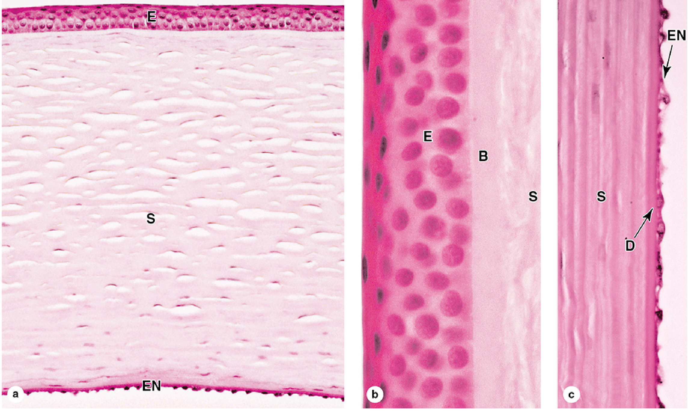
Cornea histology: (a) full cross-section showing epithelium (E), stroma (S), endothelium (EN); (b) anterior layers - epithelium (E), Bowman's membrane (B), stroma (S); (c) posterior layers - stroma (S), Descemet membrane (D), endothelium (EN)
The cornea is the most anterior structure of the eye. It has 5 layers:
- Anterior epithelium - nonkeratinized stratified squamous, 5-6 cells thick. Densely packed with free nerve endings that trigger the blink reflex. Protects the stroma from physical damage and prevents fluid evaporation.
- Bowman's membrane - thick basement membrane (8-10 µm). Adds structural stability and protection against infection.
- Stroma (substantia propria) - makes up ~90% of corneal thickness. Consists of ~60 layers of parallel type I collagen bundles arranged at right angles to each other. This precise orthogonal array is what gives the cornea its transparency. Keratocytes (fibroblast-like cells) sit between collagen lamellae. Completely avascular - nutrients arrive by diffusion from the limbus and aqueous humor.
- Descemet membrane - thick posterior basement membrane produced by the endothelium.
- Endothelium - simple squamous (internally); the most metabolically active cells of the cornea. Na+/K+-ATPase pumps maintain stromal deturgescence (correct hydration). If this pump fails, the stroma swells and becomes opaque - this is the basis of corneal dystrophies.
Key function: The cornea provides about two-thirds of the eye's total refractive power (around +43 D), more than the lens. Transparency depends on stromal deturgescence; any disruption to hydration causes opacification and vision loss. - Junqueira's Basic Histology, p. 1177-1178; Medical Physiology, p. 539
The Retina in Detail
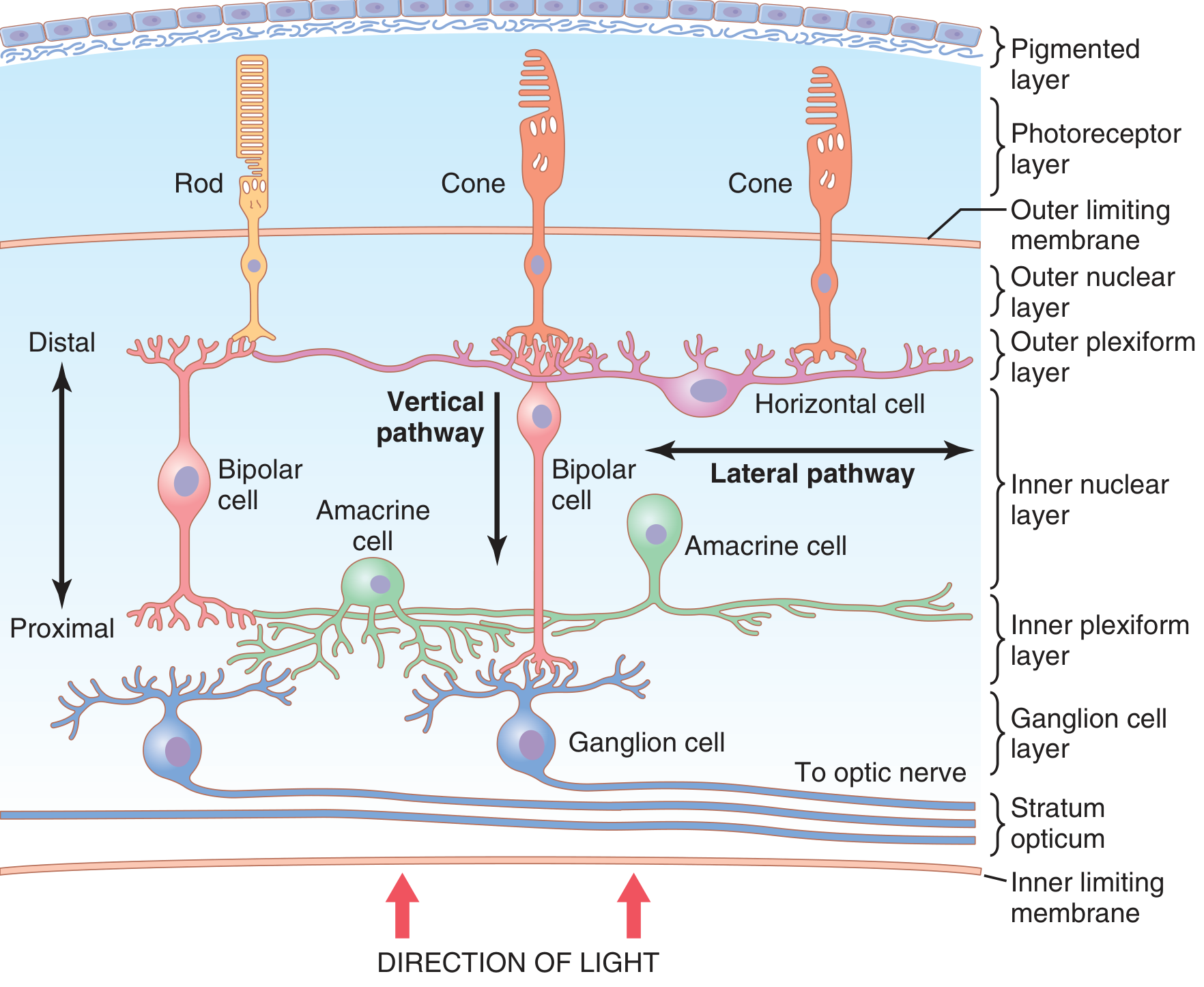
Retinal layers: light enters from below (inner surface), passes through ganglion and bipolar cell layers, and reaches photoreceptors (rods and cones) at the outer surface
The retina lines the back of the eye and is embryologically derived from the optic cup - it is a direct extension of the brain. It has 10 layers:
From innermost (vitreous side) to outermost (choroid side):
- Inner limiting membrane
- Stratum opticum (nerve fiber layer - ganglion cell axons forming optic nerve)
- Ganglion cell layer
- Inner plexiform layer
- Inner nuclear layer (bipolar, horizontal, amacrine cells)
- Outer plexiform layer
- Outer nuclear layer (photoreceptor cell bodies)
- Outer limiting membrane
- Photoreceptor layer (rods and cones outer segments)
- Retinal pigment epithelium (RPE)
Key cells:
- Rods (~120 million): peripheral vision, night vision (scotopic), extremely sensitive to light, contain rhodopsin
- Cones (~6 million): concentrated at the fovea, color vision and fine detail (photopic), contain red/green/blue opsins
- Bipolar cells: relay signals from photoreceptors to ganglion cells
- Ganglion cells: generate the sole output of the retina via the optic nerve (CN II)
- RPE: absorbs scattered light, forms the blood-retina barrier, recycles vitamin A (11-cis-retinal), and phagocytoses shed photoreceptor tips
Fovea: a 0.3 mm central pit composed almost entirely of cones, with all other layers displaced to the sides so light hits photoreceptors directly. Responsible for the sharpest vision. - Guyton & Hall Medical Physiology, p. 632; Junqueira's Basic Histology, p. 1197
Note on retinal detachment: The RPE and neural retina are not firmly joined - they can separate (retinal detachment). When separated, photoreceptors lose metabolic support from the RPE and die. Prompt laser reattachment is essential.
Summary of Key Differences
| Cornea | Retina | |
|---|---|---|
| Origin | Neural crest + surface ectoderm | Neuroectoderm (optic cup = brain) |
| Role | Refractive medium, barrier | Sensory transduction, initial visual processing |
| Layers | 5 | 10 |
| Vascularity | Avascular | Vascular (central retinal artery) |
| Cells | Keratinocytes, keratocytes, endothelial cells | Photoreceptors, bipolar, ganglion, Müller, RPE |
| Disease examples | Keratitis, corneal dystrophy, keratoconus, LASIK candidates | Retinal detachment, diabetic retinopathy, macular degeneration, retinitis pigmentosa |
| Transplantability | Easily transplanted (no vessels = low rejection risk) | Cannot be transplanted; degeneration is permanent |
The cornea is the eye's outer "window" - a structural refractive barrier. The retina is the eye's inner "film" - a neural sensory processor. Both are required for vision, but they fail in completely different ways and are treated by very different specialties (corneal surgeons vs. vitreoretinal surgeons).
This is a shared conversation. Sign in to Orris to start your own chat.