
Nocardia vs actinomycetes
I'll look this up in the medical library for an authoritative comparison.
Reading File
Now let me search the library for both organisms in parallel.
Finding Sources
Finding Sources
Excellent sources found. Let me read the most relevant sections in parallel.
Reading File
Reading File
Reading File
Great data. Let me read more of the Actinomyces/Nocardia section from Sherris and the Nocardia section from Medical Microbiology to get clinical details, plus fetch key images.
Reading File
Reading File
Loading Image
I now have comprehensive data to write a detailed comparison. Let me also fetch the Nocardia Gram stain image and the cervicofacial actinomycosis image.
Loading Image
Reading File
I now have all the information needed for a thorough comparison. Here is the full, structured answer:
Nocardia vs. Actinomyces - A Complete Comparison
Both are Gram-positive, filamentous, branching rods that were historically mistaken for fungi. The single most testable distinction: Nocardia is aerobic and weakly acid-fast; Actinomyces is anaerobic and NOT acid-fast.
Key Features at a Glance
| Feature | Nocardia | Actinomyces |
|---|---|---|
| Kingdom | Bacteria (true) | Bacteria (true) |
| Gram stain | Gram-positive (often stains poorly, beaded) | Gram-positive |
| Acid-fast | Weakly acid-fast (1% H₂SO₄ decolorizer) | NOT acid-fast |
| Oxygen requirement | Strict aerobe | Anaerobe / microaerophile |
| Morphology | Branching filamentous rods; aerial hyphae | Branching filamentous rods; no aerial hyphae |
| Cell wall | Mycolic acids (C50-62; shorter than mycobacteria) | No mycolic acids |
| Source / Habitat | Exogenous - soil (rich organic matter) | Endogenous - normal oral/GI/genital flora |
| Infection route | Inhalation or traumatic inoculation | Mucosal disruption (trauma, surgery, IUD) |
| Host | Primarily immunocompromised | Immunocompetent (low virulence) |
| Sulfur granules | Absent | Present (pathognomonic) |
| Key species | N. asteroides, N. brasiliensis | A. israelii (most common) |
| Culture speed | 3-7+ days; aerobic media | 4-10 days; anaerobic media |
| Growth on non-selective media | Yes (if incubated long enough) | Requires anaerobic conditions |
Microbiology Details
Nocardia
- Gram-positive, strictly aerobic branching rods with mycolic acids in the cell wall (C50-62 carbons - shorter chain than mycobacteria, explaining why it is only weakly acid-fast vs. strongly acid-fast in TB).
- Virulence depends on avoiding intracellular killing: catalase and superoxide dismutase neutralize the oxidative burst; a cord factor prevents phagosome-lysosome fusion.
- Colonies initially white, can become dry/waxy or orange; aerial hyphae visible on dissecting microscopy. The combination of aerial hyphae + acid-fastness is unique to Nocardia.
-
100 species identified by gene sequencing; most infections caused by a few (N. asteroides complex, N. brasiliensis).
Actinomyces
- Gram-positive, anaerobic/microaerophilic branching rods. No mycolic acids, no acid-fastness.
- Colonize the alimentary tract (oral, GI, female genital tract) - never found on normal skin.
- Disease occurs only when mucosal barriers are disrupted (dental procedure, surgery, aspiration, IUD).
- Low virulence - disease is endogenous, with no person-to-person spread.
- The hallmark histological finding is sulfur granules - macroscopic colonies that look like grains of sand, with a dense Gram-positive center and filamentous rods at the periphery.
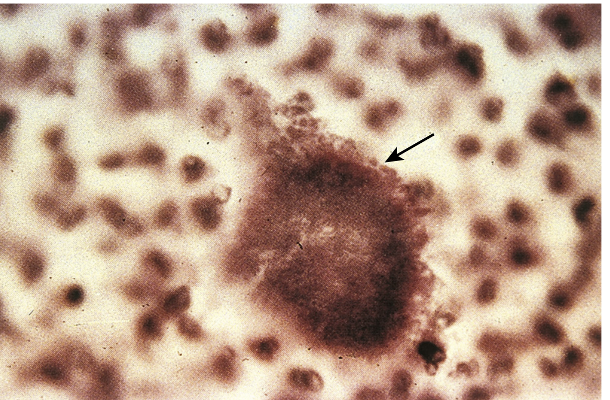
Sulfur granule from actinomycosis sinus tract - Medical Microbiology 9e, Fig. 31.3
Clinical Diseases
Nocardia - diseases
- Bronchopulmonary (most common): cavitary pneumonia, lung abscess, indolent course. Mimics TB or fungal infection on imaging.
- Cutaneous: mycetoma (chronic destructive disease of extremities), lymphocutaneous spread, cellulitis, subcutaneous abscess.
- CNS (disseminated): brain abscesses - occurs in 30% of pulmonary cases.
- Immunocompromised hosts: transplant recipients (tacrolimus/steroids), HIV, malignancy, corticosteroid use.
Actinomyces - diseases ("actinomycosis")
Follows a slow, burrowing course with induration, abscess, and draining sinus tracts opening through the skin.
- Cervicofacial ("lumpy jaw", most common, ~50%): follows dental trauma or extraction. Firm, indurated jaw mass with draining sinuses.
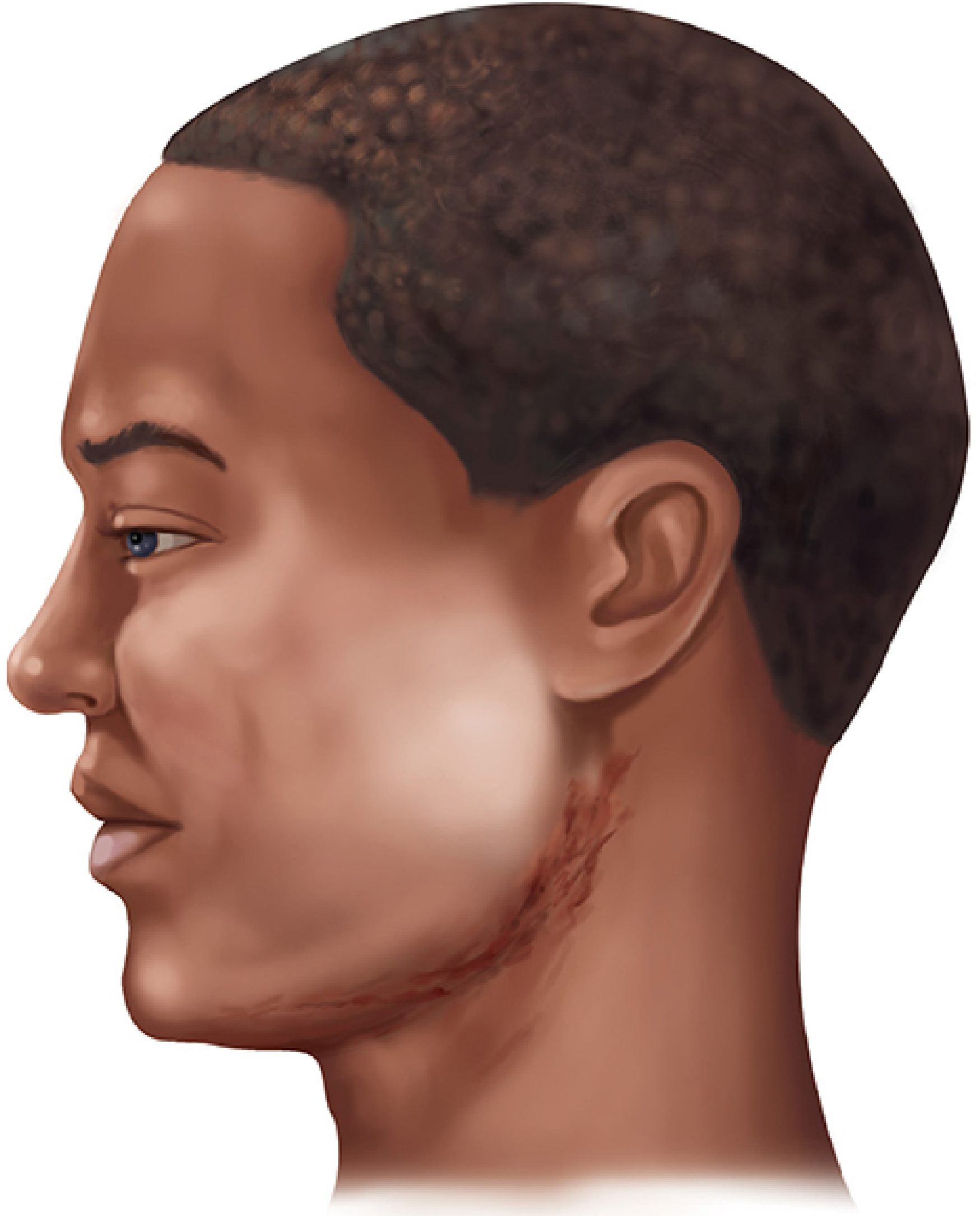
Cervicofacial actinomycosis - Sherris & Ryan's Medical Microbiology 8e, Fig. 28-4
- Thoracic: follows aspiration; erodes through pleura and chest wall; often mistaken for malignancy.
- Abdominal: follows bowel surgery or trauma; erosion through abdominal wall.
- Pelvic: associated with IUD use (chronic endometritis).
- CNS: rare.
Diagnosis
| Step | Nocardia | Actinomyces |
|---|---|---|
| Stain | Weakly acid-fast (1% H₂SO₄); beaded Gram-positive rods | Gram-positive branching rods; NOT acid-fast |
| Specimen | Sputum, BAL, pus aspirate, biopsy | Pus (look for sulfur granules), tissue biopsy |
| Key finding | Branching + weakly acid-fast = genus-level ID | Sulfur granules on direct smear or H&E biopsy |
| Culture | Aerobic; BCYE agar useful; 3-7+ days | Anaerobic; 7+ days; anaerobic blood agar |
| Species ID | Gene sequencing or mass spectrometry | Biochemical tests; genus-level usually sufficient |
| Pitfall | Slow growth - lab must be notified to hold cultures | Sinus drainage heavily contaminated with other organisms; organisms few in pus |
Treatment
| Nocardia | Actinomyces | |
|---|---|---|
| Drug of choice | TMP-SMX (trimethoprim-sulfamethoxazole) | Penicillin G (high dose, prolonged) |
| Severe/CNS disease | TMP-SMX + imipenem or cephalosporin | Penicillin IV, then amoxicillin PO |
| Cutaneous (immunocompetent) | TMP-SMX alone | Penicillin |
| Pulmonary (severe/immunocompromised) | TMP-SMX + amikacin | Penicillin |
| Duration | Up to 12 months | Weeks to 12 months (depending on extent) |
| Surgery | Drainage of abscesses as needed | Drainage of abscesses + excision of sinus tracts |
The Single Most Important Mnemonic
"NAA vs. AAA"
- Nocardia = Aerobic, Acid-fast (weak), Acquired from soil (exogenous)
- Actinomyces = Anaerobic, Acid-fast negative, Alimentary tract flora (endogenous)
Sources: Sherris & Ryan's Medical Microbiology 8e, p. 1121-1128; Medical Microbiology 9e, pp. 363-366, 269-274; The Washington Manual of Medical Therapeutics.
This is a shared conversation. Sign in to Orris to start your own chat.