
Filariasis wiki
Reading File
Now let me search the medical library and web in parallel for comprehensive filariasis coverage.
Finding Sources
Searching the Web
filariasis overview pathophysiology clinical features treatment 2024
Now let me read the key textbook sections in parallel:
Reading File
Reading File
Reading File
Let me read more of the microbiology section and the Robbins pathogenesis content:
Reading File
Reading File
Now let me also fetch the elephantiasis image and get the urology section for chyluria and genital involvement:
Reading File
Finding Sources
Let me get Park's Textbook content (preventive medicine) and the dermatology section:
Reading File
Reading File
Let me read the remaining dermatology content (treatment and diagnosis) and fetch the elephantiasis image:
Reading File
Loading Image
I now have comprehensive information from multiple authoritative textbooks. Let me compose the full wiki-style response.
Filariasis
Filariasis is a group of parasitic diseases caused by thread-like filarial nematodes (superfamily Filarioidea) transmitted to humans by mosquito vectors. The most clinically significant form is lymphatic filariasis, which infects ~120 million people across ~50 tropical and subtropical countries in Africa, South and Southeast Asia, Latin America, and the Pacific Islands.
Causative Organisms
| Species | Distribution | Vector |
|---|---|---|
| Wuchereria bancrofti | Worldwide tropics (90% of cases) | Culex, Anopheles, Aedes spp. |
| Brugia malayi | South and East Asia (rural, coastal) | Mansonia, Aedes spp. |
| Brugia timori | Eastern Indonesian archipelago | Anopheles barbirostris |
Key difference: Brugian filariasis rarely involves the genitalia, unlike Bancroftian filariasis. - Park's Textbook of Preventive and Social Medicine
Life Cycle
- An infected mosquito releases infective L3 larvae into the skin during a blood meal.
- Larvae migrate into lymphatic channels and develop into adult worms (male and female) over several months (incubation 2-18 months).
- Adult worms mate within lymphatics or lymph nodes and release sheathed microfilariae (Mf) into the bloodstream.
- Mosquitoes ingest Mf during a blood meal; Mf develop into infective larvae within the mosquito over several days.
- The cycle repeats.
W. bancrofti microfilariae show nocturnal periodicity - they peak in peripheral blood during nighttime hours, coinciding with the feeding habits of Culex vectors. - Sherris & Ryan's Medical Microbiology
Pathogenesis
Worm-mediated immune evasion
The genomes of W. bancrofti and B. malayi encode multiple molecules that facilitate immune evasion:
- Elastases and trypsin-like proteases - facilitate tissue invasion
- Surface glycoproteins with antioxidant function - protect against reactive oxygen species
- Cystatin homologues (cysteine protease inhibitors) - impair MHC class II antigen processing
- Serpins (serine protease inhibitors) - inhibit neutrophil proteases
- TGF-β and macrophage migration inhibition factor homologues - dampen host immune responses
Wolbachia endosymbionts
Filarial worms harbor obligate intracellular Wolbachia bacteria that are required for worm development, embryogenesis, and survival. Wolbachia also stimulates host innate and adaptive immune responses, including expression of vascular endothelial growth factors (VEGFs), contributing to lymphatic dilation and pathology. - Robbins Pathologic Basis of Disease
Lymphatic damage
- Acute phase: Presence of molting adolescent worms and dying adults causes lymphatic dilation, endothelial hyperplasia, infiltration by lymphocytes, plasma cells and eosinophils, and thrombus formation (acute lymphangitis), followed by granuloma formation and fibrosis.
- Chronic phase: Repeated infections lead to massive lymphatic blockade. Skin and subcutaneous tissues become edematous, thickened, and fibrotic. Dilated lymphatics may rupture, spilling lymph into tissues or body cavities.
- Bacterial and fungal superinfections frequently supervene and worsen tissue damage.
T-cell immunology
- Th1 responses drive granuloma formation around adult worms - major driver of lymphatic damage in chronic disease.
- Th2 responses (IL-4, IL-5) stimulate IgE and eosinophilia - associated with tropical pulmonary eosinophilia.
Clinical Spectrum
Stage 1 - Asymptomatic amicrofilaraemia
No detectable microfilariae and no clinical signs, despite exposure to infective larvae. Cannot be distinguished from true non-infection by current tests.
Stage 2 - Asymptomatic microfilaraemia
Blood is Mf-positive but the patient is completely asymptomatic. Important reservoir in the community; detected only by nocturnal blood examination.
Stage 3 - Acute manifestations (filarial fever)
Recurrent episodes occurring 8-12 months after exposure:
- Filarial fever - typically low-grade; occasionally up to 40°C with chills and myalgia
- Lymphadenitis - classically begins in the femoral region as a red, tender, enlarged lymph node, then spreads centrifugally down the lymphatic channels of the leg
- Lymphangitis - lymphatic vessels become enlarged and tender; overlying skin is warm, red, and edematous
- Urticaria and eosinophilia - elevated IgE levels
- In Bancroftian filariasis: lymphatics of the testicle, epididymis, and spermatic cord are frequently involved, causing painful orchitis, epididymitis, and funiculitis; inflamed retroperitoneal vessels may simulate an acute abdomen
- Acute episodes last a few days, resolve spontaneously, and recur periodically over weeks to months
Stage 4 - Chronic obstructive lesions (10-15 years after first acute attack)
Permanent lymphatic obstruction leads to:
- Lymphedema - affects legs (most common), arms, scrotum, penis, vulva, breasts (in that order of decreasing frequency)
- Elephantiasis - tough subcutaneous fibrosis with epithelial hyperkeratosis; skin shows dilated dermal lymphatics, widespread lymphocytic infiltrates, and focal cholesterol deposits; epidermis thickened and hyperkeratotic
- Hydrocele - fluid contains cholesterol crystals, red cells, and hemosiderin; causes thickening and calcification of the tunica vaginalis
- Chyluria - rupture of dilated lymphatics into the urinary tract, producing milky/white urine; usually intermittent and may spontaneously remit
Occult filariasis - Tropical Pulmonary Eosinophilia (TPE)
A hypersensitivity response to microfilariae in the lungs (Th2-mediated), NOT classic lymphatic disease:
- Paroxysmal nocturnal cough and wheeze
- Marked peripheral eosinophilia (>3000/µL)
- Elevated IgE
- Histology: dead microfilariae surrounded by stellate eosinophilic precipitates within small epithelioid granulomas (Meyers-Kouwenaar bodies)
- Responds well to DEC
Clinical Image
Filariasis of the scrotum and penis (advanced elephantiasis):
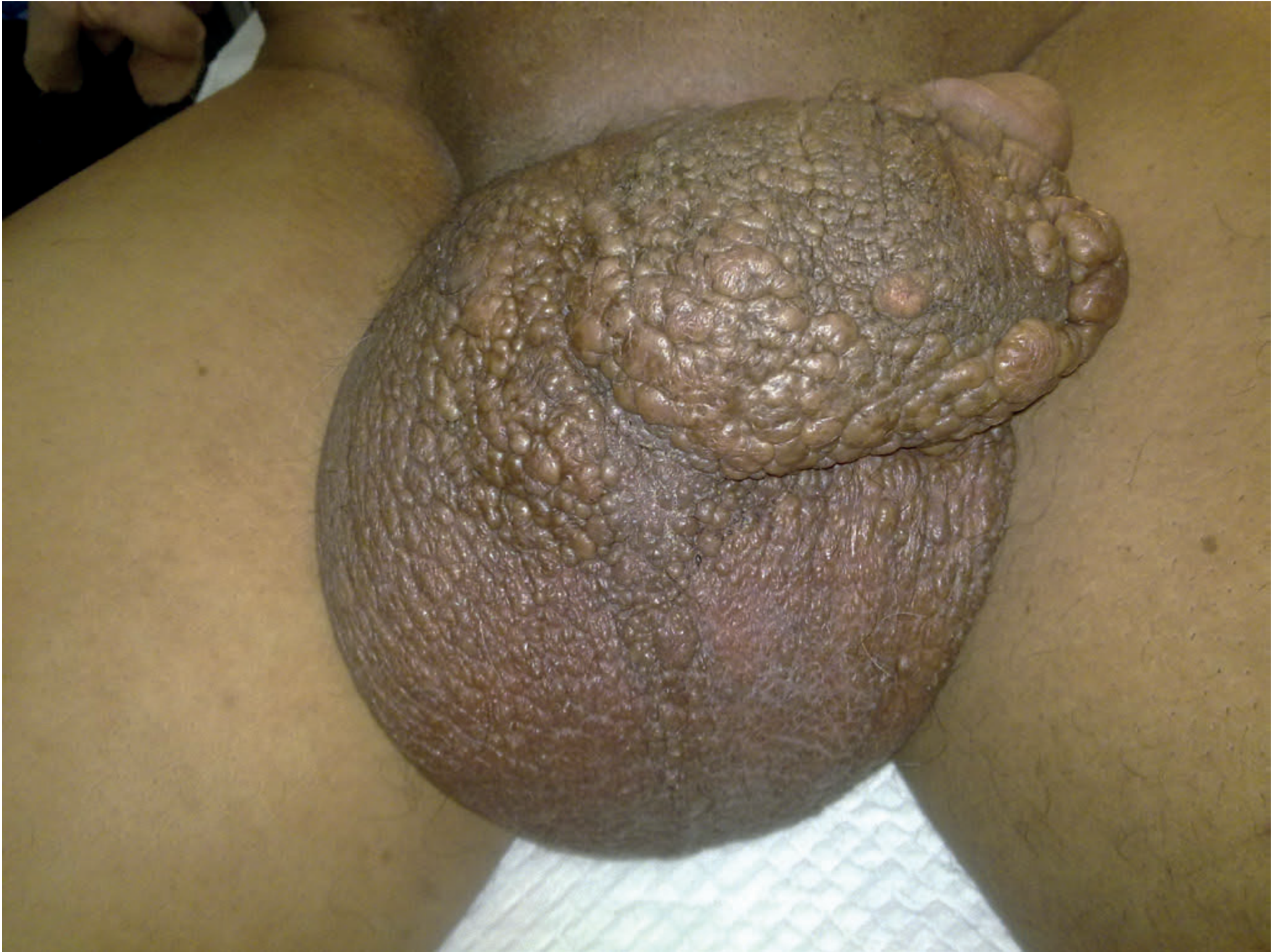
Filariasis of the scrotum and penis - Bailey and Love's Short Practice of Surgery, 28th ed.
Diagnosis
Blood smear (gold standard)
- Thick and thin blood smears stained with Giemsa or Wright stain
- Must be collected at the correct time - for W. bancrofti (nocturnal periodicity), collect blood between 10 PM and 2 AM
- If parasitemia is below detection threshold, concentrate the sample (Knott's concentration technique, membrane filtration)
- DEC provocation test: DEC stimulates migration of Mf from pulmonary to systemic circulation, increasing detection yield
Antigen detection
- Circulating filarial antigen (CFA) test - detects W. bancrofti antigen (Og4C3 ELISA or immunochromatographic card test); present in most microfilaraemic patients and some non-microfilaraemic infected individuals - useful for active infection
- Brugia Rapid test - antibody-based, for Brugian filariasis
Serology
- IgG4 ELISA - most specific serologic test; cross-reactivity with other tissue parasites is well described; limited value in endemic populations (prior exposure confounds results)
Ultrasound
- "Filarial dance sign" - characteristic movement of adult worms visible on scrotal ultrasound (W. bancrofti commonly found in intrascrotal lymphatic vessels)
- Filarial lymphadenopathy detectable in children (inguinal, crural, axillary nodes)
Eosinophilia
- Present during acute inflammatory episodes
- Marked eosinophilia (>3000/µL) in TPE
Treatment
Diethylcarbamazine (DEC) - Drug of choice
- Both microfilaricidal AND active against adult worms
- Results in long-term suppression or parasitologic cure in some cases
- Dying microfilariae trigger an allergic (Mazzotti-like) reaction - may require antihistamines and corticosteroids
- Important: Must exclude onchocerciasis before giving DEC in endemic areas - coinfection causes severe skin and eye reactions
- Used in mass drug administration (MDA) programs; sometimes added to cooking salt in highly endemic areas
Ivermectin
- Kills microfilariae only, NOT adult worms
- Single dose temporarily clears microfilaraemia
- Used in combination regimens (DEC + albendazole, or ivermectin + albendazole in areas co-endemic with onchocerciasis)
Albendazole
- Has beneficial effects on both microfilariae and adult worms
- Used in combination with DEC or ivermectin for MDA programs
Doxycycline (anti-Wolbachia strategy)
- Kills endosymbiotic Wolbachia bacteria
- With prolonged administration (4-6 weeks), impairs nematode survival and fertility
- Can ultimately kill adult worms
- May be used alone or in combination with albendazole
Management of complications
- Lymphedema: pressure bandages, elevation, limb hygiene (soap, water, proper footwear), pneumatic compression
- Bacterial/fungal superinfection: antibiotics/antifungals - essential to prevent progression
- Hydrocele: surgical excision with eversion of the sac ± excision of redundant scrotal skin
- Elephantiasis: tissue changes are often irreversible; surgical debulking is rarely performed due to poor success rates
- Chyluria: conservative management (dietary fat restriction, high fluid intake); endoscopic instillation of sclerosants; surgical - renal pedicle lymphatic disconnection (Gerota's fasciectomy) for intractable cases
Prevention and Control (WHO Global Programme)
The WHO Global Programme to Eliminate Lymphatic Filariasis (GPELF) uses a five-pronged strategy:
- Mass Drug Administration (MDA) - Annual single-dose treatment of entire at-risk populations with DEC + albendazole (or ivermectin + albendazole where onchocerciasis is co-endemic). Mass drug administration programs have greatly reduced cases in many nations.
- Vector control - Mosquito control (insecticide spraying, bed nets, larval control)
- Morbidity Management and Disability Prevention (MMDP) - Lymphedema management, hydrocele surgery
- Disease mapping - Identifying endemic areas using CFA card tests
- Surveillance - Post-MDA monitoring to verify elimination
Key Distinctions: Bancroftian vs. Brugian Filariasis
| Feature | Bancroftian (W. bancrofti) | Brugian (B. malayi/timori) |
|---|---|---|
| Distribution | Worldwide tropics | South/Southeast Asia |
| Genital involvement | Yes (hydrocele, orchitis, elephantiasis of genitalia) | Rarely (key distinguishing feature) |
| Chyluria | Yes | Rarely |
| Microfilarial periodicity | Nocturnal (mostly) | Nocturnal or subperiodic |
| Vector | Culex, Anopheles, Aedes | Mansonia, Anopheles, Aedes |
Sources: Robbins Cotran & Kumar Pathologic Basis of Disease | Sherris & Ryan's Medical Microbiology 8th ed. | Dermatology 2-Volume Set 5e | Bailey and Love's Surgery 28th ed. | Park's Textbook of Preventive and Social Medicine | Smith and Tanagho's General Urology 19th ed. | CDC Clinical Overview
This is a shared conversation. Sign in to Orris to start your own chat.