

70 year old male patient with chest pain
STEMI inferior ST elevation myocardial infarction ECG 12 lead
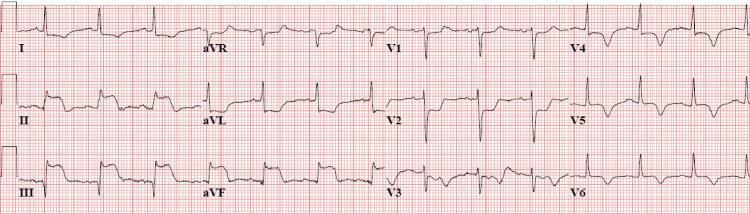
A standard 12-lead electrocardiogram (ECG) demonstrating an acute inferior ST-segment elevation myocardial infarction (STEMI). The tracing reveals significant ST-segment elevation in the inferior leads (II, III, and aVF), characterized by concave or 'tombstoning' morphology and associated with prominent, hyperacute T-waves. Reciprocal ST-segment depression is clearly visible in the high lateral lead (aVL) and the precordial leads (V1, V2, and V3). The precordial leads also show deep S-waves and evolving R-wave transitions. The heart rhythm indicates a regular sinus tachycardia. This ECG is a critical diagnostic tool in emergency medicine and cardiology for identifying transmural ischemia in the distribution of the right coronary artery (RCA) or left circumflex artery, requiring immediate clinical correlation and reperfusion therapy.
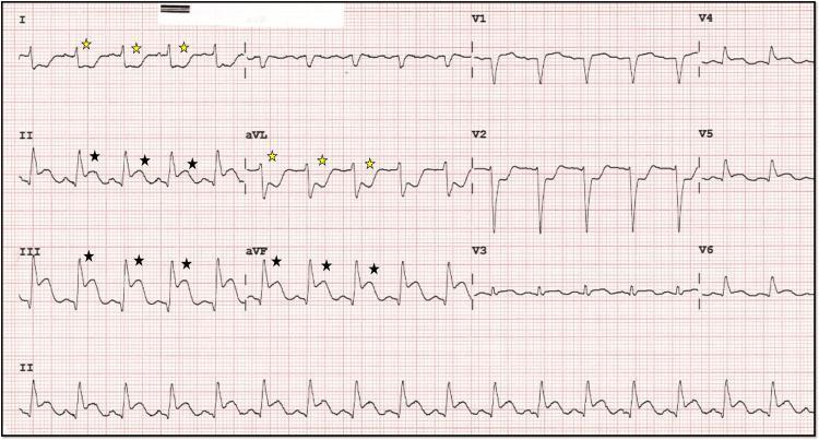
A 12-lead electrocardiogram (ECG) demonstrating diagnostic findings of an acute inferior wall myocardial infarction (ST-elevation myocardial infarction, or STEMI). The primary findings include significant ST-segment elevation in the inferior leads (II, III, and aVF), marked with black stars. Concurrently, there is evidence of reciprocal ST-segment depression in the high lateral leads (I and aVL), indicated by yellow stars. The precordial leads (V1-V3) also exhibit prominent ST-segment depression, which can suggest posterior wall involvement or reciprocal changes from the inferior injury. The inferior leads show a 'shark fin' morphology, characterized by a fusion of the QRS complex, ST segment, and T wave, typically indicating a large area of transmural ischemia. The rhythm appears to be a junctional or sinus bradycardia/tachycardia depending on the clinical phase, with evident electrical instability. This ECG is a critical educational example for identifying acute coronary syndrome and understanding the relationship between anatomical lead groups and reciprocal changes.
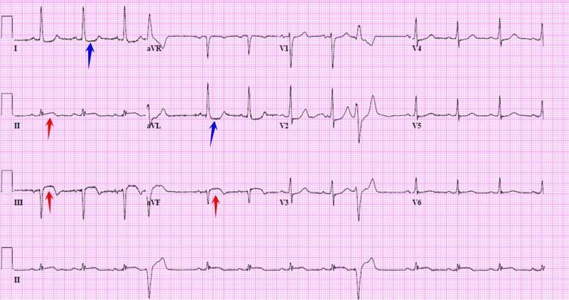
A standard 12-lead diagnostic electrocardiogram (ECG) demonstrating an acute ST-elevation myocardial infarction (STEMI) in the inferior wall distribution. The tracing shows a regular sinus rhythm with narrow QRS complexes. Key diagnostic features include significant ST-segment elevation in the inferior leads (II, III, and aVF), highlighted by red arrows, which is highly suggestive of an acute right coronary artery (RCA) or left circumflex (LCx) artery occlusion. Reciprocal ST-segment depression and T-wave inversion are clearly visible in the high lateral leads (I and aVL), marked by blue arrows. The precordial leads (V1-V6) show relatively stable baselines without prominent acute ST-T changes. This image serves as a classic educational example of inferior myocardial injury with characteristic reciprocal changes, appropriate for teaching ECG interpretation, cardiology diagnostics, and emergency cardiovascular care protocols.
ECG Interpretation: 70-Year-Old Male with Chest Pain
Machine-Read Parameters (from ECG header)
- Paper speed: 25 mm/s | Gain: 10 mm/mV (standard)
- PR interval: 146 ms | QRS: 65 ms | QT: 366 ms | QTc: 422 ms
- QRS Axis: 44° (normal)
- ST interval: 130 ms
Systematic Lead-by-Lead Analysis
| Feature | Findings |
|---|---|
| Rhythm | Regular sinus rhythm |
| Rate | ~67 bpm (RR interval ~900 ms) |
| Axis | ~44° - Normal |
| P waves | Present, upright in II, inverted in aVR - normal sinus |
| PR interval | 146 ms - normal |
| QRS duration | 65 ms - narrow, no bundle branch block |
| ST changes | ST elevation in II, III, aVF (inferior leads) |
| Reciprocal changes | ST depression in I, aVL (high lateral leads) |
| Precordial | ST depression in V1-V3 (reciprocal / possible posterior extension) |
| V3 morphology | Prominent/deep morphology - possible reciprocal to inferior injury |
Diagnosis: INFERIOR STEMI
- ST elevation in leads II, III, and aVF - the hallmark of inferior STEMI
- ST elevation greater in lead III than in lead II - strongly suggests Right Coronary Artery (RCA) occlusion (vs. left circumflex, where elevation is greater in II)
- Reciprocal ST depression in leads I and aVL - confirms the inferior injury pattern; this reciprocal change is a hallmark of inferior STEMI and increases diagnostic specificity
- ST depression in V1-V3 - raises concern for concurrent posterior wall involvement and should prompt right-sided leads (V3R-V6R) to exclude Right Ventricular (RV) infarction
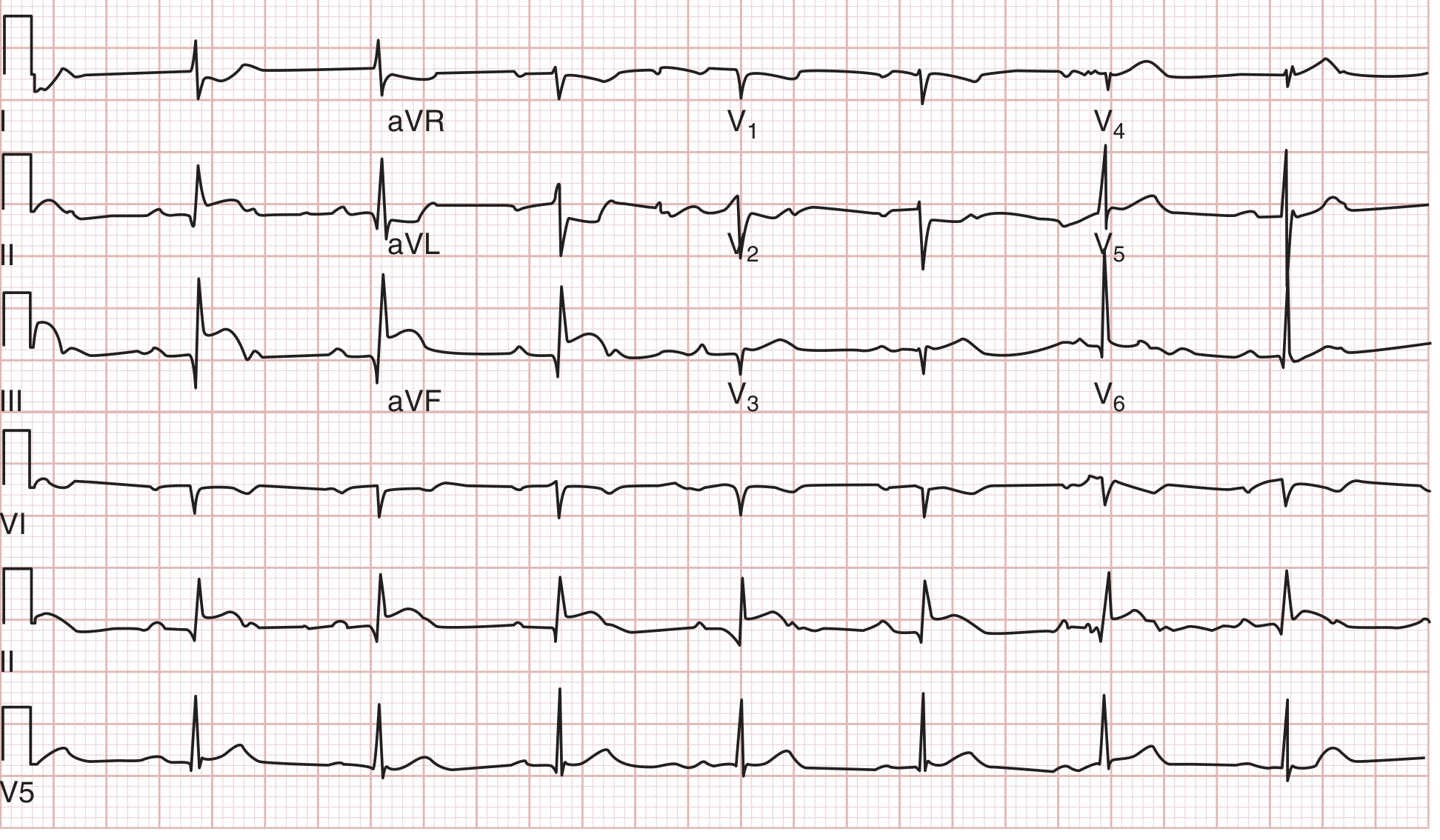
Comparison with a Classic Inferior STEMI ECG
Critical Warning: Rule Out Right Ventricular Infarction
- RV infarction presents with the Bezold-Jarisch triad: hypotension, bradycardia, clear lungs
- Nitrates are CONTRAINDICATED - can cause catastrophic hypotension (preload-dependent state)
- Diuretics are CONTRAINDICATED
- Treatment requires IV fluid loading (normal saline boluses) to maintain RV preload
- Avoid agents that reduce preload or afterload precipitously
Immediate Management
Activate STEMI Protocol NOW
| Priority | Action |
|---|---|
| A | Activate catheterization lab / STEMI alert |
| B | Aspirin 300 mg PO (loading dose) immediately |
| C | P2Y12 inhibitor loading: Ticagrelor 180 mg OR Clopidogrel 600 mg |
| D | Anticoagulation: Unfractionated heparin or enoxaparin |
| E | IV access x2, continuous cardiac monitoring, defibrillator ready |
| F | Supplemental O2 if SpO2 < 90% |
| G | Right-sided ECG (V4R) to exclude RV infarction |
| H | Nitrates: USE WITH CAUTION - hold until RV infarction excluded; if BP is adequate and no RV involvement, can use for pain |
| I | Morphine/fentanyl for refractory pain |
| J | Stat cardiac biomarkers: Troponin I/T, CK-MB, CBC, BMP, coagulation panel |
| K | Primary PCI is the preferred reperfusion strategy (if available within 120 min) |
| L | If PCI not available within 120 min: fibrinolytic therapy if no contraindications |
Watch for Complications (common in inferior STEMI)
- Bradycardia / AV block (Mobitz I / complete heart block) - RCA supplies the AV node in ~90% of people; atropine 0.5-1 mg IV, temporary pacing if needed
- Hypotension - assess for RV infarction vs. cardiogenic shock
- Ventricular arrhythmias - reperfusion arrhythmias are common
Summary
| Diagnosis | Acute Inferior STEMI |
| Likely culprit | Right Coronary Artery (RCA) occlusion |
| Concurrent concern | RV infarction (get right-sided leads immediately) |
| Critical action | Emergency primary PCI - activate STEMI protocol now |
| Drug caution | NO nitrates until RV involvement excluded |
This is a medical emergency. Time is myocardium. Every minute of delay costs ~2 million cardiomyocytes.