

Report case
foot fractures radiography
ACR Appropriateness Criteria acute trauma foot radiography Ottawa foot rules
Single oblique radiograph provided. No obvious acute fracture or dislocation is identified on this image. Alignment of the visible tarsometatarsal joints is maintained. Mild hallux valgus configuration with mild first metatarsophalangeal osteoarthritic change. No focal aggressive osseous lesion seen.
- No acute osseous abnormality evident on this single oblique view.
- Mild hallux valgus and first MTP osteoarthrosis.
- Limited assessment because only one photographed oblique view is available. If there is focal pain, inability to bear weight, or concern for Lisfranc/midfoot injury, obtain a standard 3-view foot series with weight-bearing views where appropriate, and consider CT or MRI if radiographs remain negative but clinical suspicion persists.

Radilogist review twisting of rt foot eversion
Lisfranc injury foot radiograph tarsometatarsal alignment fleck sign
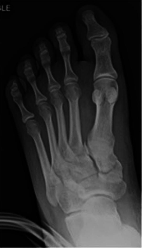
Anteroposterior (AP) radiograph of a human left foot illustrating a Lisfranc joint injury. The image displays a disruption of the normal alignment at the tarsometatarsal (TMT) joints. A key finding is the pathognomonic 'fleck' sign, characterized by a small bony avulsion fragment located in the space between the base of the second metatarsal and the medial cuneiform, signifying an avulsion of the Lisfranc ligament. There is a visible widening of the interval (diastasis) between the first and second metatarsal bases, alongside lateral displacement of the second through fifth metatarsals relative to the tarsal bones. The diagnostic focus is the evaluation of midfoot stability and the integrity of the tarsometatarsal complex. This radiographic view is essential for identifying fracture-dislocations of the midfoot columns, often resulting from high-energy axial loading or hyper-plantarflexion injuries.
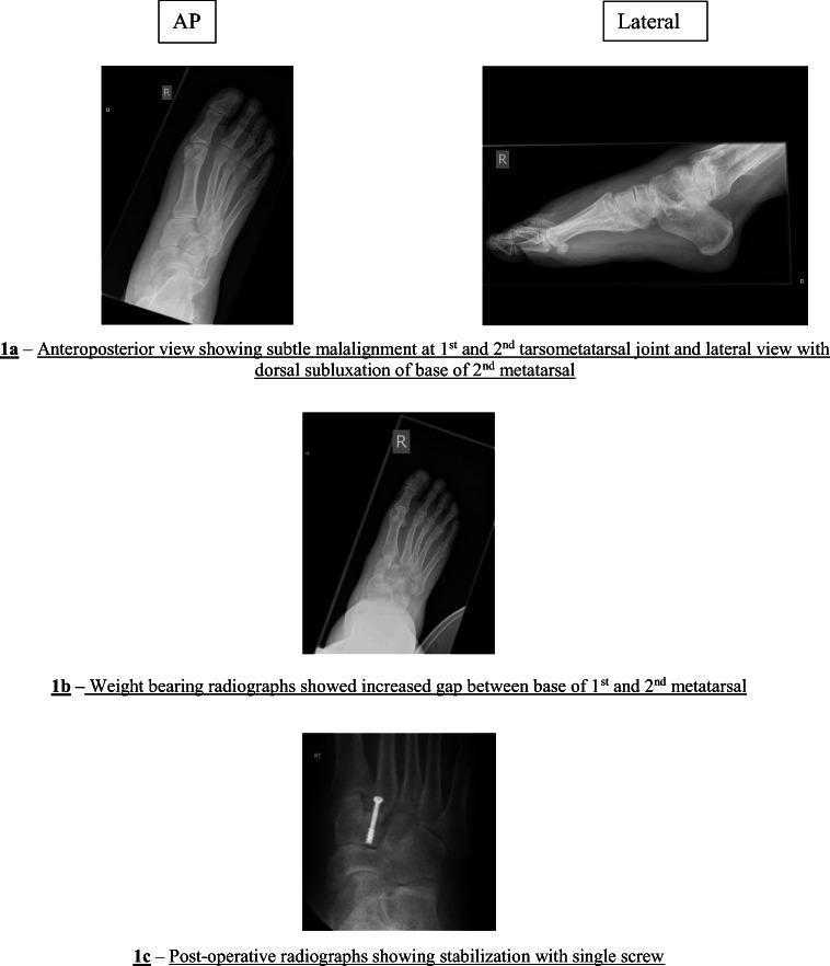
This set of diagnostic radiographs illustrates the clinical presentation and surgical management of a Lisfranc (tarsometatarsal) joint injury in a human foot. Image 1a provides pre-operative anteroposterior (AP) and lateral views. The AP view shows subtle malalignment at the 1st and 2nd tarsometatarsal joints, while the lateral view demonstrates dorsal subluxation of the 2nd metatarsal base. Image 1b is a weight-bearing AP radiograph, which emphasizes the pathology by revealing a pronounced 'fleck sign' or diastasis (increased gap) between the bases of the 1st and 2nd metatarsals, a key diagnostic feature of Lisfranc instability. Image 1c is a post-operative AP view showing surgical stabilization. A single cannulated or cortical screw has been placed across the tarsometatarsal joint, specifically bridging the medial cuneiform and the base of the second metatarsal to restore anatomical alignment and stability. This series serves as an educational tool for identifying subtle orthopedic injuries that can be easily missed on non-weight-bearing films and demonstrates standard internal fixation techniques in podiatric or orthopedic surgery.
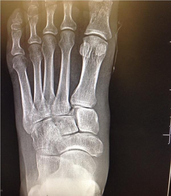
Anteroposterior (AP) radiograph of the left foot demonstrating multi-focal orthopedic injuries. Key diagnostic features include a non-displaced fracture of the proximal phalanx of the first toe (hallux) and significant pathology at the midfoot. There is a subluxation and fracture at the base of the second metatarsal, characterized by a visible 'fleck sign'—a small bony fragment located in the space between the first and second metatarsal bases. This avulsion fracture is a pathognomonic radiographic indicator of a Lisfranc ligament injury, representing a disruption of the tarsometatarsal joint complex. The image provides clinical relevance for identifying unstable midfoot injuries that may require surgical reduction and fixation. Other visible structures include the sesamoid bones beneath the first metatarsal head and the tarsal bones (cuneiforms and navicular), which serve as landmarks for assessing alignment across the Lisfranc joint line.
Lisfranc injury radiography
ACR Appropriateness Criteria acute trauma foot suspected Lisfranc injury radiographs CT MRI
- No visible acute fracture or dislocation on the supplied image.
- Tarsometatarsal alignment appears maintained. No clear widening between the first and second metatarsal bases and no definite Lisfranc avulsion fragment is seen.
- Fifth metatarsal base appears intact.
- Mild hallux valgus with mild first MTP degenerative change.
- Soft-tissue assessment is limited.
- No acute osseous abnormality identified on this limited single-view photographed radiograph.
- If there is focal midfoot tenderness, plantar bruising, marked swelling, or inability to bear weight after the twisting injury, a subtle Lisfranc ligament injury or occult fracture cannot be excluded. Obtain a complete 3-view foot series, preferably weight-bearing AP comparison if tolerated. CT or MRI is appropriate when clinical suspicion remains despite normal radiographs.
- With an eversion mechanism, assess the medial ankle and syndesmosis clinically. If there is medial malleolar tenderness or ankle pain, ankle radiographs should also be obtained.