
Multiparametric mri in prostate malignancy Trans rectal usg vs mri of prostate gland Development ,imaging modalities of prostate MRI prostate Answer each of these radiology questions for 10 marks each for radiology pg exam use standard radiology textbooks or radio Pedia also collect diagrams from internet which can be easily drawn for respective ans
Reading File
Now let me run parallel searches across the medical library and web simultaneously:
Finding Sources
Finding Sources
Finding Sources
Searching the Web
multiparametric MRI prostate PI-RADS zones diagram radiology
Finding Sources
Finding Sources
Finding Sources
Searching the Web
PI-RADS v2 scoring prostate MRI zones diagram radiology exam
Good - I have key sources. Now let me read the main textbook sections:
Reading File
Reading File
Excellent content from the textbooks. Let me now read the Grainger & Allison radiology section and search for prostate development/TRUS content:
Reading File
Finding Sources
Finding Sources
Reading File
Reading File
I found the PI-RADS flowchart image. Let me fetch it and also read more content about prostate anatomy/MRI sequences:
Loading Image
Reading File
Reading File
Excellent! I have the PI-RADS flowchart image and comprehensive textbook content. Let me fetch the prostate MRI image and read the DWI/DCE sections:
Loading Image
Reading File
I now have more than enough content. Let me also read DWI and DCE sections from the same block:
Reading File
Loading Image
Reading File
I now have extensive, high-quality content from Grainger & Allison's Diagnostic Radiology and Campbell-Walsh Urology. Let me update the todos and compile the complete answers:Here are four comprehensive 10-mark answers for your Radiology PG exam, based on Grainger & Allison's Diagnostic Radiology, Campbell-Walsh Urology, and standard radiological practice:
RADIOLOGY PG EXAM ANSWERS - PROSTATE GLAND
Q1. MULTIPARAMETRIC MRI (mpMRI) IN PROSTATE MALIGNANCY (10 Marks)
Introduction
Multiparametric MRI (mpMRI) of the prostate combines anatomical and functional sequences to optimise prostate cancer (PCa) detection, localisation, local staging, risk stratification, biopsy guidance, and treatment planning. It has become the gold-standard imaging tool in the prostate cancer diagnostic pathway.
Sequences in mpMRI (the "Multi" Parameters)
mpMRI comprises THREE essential components:
| Sequence | Abbreviation | Role |
|---|---|---|
| T2-weighted imaging | T2WI | Zonal anatomy, lesion detection, staging |
| Diffusion-weighted imaging | DWI/ADC | Tumour cellularity (primary sequence for PZ) |
| Dynamic contrast-enhanced MRI | DCE | Tumour vascularity/angiogenesis |
| T1-weighted imaging | T1WI | Post-biopsy haemorrhage detection |
(Source: Grainger & Allison's Diagnostic Radiology)
1. T1-Weighted Imaging (T1WI)
- Prostate parenchyma is isointense on T1 - no anatomical detail
- Primary use: detect post-biopsy haemorrhage (T1 hyperintense foci)
- Larger FOV up to aortic bifurcation: assess lymph nodes and bone metastases
- A minimum 6-week interval between biopsy and MRI is advised to reduce false-positive T2 staging due to haemorrhage
2. T2-Weighted Imaging (T2WI)
- High-resolution, multiplanar (axial, sagittal, coronal), slice thickness 3 mm, no gap
- Depicts zonal anatomy of the prostate, seminal vesicles, capsule
- Peripheral Zone (PZ): Normal = homogeneous intermediate-to-high signal; thin T2-hypointense rim = capsule
- PCa appears as focal low signal intensity area
- High-grade tumours (Gleason 4/5): lower SI; Low-grade (Gleason 2/3): mildly hypointense/isointense
- Transition Zone (TZ): Heterogeneous due to BPH ('organised chaos')
- PCa sign: ill-defined homogeneous T2 hypointense lesion = 'Erased Charcoal Drawing Sign'
- Also: lenticular/fusiform anterior lesion, spiculated margins, loss of pseudocapsule
- T2WI is the dominant (primary) sequence for Transition Zone in PI-RADS v2
3. Diffusion-Weighted Imaging (DWI) + ADC Map
- Shows random Brownian motion of water molecules
- PCa has high cell density → restricted diffusion → bright on high b-value DWI, dark on ADC map
- ADC values are inversely proportional to diffusion restriction; PCa has significantly lower ADC than normal tissue
- High-grade tumours (Gleason 4/5) show lower ADC than low-grade tumours
- Recommended b-values: 50-100 s/mm², 800-1000 s/mm², and computed/acquired ≥1400 s/mm²
- DWI is the dominant (primary) sequence for Peripheral Zone in PI-RADS v2
- Slice thickness ≤4 mm, in-plane dimension ≤2.5 mm
4. Dynamic Contrast-Enhanced MRI (DCE)
- Gadolinium-based contrast: exploits tumour angiogenesis (VEGF-driven)
- PCa shows early, intense enhancement + early washout compared to normal parenchyma
- Role: minor/adjunct when T2WI and DWI quality is good
- Key role: upgrades a PI-RADS 3 peripheral zone lesion to PI-RADS 4 if DCE is positive
- Does NOT influence scoring at PI-RADS 1, 2, 4, or 5
- Clinically significant cancer cannot be excluded solely based on absent early enhancement
PI-RADS v2 Scoring System
Developed jointly by ESUR + ACR + AdMeTech Foundation (2015), PI-RADS v2 standardises mpMRI reporting on a 5-point scale:
| PI-RADS | Likelihood | Action |
|---|---|---|
| 1 | Very low | No biopsy |
| 2 | Low | No biopsy |
| 3 | Equivocal | Consider biopsy (DCE role here) |
| 4 | High | Biopsy recommended |
| 5 | Very high | Biopsy recommended |
Clinically significant PCa = Gleason ≥7 (including 3+4), and/or volume ≥0.5 cc, and/or extraprostatic extension
Scoring rule:
- Peripheral Zone: DWI is dominant; DCE can upgrade DWI 3 → PI-RADS 4
- Transition Zone: T2WI is dominant; DWI 5 can upgrade T2 score 3 → PI-RADS 4
PI-RADS v2 Flowchart (Grainger & Allison, Fig. 32.3)
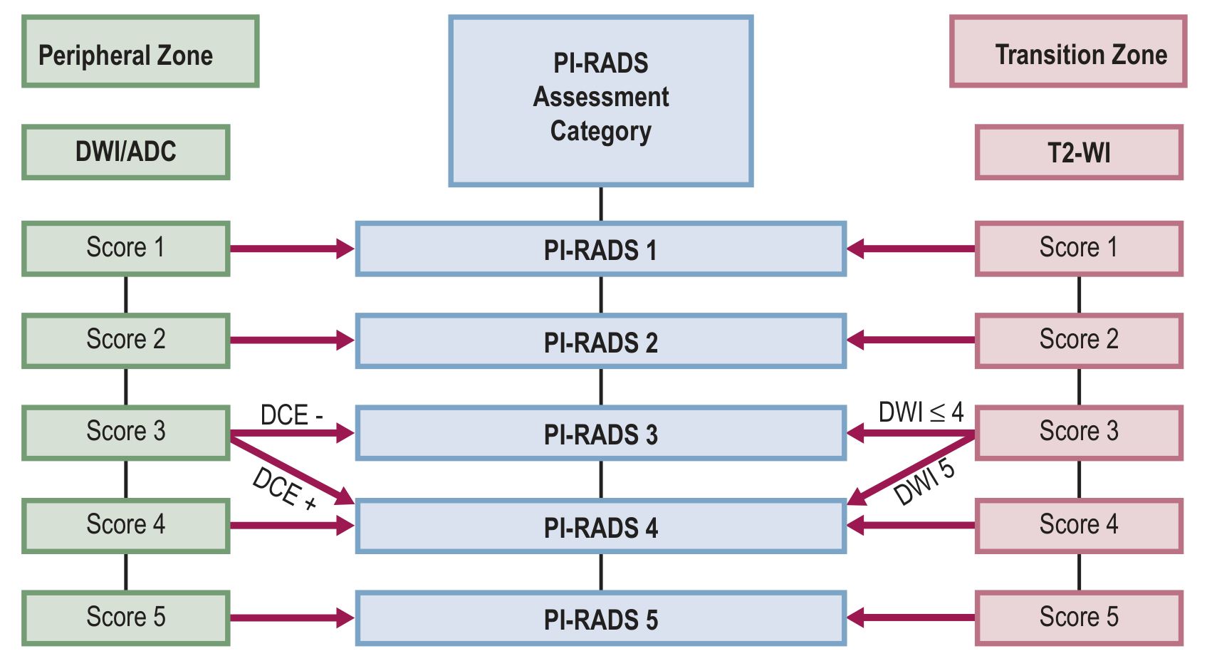
Diagram to draw in exam:
                    PI-RADS Assessment Category
                           |
    PERIPHERAL ZONE        |          TRANSITION ZONE
    (DWI dominant)         |          (T2WI dominant)
                           |
    DWI Score 1 ---------> PI-RADS 1 <--------- T2 Score 1
    DWI Score 2 ---------> PI-RADS 2 <--------- T2 Score 2
    DWI Score 3 + DCE- --> PI-RADS 3 <--------- T2 Score 3 + DWI ≤4
    DWI Score 3 + DCE+ --> PI-RADS 4 <--------- T2 Score 3 + DWI 5
    DWI Score 4 ---------> PI-RADS 4 <--------- T2 Score 4
    DWI Score 5 ---------> PI-RADS 5 <--------- T2 Score 5
Technical Requirements
- Field strength: 3 Tesla preferred (better SNR, spatial/temporal resolution)
- 1.5T acceptable without endorectal coil
- Endorectal coil: Optional - improves SNR but causes prostate deformation, artefacts, patient discomfort
- External phased-array coil recommended
- Protocol: T2WI + DWI + DCE (mandatory); T1WI (optional)
Clinical Applications
- Primary detection - triage to avoid unnecessary biopsy (PROMIS trial: 27% of patients avoided biopsy)
- Targeted biopsy guidance - via cognitive fusion, MRI/TRUS software fusion, or "in-bore" MRI biopsy
- Local staging - extracapsular extension, seminal vesicle invasion
- Active surveillance - monitor low-risk disease
- Treatment planning - radiation therapy targeting (MRI volumes 10% smaller than CT)
- Post-treatment recurrence - T2WI for biochemical recurrence localisation
(Source: Grainger & Allison's Diagnostic Radiology, Chapter 32; Campbell-Walsh Urology, Chapter 150)
Q2. TRANSRECTAL USG vs MRI OF THE PROSTATE GLAND (10 Marks)
Introduction
Both TRUS and MRI are established imaging modalities for prostate evaluation. The choice depends on indication, availability, and clinical context.
Comparison Table
| Parameter | TRUS | MRI |
|---|---|---|
| Modality | High-frequency ultrasound (7-10 MHz) | Magnetic resonance (1.5T / 3T) |
| Radiation | None | None |
| Cost | Low, widely available | High, limited availability |
| Real-time | Yes | No |
| Guidance for biopsy | Direct, real-time | Indirect (fusion) or "in-bore" |
| Soft tissue contrast | Moderate | Excellent |
| Zonal anatomy | Limited | Excellent (T2WI) |
| Cancer detection sensitivity | Low (echogenicity overlap) | High (mpMRI) |
| Functional imaging | Limited (Doppler, elastography) | Full (DWI, DCE) |
| Staging accuracy | Lower | Higher (capsule, ECE, SVI) |
| Anterior prostate | Poor access | Excellent |
| Patient comfort | Mild discomfort | Claustrophobia possible |
| Operator dependence | High | Moderate |
TRUS - Details
Technique:
- 7-10 MHz bi-planar transducer inserted transrectally
- Sagittal + transverse planes
- Volume calculation: prolate ellipsoid formula = L × W × H × 0.523
Normal Sonographic Anatomy:
- Peripheral zone: uniform hypoechoic (central gland echogenic)
- Surgical capsule: echogenic line
- Seminal vesicles: symmetrical paired structures superior-posterior
Uses of TRUS:
- Prostate volume measurement (essential for PSA density calculation)
- TRUS-guided systematic biopsy - 12-core extended sextant biopsy (gold standard access route for random biopsy)
- Colour Doppler - increased vascularity at tumour sites
- TRUS + Elastography - tissue stiffness mapping
- Contrast-enhanced TRUS - emerging technique
- Guidance for brachytherapy seed implantation
- Drainage of prostatic abscess
Limitations of TRUS:
- 60-70% of PCa is isoechoic to background (invisible)
- Cannot assess anterior prostate, apex, or periurethral zone adequately
- Cannot assess extracapsular extension reliably
- Operator-dependent
- Cannot differentiate cancer from prostatitis, BPH nodules
MRI - Details (See also Q1 and Q4)
Advantages of MRI over TRUS:
- Superior soft tissue contrast
- Better zonal anatomy depiction
- Accurate capsular assessment - extracapsular extension (ECE), seminal vesicle invasion (SVI)
- Identifies anterior and transition zone tumours
- Multi-parametric functional data (DWI, DCE)
- Systematic staging via PI-RADS
- Nodal and bone marrow assessment on T1WI
- RDOG study: MRI shows higher sensitivity and specificity than endorectal ultrasound for ECE detection
MRI-TRUS Fusion Biopsy:
- Best of both worlds: MRI identifies suspicious lesion, TRUS guides real-time needle
- Software-based image co-registration
- Three techniques: (a) In-bore MRI biopsy, (b) Cognitive fusion, (c) MRI/TRUS software fusion
- Allows targeted biopsy of PI-RADS 3-5 lesions
Diagram to Draw in Exam:
PROSTATE IMAGING COMPARISON

TRUS                              MRI
├─ 7-10 MHz transducer            ├─ 1.5T / 3T magnet
├─ Transrectal approach           ├─ External phased-array coil
├─ Real-time guided biopsy        ├─ mpMRI protocol
├─ Volume measurement             ├─ T2WI: anatomy + staging
├─ Doppler vascularity            ├─ DWI: cancer detection
└─ Limited cancer detection       └─ DCE: vascularity

MRI-TRUS FUSION BIOPSY:
[MRI lesion map] + [Real-time TRUS] = Targeted needle placement
(Source: Grainger & Allison's Diagnostic Radiology, Chapters 32 & 81; Bailey and Love's Surgery)
Q3. DEVELOPMENT AND IMAGING MODALITIES OF PROSTATE GLAND (10 Marks)
A. EMBRYOLOGICAL DEVELOPMENT
Origin:
- The prostate develops from the urogenital sinus (endodermal origin)
- Outgrowths of urogenital sinus epithelium invade the surrounding urogenital mesenchyme
- Begins at 10-12 weeks of gestation
- Development is androgen-dependent (dihydrotestosterone / DHT via 5-alpha reductase)
- Full secretory function only at puberty under testosterone influence
Key structures:
| Structure | Embryological Origin |
|---|---|
| Prostatic epithelium | Endodermal urogenital sinus |
| Prostatic stroma | Urogenital mesenchyme |
| Bladder trigone | Mesonephric (Wolffian) duct |
| Seminal vesicles | Mesonephric duct |
(Source: Campbell-Walsh Urology; Smith & Tanagho's General Urology)
B. ANATOMY (McNeal's Zonal Anatomy - 1981)
McNeal described four zones:
| Zone | % of prostate | Location | Significance |
|---|---|---|---|
| Peripheral Zone (PZ) | 70% | Posterior-lateral; palpable on DRE | 70-75% of PCa arise here |
| Transition Zone (TZ) | 5-10% | Periurethral; surrounds urethra | Site of BPH; 25% of PCa |
| Central Zone (CZ) | 20-25% | Surrounds ejaculatory ducts, base | Rarely involved by PCa |
| Anterior Fibromuscular Stroma | - | Anterior prostate | No glandular tissue |
Diagram to Draw in Exam (Zonal Anatomy - ESSENTIAL):
PROSTATE ZONAL ANATOMY - Axial View (Mid-gland)
         ANTERIOR
    ___________________
   |  ANTERIOR        |
   |  FIBROMUSCULAR   |
   |  STROMA (AFS)    |
   |___________________|
   |  TRANSITION  |CZ |
   |  ZONE  (TZ)  |   |  <- Ejaculatory ducts
   |______________|___|
   |                   |
   |  PERIPHERAL ZONE (PZ)       |
   |___________________________|
         POSTERIOR
         (Palpable on DRE)

Urethra runs through centre of TZ
C. IMAGING MODALITIES FOR PROSTATE
1. Plain X-Ray
- Not useful for prostate itself
- May show prostatic calcifications (corpora amylacea)
- IVU: Indentation on bladder base in BPH
2. Ultrasound (TRUS)
- First-line imaging for prostate
- Volume measurement, biopsy guidance
- Normal appearance: PZ hypoechoic, CG echogenic
- Colour Doppler: vascularity in PCa
- Limitations: PCa often isoechoic, operator-dependent
3. CT Scan
- NOT useful for primary tumour detection - poor soft tissue contrast
- Staging role: lymph node assessment (N staging), osseous metastases
- CT is primary for N/M staging in high-risk PCa
- Cannot distinguish tumour from BPH within prostate
4. MRI (mpMRI - See Q1 and Q4 for details)
- Gold standard for local staging
- T2WI: anatomy, capsular integrity
- DWI: tumour detection
- DCE: vascularity
- PI-RADS v2 reporting
- 3T preferred; no endorectal coil needed at 3T
5. Bone Scintigraphy (Tc-99m MDP)
- Standard for bone metastases detection
- Recommended for: PSA >20, Gleason >7, T3/T4 disease
- Hot spots at sites of osteoblastic metastases
- Superscan in widespread disease
6. PET-CT
- Choline PET-CT: Early PCa, biochemical recurrence
- PSMA PET-CT (Ga-68 PSMA): Highly sensitive for PCa; PCa cells overexpress PSMA
- Superior to bone scan + CT for staging and restaging
- Increasingly preferred over choline PET
- Detects disease at PSA levels as low as 0.2 ng/mL
- NaF PET-CT: Bone-specific, more sensitive than bone scan for skeletal metastases
7. TRUS-Guided Biopsy
- 12-core extended sextant systematic biopsy
- MRI-TRUS fusion for targeted biopsy
8. Multiparametric TRUS (mpTRUS)
- Analogous to mpMRI: combines B-mode + Doppler + contrast enhancement + elastography
- Emerging alternative/complement to mpMRI
Q4. MRI OF THE PROSTATE GLAND (10 Marks)
Introduction
MRI of the prostate is the most accurate imaging modality for local staging of prostate cancer. The development of multiparametric MRI (mpMRI) has transformed the prostate cancer diagnostic pathway.
Technical Requirements
Scanner:
- 3T preferred (higher SNR, better spatial and temporal resolution)
- 1.5T is acceptable without endorectal coil
- 3T without endorectal coil = diagnostic quality equivalent to 1.5T with endorectal coil
Coils:
- External phased-array pelvic coil - standard of care
- Endorectal coil (optional): improves SNR but deforms prostate, increases cost, patient discomfort, artefacts
Contraindications:
- Metallic implants, pacemakers, claustrophobia
- Post-biopsy waiting: 6 weeks (haemorrhage resolution)
MRI Sequences and Protocols
1. T1-Weighted Imaging
- Prostate isointense: no zonal anatomy
- Detects post-biopsy haemorrhage (T1 hyperintense)
- Large FOV: nodal + bone marrow assessment
- Fat-saturation versions: improves nodal detection
2. T2-Weighted Imaging (Anatomical Sequence)
- Key sequence for anatomy and staging
- Multiplanar: axial (mandatory), coronal, sagittal
- Slice thickness 3 mm, no gap; in-plane ≤0.7 mm × 0.4 mm
- FOV 12-20 cm, covering prostate + seminal vesicles
Normal Zonal Anatomy on T2WI:
| Zone | T2 Signal |
|---|---|
| Peripheral zone | Homogeneous high signal (bright) |
| Transition zone | Heterogeneous intermediate (BPH = 'organised chaos') |
| Central zone | Low-intermediate signal |
| Capsule | Thin T2-hypointense rim |
| Seminal vesicles | High signal (fluid-filled) |
T2WI: PCa Features:
Peripheral Zone (PZ):
- Focal low signal intensity area
- High-grade: markedly low SI
- Low-grade: mildly hypointense or isointense
Transition Zone (TZ):
- 'Erased charcoal drawing' sign - ill-defined homogeneous T2 hypointense lesion
- Lenticular/fusiform anterior lesion
- Spiculated/irregular margins
- Loss of pseudocapsule
T2WI PI-RADS Categories (Peripheral Zone) - Fig. 32.5, Grainger & Allison
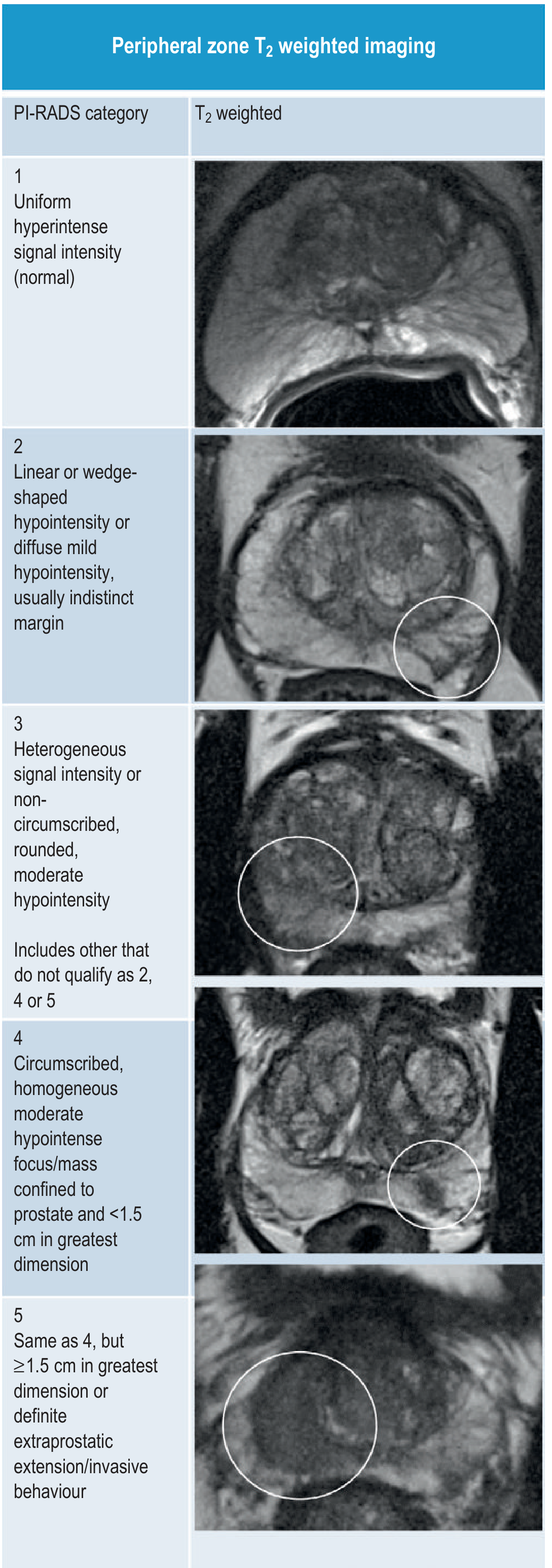
Peripheral Zone T2WI PI-RADS:
- 1: Uniform hyperintense (normal)
- 2: Linear/wedge-shaped hypointensity, indistinct margin
- 3: Heterogeneous, non-circumscribed, rounded moderate hypointensity
- 4: Circumscribed, homogeneous, moderate hypointensity, <1.5 cm
- 5: Same as 4 but ≥1.5 cm OR definite extraprostatic extension
3. Diffusion-Weighted Imaging (DWI)
Physics: Brownian motion of water molecules
- High b-value images: Restricted diffusion = bright signal (cancer)
- ADC map: Restricted diffusion = dark (low ADC value); inverse relationship
- Cancer has HIGH cell density → RESTRICTED diffusion → HIGH signal on DWI, LOW ADC
ADC values:
- Normal PZ: high ADC (fluid-rich tubular structures, especially >50 years)
- PCa: significantly lower ADC than normal tissue
- High-grade PCa (Gleason 4/5): lower ADC than low-grade (Gleason 3)
Technique: Free-breathing spin-echo, spectral fat saturation; b-values: 50-100, 800-1000, and ≥1400 s/mm²; slice thickness ≤4 mm
DWI PI-RADS Categories (PZ):
- 2: Non-focal hypointense ADC
- 3: Focal, mildly/moderately hypointense ADC + iso/mildly hyperintense high b-value DWI
- 4: Focal, markedly hypointense ADC + marked hyperintensity on high b-value, <1.5 cm
- 5: Same as 4 but ≥1.5 cm or extraprostatic extension
4. Dynamic Contrast-Enhanced MRI (DCE)
Principle: Gadolinium-based CA → exploits tumour angiogenesis (VEGF-driven neovascularisation)
PCa kinetics:
- Early, intense enhancement + early washout (type 3 curve)
- Due to increased vascular permeability and AV shunting
Role in PI-RADS v2:
- Positive DCE = focal area of earlier onset enhancement than surrounding normal tissue
- DCE upgrades PZ PI-RADS 3 to PI-RADS 4 (the only upgrade role)
- Does NOT influence PI-RADS 1, 2, 4, or 5 scoring
- Visual assessment of early enhancement phase is recommended
- Gadolinium: dose 0.1 mmol/kg, temporal resolution <10 seconds per dynamic phase
Staging with MRI - TNM
| Stage | MRI Finding |
|---|---|
| T2a/b | Tumour confined to one/both lobes; intact capsule |
| T3a | Extracapsular extension (ECE) - irregular capsular bulge, obliteration of rectoprostatic angle |
| T3b | Seminal vesicle invasion - T2 low signal, non-tapering, asymmetric enlargement |
| T4 | Invasion of adjacent structures (bladder, rectum) |
Signs of ECE on MRI:
- Irregular or spiculated margin
- Asymmetric neurovascular bundle
- Obliteration of rectoprostatic angle
- Direct tumour extension beyond capsule
MRI in Prostate Cancer Pathway
Elevated PSA / Abnormal DRE
          |
    mpMRI (PI-RADS scoring)
          |
    PI-RADS 1-2        PI-RADS 3         PI-RADS 4-5
    No biopsy      Consider biopsy    MRI-TRUS fusion
    (monitor)      (clinical factors)  targeted biopsy
                                           |
                                   Histological diagnosis
                                           |
                               Staging MRI (T/N/M staging)
                                           |
                      Treatment planning (surgery / RT / surveillance)
MRI in Active Surveillance
- Serial mpMRI monitoring for low-risk PCa on active surveillance
- Detects disease upgrading or progression
- Guides repeat biopsy only when PI-RADS changes
MRI vs CT vs Bone Scan for Staging
| Modality | T staging | N staging | M (bone) |
|---|---|---|---|
| mpMRI | Excellent | Moderate | Poor |
| CT | Poor | Good | Moderate |
| Bone scan | Not applicable | Not applicable | Good |
| PSMA PET-CT | Good | Excellent | Excellent |
Prostate MRI - Summary Box (for quick revision)
Protocol: T2WI + DWI + DCE (+ T1WI optional)
PI-RADS v2: 5-point scale, PZ = DWI dominant, TZ = T2WI dominant
3T without endorectal coil: Standard recommendation
Key cancer sign T2WI PZ: Focal low signal intensity
Key cancer sign DWI: Bright on high b-value + dark on ADC map
Key cancer sign DCE: Early enhancement + early washout
Clinical significance threshold: Gleason ≥7, volume ≥0.5 cc, or extraprostatic extension
Sources: Grainger & Allison's Diagnostic Radiology (9780323760751), Chapter 32; Campbell-Walsh Urology (9780323546423), Chapter 150; Smith & Tanagho's General Urology; Bailey & Love's Short Practice of Surgery.
Imaging of bronchogenic ca Rguhs
Reading File
Now let me run parallel searches for bronchogenic carcinoma imaging content:
Finding Sources
Finding Sources
Finding Sources
Excellent - found the dedicated "IMAGING FEATURES OF BRONCHOGENIC CARCINOMA" section in Grainger & Allison. Let me read it comprehensively, plus search for more content in parallel:
Reading File
Finding Sources
Finding Sources
Found rich sources. Let me read the next section of the Grainger & Allison bronchogenic carcinoma chapter and the staging protocol section simultaneously:
Reading File
Reading File
Reading File
Reading File
Reading File
Reading File
Reading File
Finding Sources
Reading File
Finding Sources
Now let me fetch some of the key images from this chapter and read the PET/CT and TNM staging sections:
Loading Image
Loading Image
Reading File
I now have comprehensive, high-quality content from Grainger & Allison's Diagnostic Radiology. I have all the material needed for a thorough RGUHS-style answer. Let me compile the complete answer now.
IMAGING OF BRONCHOGENIC CARCINOMA
RGUHS Radiology PG Exam - 10 Marks
(Source: Grainger & Allison's Diagnostic Radiology; Fishman's Pulmonary Diseases; Murray & Nadel's Respiratory Medicine)
INTRODUCTION
Bronchogenic carcinoma (lung cancer) is the leading cause of cancer-related death worldwide. Imaging plays a central role in its:
- Detection and characterization
- Local staging (T descriptor)
- Nodal staging (N descriptor)
- Distant metastasis staging (M descriptor)
- Treatment planning and response assessment
Approximately 25% of patients are asymptomatic at diagnosis, detected on chest radiograph or CT screening. The remaining present with cough, haemoptysis, wheeze, pneumonia, hoarseness (RLN involvement), SVC syndrome, Horner's syndrome (Pancoast), chest pain, or paraneoplastic syndromes.
IMAGING MODALITIES USED
| Modality | Primary Role |
|---|---|
| Chest X-ray (CXR) | First-line investigation, initial detection |
| CT (MDCT) | Primary staging modality |
| PET-CT (¹⁸F-FDG) | Nodal + distant staging |
| MRI | Superior sulcus / chest wall / mediastinal invasion |
| Bone scintigraphy | Skeletal metastases (largely replaced by PET-CT) |
| Ultrasound | Chest wall invasion, supraclavicular nodal assessment |
| EBUS | Mediastinal nodal biopsy guidance |
A. CHEST RADIOGRAPHY (CXR)
The initial and universal first investigation for suspected bronchogenic carcinoma.
The thoracic imaging features are classified under three headings:
- Peripheral tumours
- Central tumours
- Intrathoracic staging features
1. PERIPHERAL TUMOURS (~40% of bronchogenic Ca)
Shape and Margins:
- Most are approximately spherical or oval
- Lobulation - common; indicates uneven growth in different parts
- Dumb-bell shape or two adjacent nodules may be seen
- Pancoast (superior sulcus) tumour may resemble apical pleural thickening
Key Radiological Signs:
| Sign | Description | Significance |
|---|---|---|
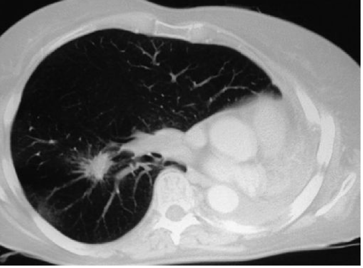
| Corona radiata | Fine strands radiating from a central mass into the lung | Highly suggestive of bronchogenic Ca |
| Pleural tail sign | Peripheral line/tail between mass and pleura | Seen in benign and malignant lesions |
| Lobulated margin | Uneven contour | Common in carcinoma |
| Spiculated margin | Irregular, spiky edges | Strong predictor of malignancy |
CT image showing corona radiata sign (spiculated lung carcinoma):
Cavitation:
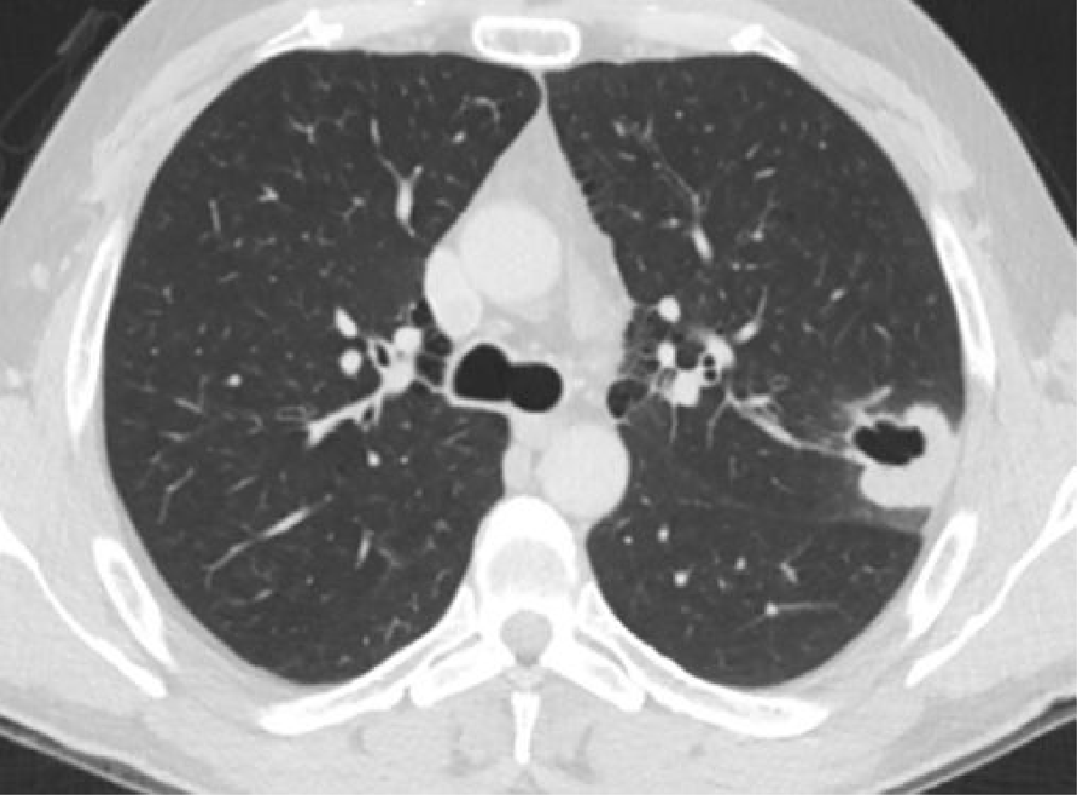
- Seen in tumours of any size; best demonstrated on CT
- Squamous cell carcinoma - most commonly cavitates
- Wall is thick and irregular (unlike thin-walled benign cysts)
- CT distinguishes malignant from benign cavities by wall thickness (>15 mm = malignant)
CT showing cavitating squamous cell carcinoma:
Calcification:
- Rare on CXR; seen on CT in 6-10% of cases
- May represent pre-existing calcified granuloma engulfed by tumour
- Amorphous/cloud-like = dystrophic tumour calcification (<10%)
- Calcified tumours are usually large (≥5 cm)
Other CT findings:
- Air bronchogram / bubble lucencies / pseudo-cavitation: adenocarcinoma
- Ground-glass opacity (GGO): more common in adenocarcinoma; may be pure GGO
- Mucocele / mucoid impaction: dilated mucus-filled bronchi distal to obstruction
- Ill-defined edges resembling pneumonia: some adenocarcinomas
2. CENTRAL TUMOURS
Arise in large bronchi at or close to the hilum. Cardinal signs:
a) Hilar Enlargement:
- Common presenting feature
- May reflect: proximal tumour mass, lymphadenopathy, consolidated lung, or combination
- A mass superimposed on hilum → increased hilar density (summation sign)
- "Dense hilum sign": only radiographic finding in some cases - must inspect lateral film
- Lateral CXR is essential when dense hilum is suspected
b) Collapse/Consolidation (Post-obstructive):
- Obstruction of major bronchus → combination of atelectasis + secretion retention + opacity
- Collateral air drift may partially/completely prevent collapse
Features suggesting post-obstructive neoplasm:
- Golden S sign (Reverse S sign): When RUL collapses due to a central mass, the minor fissure bulges outward (over the hilar mass) while collapsing medially - creating an S-shape. Classic for right upper lobe bronchial carcinoma
- Pneumonia confined to one lobe persisting >2-3 weeks without resolution
- Recurring pneumonia in the same lobe - until proven otherwise = underlying carcinoma
- Absent air bronchograms within the collapsed lobe (secretions fill the bronchi)
- Visible mass with bronchial stenosis on CT
- Mucus-filled dilated bronchi (branching tubular low-density structures = "finger in glove") on CT
- Drowned lobe: opacified lobe larger than normal (build-up of infected secretions)
Diagram to Draw in Exam (ESSENTIAL):
GOLDEN S SIGN - Right Upper Lobe Collapse with Central Mass

Normal position                  RUL collapse + central mass
of minor fissure:                (Golden S sign):

_____Trachea_____               _____Trachea_____
  |               |               |               |
  |               |               |        ___/   |
  |    RUL        |               |  RUL  /       |
  |_______________|               |______/ Mass   |
  |  Right hilum  |               |  Central |    |
  |               |               |  mass    |    |
  |               |               |          |    |

Minor fissure =                  Minor fissure bulges
horizontal line                  outward over mass = S shape
B. CT SCAN (Primary Staging Modality)
Standard protocol: Multi-detector CT (MDCT, ≥64 rows), submillimetre collimation, contrast-enhanced; coverage from skull base to upper thigh. Arms elevated; supine position.
CT remains the mainstay of staging supplemented by PET-CT and MRI.
CT Features for T-Staging (8th edition AJCC/UICC TNM):
| T Category | CT Criteria |
|---|---|
| Tx | Primary tumour cannot be assessed (cytology positive, not visualised) |
| Tis | Carcinoma in situ |
| T1a(mi) | Minimally invasive adenocarcinoma |
| T1a | ≤1 cm, surrounded by lung/visceral pleura |
| T1b | >1 cm but ≤2 cm |
| T1c | >2 cm but ≤3 cm |
| T2a | >3 cm but ≤4 cm OR involves main bronchus (no carina) OR visceral pleura invaded OR atelectasis to hilum |
| T2b | >4 cm but ≤5 cm |
| T3 | >5 cm but ≤7 cm OR chest wall / pericardium / phrenic nerve invasion OR satellite nodule in same lobe |
| T4 | >7 cm OR invasion of mediastinum, heart, great vessels, trachea, carina, vertebra, oesophagus OR satellite nodule in different ipsilateral lobe |
CT Features for N-Staging:
| N Stage | Description |
|---|---|
| N0 | No regional node involvement |
| N1 | Ipsilateral peribronchial/hilar nodes |
| N2 | Ipsilateral mediastinal/subcarinal nodes |
| N3 | Contralateral mediastinal/hilar nodes; supraclavicular nodes |
Criteria for nodal involvement on CT:
- Short-axis diameter >10 mm = enlarged (95th percentile)
- Simple but imperfect criterion - many causes of benign node enlargement (TB, sarcoid, reactive hyperplasia)
- Sensitivity 55-60%, Specificity 80% for CT nodal staging
CT Signs of Mediastinal Invasion:
- Visible tumour deep within mediastinal fat
- Tumour surrounds mediastinal vessels (aorta, pulmonary artery, SVC, oesophagus, carina)
- Glazer criteria: <3 cm contact with mediastinum + preserved mediastinal fat plane = likely no invasion
- >3 cm contact / >90° circumference of vessel involvement = likely invasion
CT Signs of Chest Wall Invasion:
- Bone destruction - definitive for invasion
- Large soft-tissue mass extending into chest wall - definitive
- Contact with pleura alone: does NOT confirm invasion
- Obliteration of extrapleural fat plane = strongly suggests invasion
- Preserved extrapleural fat plane (Fig. 8.23) = likely no invasion
- CT sensitivity for chest wall invasion = ~60%
C. PET-CT (¹⁸F-FDG)
¹⁸F-FDG (fluorodeoxyglucose) PET-CT is routinely utilised for staging lung carcinoma.
Principle: Cancer cells have increased glycolytic activity → uptake of FDG (glucose analogue) → PET-avid lesions. Measured as Standardised Uptake Value (SUV).
Indications:
- Nodal staging (superior to CT alone)
- Detection of distant metastases
- Identification of occult disease before planned surgery
- Recurrence detection
- Response assessment
Advantages over CT for staging:
- Higher sensitivity and specificity for nodal disease
- Detects metabolically active disease in normal-sized nodes
- Identifies distant metastases (skeleton, adrenal, liver, contralateral lung)
- Decision analysis: identifies inoperable patients, avoiding unnecessary thoracotomy
- PET/CT = single study covering skull base to upper thigh
PET-CT Protocol:
- CT acquisition: usually low-dose unenhanced (for co-registration)
- PET acquisition: 5-7 bed positions, 3-5 minutes each (~35 minutes total)
- Modern systems: significantly faster acquisition
Limitations:
- False positives: active inflammation, granulomas (TB, sarcoid), reactive hyperplasia
- Brain metastases: high background FDG activity - MRI preferred
- Small lesions <8 mm: limited resolution
- Carcinoid tumours: may be FDG-negative (low metabolic activity)
M-Staging by Modality:
| Site of Metastasis | Best Modality |
|---|---|
| Adrenal | CT/PET-CT |
| Liver | CT/PET-CT |
| Bone | PET-CT > Bone scan |
| Brain | MRI (PET unreliable) |
| Contralateral lung | CT/PET-CT |
| Pleural | CT/PET-CT |
D. MRI
MRI is a problem-solving tool for lung cancer staging, not routinely used.
Specific indications:
| Indication | Why MRI? |
|---|---|
| Superior sulcus / Pancoast tumour | Best assessment of brachial plexus, subclavian vessels, vertebral body, neural foramina involvement |
| Mediastinal invasion | Better soft tissue contrast |
| Chest wall invasion | Better than CT for soft tissue planes |
| Diaphragm integrity | Dynamic cine MRI |
| SVC/aortic wall involvement | T1WI shows vessel wall disruption |
| Brain metastases | Most sensitive modality |
| Post-contrast CT contraindicated | Allergy, renal failure |
MRI Protocol for Pancoast tumour:
- T1WI axial: Shows anatomy, tumour vs. fat planes
- T2WI coronal: Extension into root of neck, brachial plexus
- Gadolinium-enhanced T1WI: Vessel/nerve enhancement
- Respiratory-gated sequences
- Advantages of current MDCT (multiplanar reformats) have reduced need for MRI
MRI for Nodal Staging:
- Similar accuracy to CT using size criteria alone
- STIR imaging: 93% sensitivity, 87% specificity for malignant nodes (signal difference between normal and pathological nodes)
- Limited advantage over modern MDCT for routine nodal staging
E. SKELETAL (BONE) SCINTIGRAPHY
- Tc-99m MDP bone scan: sensitive for skeletal metastases; positive before X-ray changes
- Largely replaced by ¹⁸F-FDG PET-CT for whole-body staging
- NaF PET-CT: more sensitive than bone scan for bone metastases
F. ULTRASOUND
- Chest wall: detects invasion by showing fixity of tissue to pleura during respiration
- Supraclavicular lymph nodes: USG ± FNA improves sensitivity for N3 detection
- Routine use recommended by some centres to improve preoperative staging
G. ENDOBRONCHIAL ULTRASOUND (EBUS)
- Assesses size, morphology of mediastinal nodes
- Guides fine-needle aspiration of aortopulmonary, subcarinal, posterior mediastinal nodes
- Greater sensitivity and specificity than CT/PET-CT in some series
- Now first-line for mediastinal node biopsy (replaced mediastinoscopy)
STAGING SUMMARY TABLE (8th Edition TNM)
| Stage | TNM | Typical findings |
|---|---|---|
| I | T1-2, N0, M0 | Solitary peripheral nodule, no nodes, no metastasis |
| II | T1-2, N1, M0 | Peripheral + ipsilateral hilar nodes |
| IIIA | T3, N1; or T1-3, N2, M0 | Local invasion or ipsilateral mediastinal nodes |
| IIIB | T3-4, N3; or any T, N3, M0 | Contralateral nodes, extensive local invasion |
| IV | Any T, any N, M1 | Distant metastasis (liver, bone, adrenal, brain) |
OVERALL IMAGING ALGORITHM (RGUHS - Exam Diagram)
SUSPECTED BRONCHOGENIC CARCINOMA
             |
    CHEST X-RAY (CXR) - First investigation
             |
    Peripheral mass / Central opacity / Hilar enlargement
             |
    CONTRAST-ENHANCED CT THORAX + UPPER ABDOMEN
    (T + N staging, assess local invasion)
             |
    ┌─────────────────────────────────────┐
    |         PET-CT (¹⁸F-FDG)           |
    |   Nodal staging + distant mets      |
    |   (except brain)                    |
    └─────────────────────────────────────┘
             |
    ┌─────── SPECIFIC INDICATIONS ────────┐
    | MRI: Pancoast, chest wall, brain    |
    | EBUS: Mediastinal node biopsy       |
    | USG: Chest wall, supraclavicular    |
    | Bone scan: If PET-CT unavailable    |
    └─────────────────────────────────────┘
             |
    TISSUE DIAGNOSIS (bronchoscopy/CT-guided biopsy)
             |
    MULTIDISCIPLINARY TEAM DISCUSSION
             |
    Surgery / Radiotherapy / Chemotherapy /
    Targeted therapy / Palliative care
SPECIAL PATTERNS
| Pattern | Association |
|---|---|
| Pancoast / Superior sulcus tumour | Apical mass; Horner's syndrome (ptosis, miosis, anhydrosis); brachial plexus neuropathy; rib destruction - MRI for extent |
| SVC syndrome | Central tumour encasing SVC - CT/PET |
| Phrenic nerve palsy | Elevated hemidiaphragm on CXR (caution: also from collapse) |
| Pleural effusion | M1a - cytological confirmation needed unless clearly unrelated cause |
| Lymphangitis carcinomatosa | Irregular, beaded thickening of bronchovascular bundles and interlobular septa on HRCT |
| Miliary pattern | Haematogenous spread to lung |
| Pancoast (MRI) | T1WI: tumour vs. fat planes; T2WI coronal: neural structures |
SUMMARY BOX (For Last-Minute Revision)
- First investigation: CXR
- Primary staging tool: CECT thorax (MDCT)
- Best for nodal staging: PET-CT
- Best for Pancoast/chest wall invasion: MRI
- Best for brain metastases: MRI
- Best for bone metastases: PET-CT > Bone scan
- Key CXR signs: Corona radiata, Golden S sign, dense hilum, pleural tail, lobar collapse
- Cavitation: Squamous cell carcinoma most common; thick irregular wall
- Nodal criterion: Short-axis >10 mm on CT = enlarged
- Mediastinal invasion on CT: <3 cm contact + preserved fat = likely no invasion
- Pleural effusion = M1a (confirm by cytology unless another cause proven)
- TNM system: 8th edition AJCC/UICC used for NSCLC and SCLC
Sources: Grainger & Allison's Diagnostic Radiology (Chapter 8); Fishman's Pulmonary Diseases & Disorders; Murray & Nadel's Textbook of Respiratory Medicine; Harrison's Principles of Internal Medicine 22e.
This is a shared conversation. Sign in to Orris to start your own chat.