
TM joint
Reading File
The temporomandibular (TM) joint is the synovial joint between the mandibular condyle and the temporal bone, unique for having an articular disc. Let me pull comprehensive textbook content on it.
Searching sources...
Finding Sources
Finding Sources
Good results. Let me read the most comprehensive anatomy and clinical sections in parallel.
Reading File
Reading File
Reading File
Good anatomy content. Let me read more of the Scott-Brown's chapter (muscles, disc, movements, disorders) and the emergency medicine / clinical section.
Reading File
Reading File
Excellent content. Let me now fetch the TMJ anatomy diagram and also read the TMJ disorders/clinical section from Scott-Brown's.
Loading Image
Reading File
I now have rich, comprehensive content. Let me compile the full answer.
Temporomandibular Joint (TMJ)
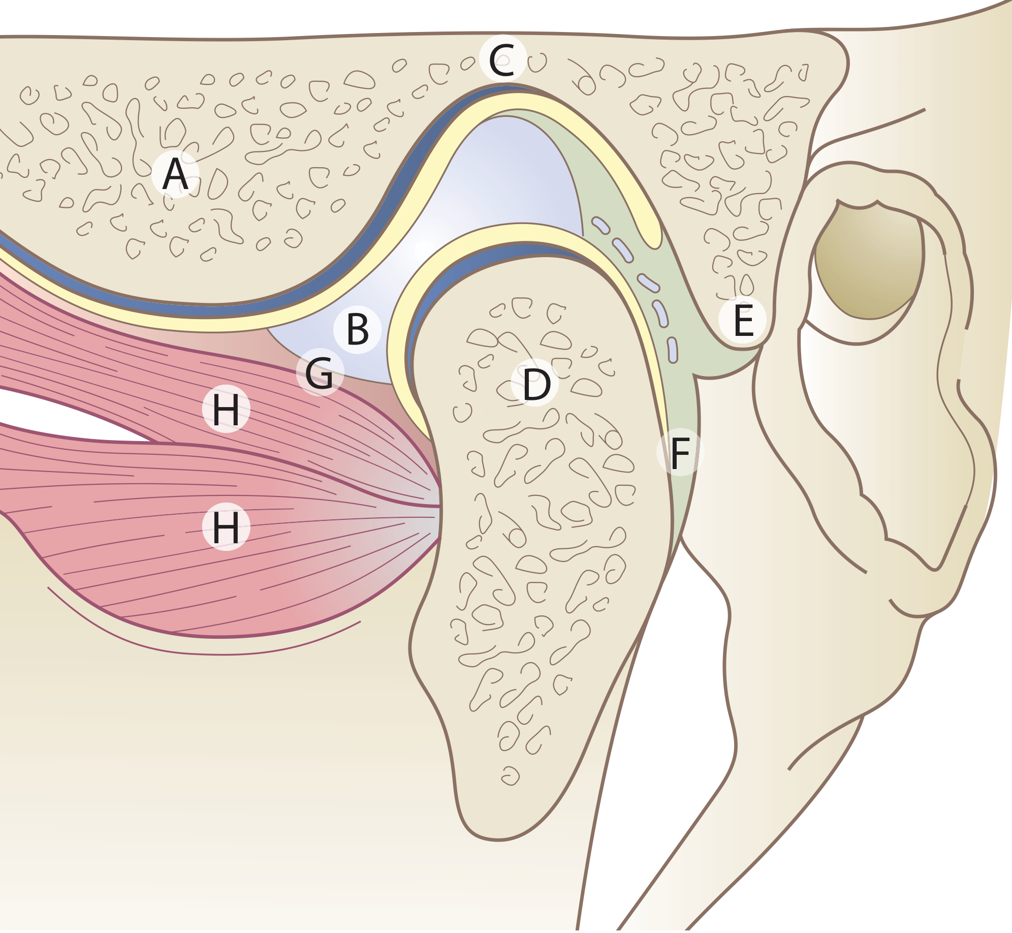
Figure: The TMJ - A = Articular eminence; B = Articular disc; C = Mandibular fossa; D = Condyle; E = Upper lamina (fibro-elastic); F = Lower lamina (non-elastic); G = Capsule; H = Lateral pterygoid muscle
Type and Formation
The TMJ (craniomandibular joint) is a synovial joint formed by the mandibular condyle articulating with the mandibular (glenoid) fossa of the temporal bone, just anterior to the external auditory canal. - Scott-Brown's Otorhinolaryngology, p. 693
Unique Features
The TMJ has several unusual characteristics that distinguish it from other synovial joints:
- Divided joint space - An intra-articular disc divides the joint into upper and lower cavities
- Fibrocartilage, not hyaline - Articular surfaces are fibrous tissue, reflecting the joint's intramembranous development
- Secondary condylar cartilage - Present in the condyle head until adolescence
- Tooth influence - Movements are influenced by the occlusion of teeth
Articular Disc
- A biconcave fibrocartilaginous disc sits between the condyle and temporal fossa
- Divides the joint into:
- Upper compartment (disc-temporal): permits gliding/translatory movement
- Lower compartment (disc-condylar): permits hinge/rotatory movement
- Anteriorly attached to the superior head of the lateral pterygoid muscle (in ~60% of people, fibres insert directly into the medial aspect of the disc's anterior border)
- Posteriorly, the bilaminar zone connects the disc to the retrodiscal tissue: the upper lamina is fibro-elastic (aids disc return), the lower lamina is non-elastic
Joint Capsule
- Attached above to the mandibular fossa (extending to just anterior to the articular eminence crest and posteriorly to the squamotympanic and petrotympanic fissures)
- Attached below to the neck of the condyle
- Thin and lax capsule
Ligaments
| Ligament | Role |
|---|---|
| Lateral (temporomandibular) ligament | Main ligament; limits lateral movement; runs from articular tubercle downward/backward to neck of condyle |
| Sphenomandibular ligament | Accessory; most influential on mandibular movement |
| Stylomandibular ligament | Reinforced band of deep cervical fascia; less functionally significant |
Muscles of Mastication
The muscles that move the TMJ:
| Muscle | Action |
|---|---|
| Masseter | Elevation (jaw closing) |
| Temporalis | Elevation + retraction |
| Medial pterygoid | Elevation + protrusion |
| Lateral pterygoid | Depression/opening + protrusion; both heads act together with functional heterogeneity; the superior head is active during opening |
| Digastric, mylohyoid, geniohyoid | Depression (jaw opening) |
Movements
- Mouth opening: combined rotation (lower compartment) + anterior translation of condyle down the articular eminence (upper compartment)
- Normal interincisal opening: 3-6 cm
- Protrusion / retrusion: gliding in upper compartment
- Lateral excursion: working side condyle rotates, balancing side condyle translates
Nerve and Blood Supply
- Nerve: Auriculotemporal nerve (branch of V3) - the main sensory supply; also masseteric and deep temporal branches
- Blood supply: Superficial temporal and maxillary arteries
Clinical Examination
- Palpate just anterior to the external auditory canal with jaw opening/closing and lateral excursion
- Assess for clicking, crepitus, tenderness, and range of motion
- Measure interincisal distance (normal 3-6 cm) - Firestein & Kelley's Rheumatology, p. 4608
TMJ Disorders
1. Internal Derangements
- Disc displacement (most common): anterior displacement with or without reduction
- With reduction: audible/palpable click on opening and closing
- Without reduction ("closed lock"): inability to fully open the mouth
2. TMJ Syndrome / Temporomandibular Disorder (TMD)
- Presents with: jaw pain, limited opening, clicking/crepitus, referred ear pain (close anatomical proximity to EAC)
- Thought to be related to bruxism and is considered a form of myofascial pain, similar to fibromyalgia
- Inflammatory cytokines (IL, TNF), matrix metalloproteinases, and VEGF may be elevated in synovial fluid
3. Arthritis
- Multiple arthritides affect the TMJ including juvenile and adult RA, osteoarthritis, psoriatic arthritis
- In children, RA of the TMJ can cause micrognathia (arrested mandibular bone growth)
4. Anterior Dislocation
- Condyle slips anteriorly over the articular eminence; muscle spasm prevents spontaneous reduction
- Unilateral: jaw deviates away from affected side
- Bilateral: jaw protrudes, mouth locked open, drooling, garbled speech, depression palpable over the TMJ
- Reduction technique: patient seated upright; thumbs/fingers in buccal sulcus pressing angle of jaw downward while rotating the chin upward and backward; gauze protection advised
- Rule out fracture-dislocation with Panorex/CT before blind reduction if trauma suspected - Rosen's Emergency Medicine, p. 194
Imaging
| Modality | Use |
|---|---|
| Panorex / Plain X-ray | Fractures, condyle position |
| CT scan | Bony architecture, condylar morphology, fracture detail |
| MRI | Gold standard for disc position, internal derangements, soft tissue pathology |
Key Clinical Pearl
The TMJ's close anatomical proximity to the external acoustic meatus means TMJ pathology commonly presents as otalgia (ear pain), which must be distinguished from primary ear disease. - Scott-Brown's, p. 693
Photo
I'd be happy to find or generate a photo/image related to the TMJ. Could you clarify what you're looking for?
Asking for Preferences
What type of TMJ photo/image do you need?
Finding Sources
Let me search the textbooks for anatomy diagram images of the TMJ.
Reading File
Reading File
I found the main anatomy diagram already in the library. Let me also search for additional TMJ diagrams in other textbook blocks.
Searching Content
"cdn.orris.care" in /textbooks/9781138094642
Reading File
Now let me fetch additional TMJ diagrams from the imaging atlas textbook.
Finding Sources
I have multiple diagrams. Let me fetch the key anatomy diagrams now.
Loading Image
Loading Image
Loading Image
Here are the TMJ anatomy diagrams sourced directly from medical textbooks:
TMJ Anatomy Diagrams
1. Sagittal Cross-Section - Articular Disc & Lateral Pterygoid (Scott-Brown's Otorhinolaryngology)
Key: A = Articular eminence | B = Articular disc | C = Mandibular fossa | D = Condyle | E = Upper lamina (fibro-elastic) | F = Lower lamina (non-elastic) | G = Capsule | H = Lateral pterygoid muscle (two heads)
2. MRI - TMJ Disc Translation During Mouth Opening (Imaging Anatomy Atlas)
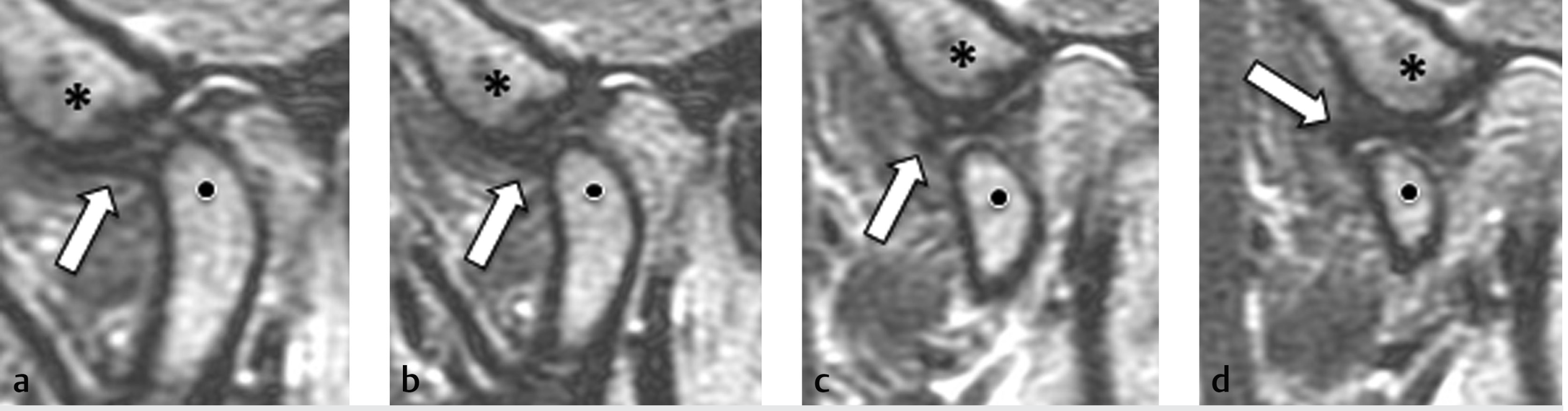
High-resolution proton-density MRI of a cadaveric TMJ in the open-mouth position. The articular disc (arrows) translates anteriorly as the condyle (dots) moves forward under the articular eminence (*). Panels a-d show progressive opening. Note the retrodiscal layers separating as the mouth opens.
3. UTE MRI - Condylar Fibrocartilage & Bone (Imaging Anatomy Atlas)
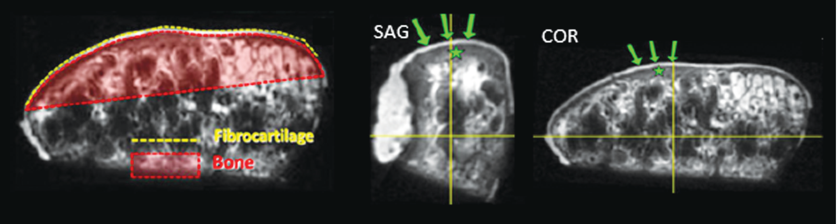
3D ultrashort time-to-echo (UTE) MRI of the harvested condyle in sagittal (SAG) and coronal (COR) planes. Green arrows point to the fibrocartilage surface of the condyle; the red-shaded zone = bone; yellow dashed = bone-cartilage interface. This confirms that TMJ articular surfaces are fibrocartilage, not hyaline cartilage.
This is a shared conversation. Sign in to Orris to start your own chat.