
Hydrocoele all surgery possible, hernia of hydrocoele and hydrocoele of hernia, all surgeries possible of hernia, why cant we aspirate hydrocoele, female hydrocoele
hydrocele types communicating non-communicating cord diagram
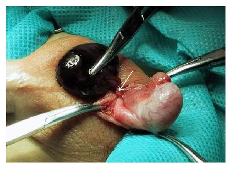
This clinical intraoperative photograph shows a surgical exploration of the left scrotum in a pediatric patient. The image features a large, dark, globular mass identified as an encysted hydrocele of the cord. The deep purple-to-black coloration of the hydrocele sac indicates hemorrhagic contents (bloody fluid), likely due to the torsion of the hydrocele. A white arrow points to the site of a 360-degree rotation (torsion) at the distal end of the sac. Adjacent to the hydrocele, the testis and epididymis are visible, appearing as pinkish-white, smaller, and morphologically normal structures with preserved vascularization, distinct from the pathology of the hydrocele. Several surgical hemostats are seen retracting the scrotal tissues to provide exposure. The image demonstrates a rare case of acute scrotum caused by the torsion of a non-communicating hydrocele, a critical differential diagnosis for testicular torsion.
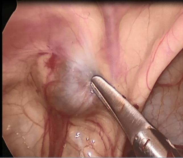
This endoscopic clinical photograph displays a laparoscopic view of the internal inguinal ring (IIR) in a pediatric patient. The image illustrates a Type I spermatic cord hydrocele, characterized by a rounded, grayish, translucent cystic structure located within the inguinal canal area. A metallic surgical grasper is shown clamped onto the cyst wall, demonstrating its location relative to the peritoneum. Crucially, the internal inguinal ring is visually closed, with a smooth peritoneal surface indicating no communication between the hydrocele and the peritoneal cavity. Surrounding tissues show normal surgical anatomy, including pinkish-red membranes, fine vascularization, and the proximity of the spermatic cord structures. This visual serves as a diagnostic reference for classifying non-communicating hydroceles where the processus vaginalis has successfully obliterated at the level of the internal ring.
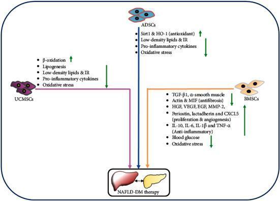
A pathophysiology diagram illustrating the therapeutic mechanisms of three mesenchymal stem cell (MSC) types in the treatment of Non-alcoholic Fatty Liver Disease (NAFLD) and Diabetes Mellitus (DM). The diagram highlights Umbilical Cord-derived MSCs (UCMSCs), Adipose-derived MSCs (ADSCs), and Bone Marrow-derived MSCs (BMSCs). Each cell type is linked to specific molecular and cellular effects indicated by green up and down arrows. UCMSCs are shown to increase beta-oxidation while decreasing lipogenesis, low-density lipids, insulin resistance (IR), pro-inflammatory cytokines, and oxidative stress. ADSCs increase Sirt1 and HO-1 (antioxidant pathways) and decrease lipids, IR, cytokines, and oxidative stress. BMSCs exhibit comprehensive effects: decreasing TGF-beta1 and alpha-SMA (antifibrosis), increasing proliferation and angiogenesis factors (HGF, VEGF, EGF, MMP-2, CXCL5), promoting anti-inflammatory cytokines (IL-10), and reducing blood glucose and oxidative stress. All three pathways converge on 'NAFLD-DM therapy,' represented by a liver and pancreas icon, emphasizing a multi-targeted regenerative approach to metabolic and hepatic restoration.
inguinal hernia repair Lichtenstein mesh Shouldice Bassini surgery
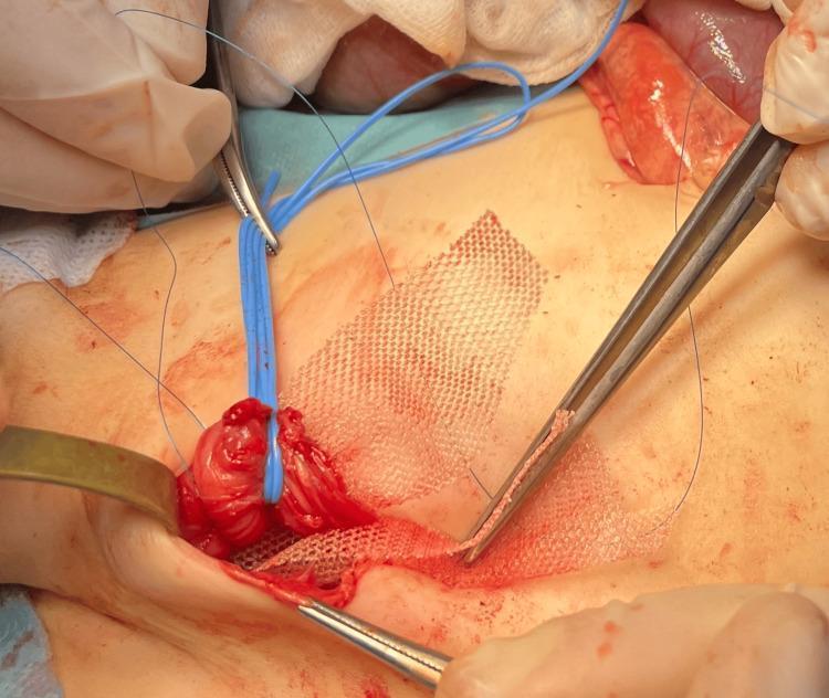
An intraoperative clinical photograph showing a step in the repair of a recurrent inguinal hernia using the Lichtenstein technique. The image features a rectangular, white, woven synthetic mesh (identified as a Vicryl patch) being positioned over the floor of the inguinal canal. A surgical forceps is visible on the right, grasping the edge of the mesh for placement. To the left, a blue vessel loop is used to retract a reddish, fleshy tissue mass, which likely represents a segment of the hernia sac or the spermatic cord. Blue non-absorbable sutures are also present within the surgical field for securing the mesh to the inguinal ligament and surrounding fascia. The surgical site is exposed via an open incision, with metal retractors maintaining visibility of the underlying anatomical structures. This visual demonstrates the tension-free mesh reinforcement used in pediatric surgery to prevent hernia recurrence, particularly in complex cases such as Loeys-Dietz syndrome.
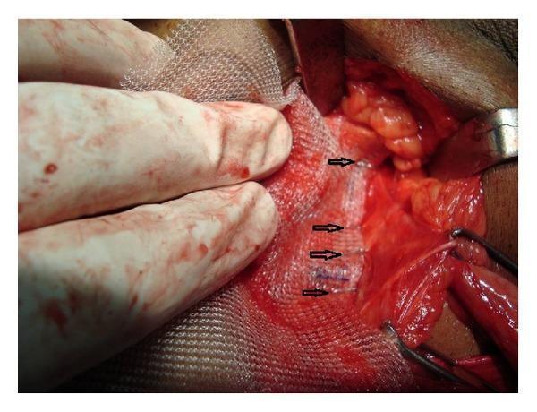
This intraoperative clinical photograph demonstrates a surgical field during an inguinal hernia repair. The image captures the fixation of a white, macroporous polypropylene mesh to the underlying anatomical structures. Black arrowheads highlight a linear series of stainless steel surgical staples, spaced approximately 1-2 cm apart, which anchor the mesh inferiorly along the inguinal ligament. A gloved hand and surgical retractors are visible, maintaining exposure and tension on the mesh during the procedure. The surrounding tissues show typical surgical hyperemia and exposure of the inguinal canal posterior wall. This visual illustrates the tension-free hernioplasty technique (Lichtenstein repair) specifically focusing on the mechanical fixation method using a skin stapler as an alternative to traditional polypropylene sutures for mesh stabilization. The target audience includes surgical residents and medical students learning about inguinal anatomy and prosthetic mesh fixation techniques in general surgery.
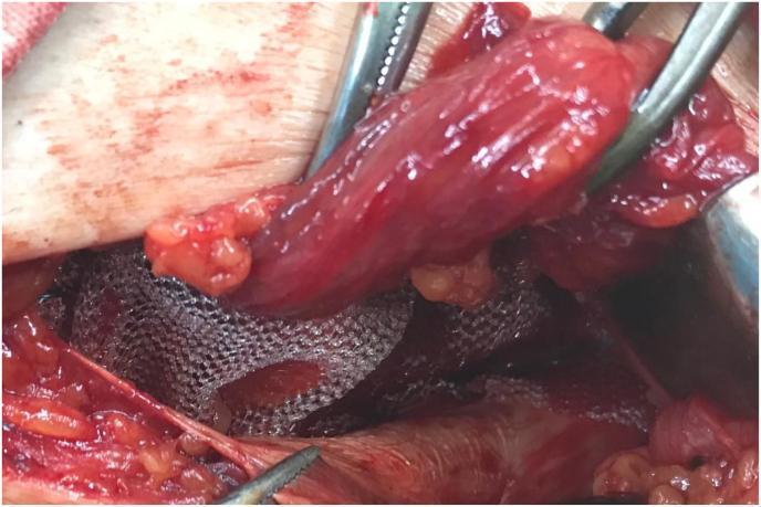
This intraoperative clinical photograph demonstrates a Lichtenstein-style inguinal hernia repair (hernioplasty). The visual focus is on the reinforcement of the posterior wall of the inguinal canal. A silver-gray, woven polypropylene mesh is visible, positioned deep to the spermatic cord. The cord, appearing as a reddish-pink, tubular, fleshy structure, is being retracted superiorly and laterally by surgical instruments to allow for the proper placement of the mesh. The mesh is shown sutured to the surrounding musculoskeletal landmarks, including the conjoined tendon superiorly and the reflection of the inguinal ligament inferiorly. A central aperture is created in the mesh to accommodate the passage of the spermatic cord through the internal ring. This image serves as an educational reference for surgical residents and medical students studying the second stage of open inguinal hernia mesh repair, emphasizing anatomical orientation and the relationship between prosthetic material and the cord structures.
Hydrocele and Hernia: Complete Surgical Overview
PART 1: HYDROCELE - DEFINITION AND TYPES
Structural Classification (4 Types)
| Type | Description |
|---|---|
| 1. Communicating (Congenital) Hydrocele | Patent processus vaginalis allows peritoneal fluid to flow into the tunica vaginalis. Intermittent - may reduce when child lies down. Associated with indirect inguinal hernia. |
| 2. Non-communicating (Vaginal) Hydrocele | Processus vaginalis is closed. Fluid accumulates due to excessive production or defective absorption by tunica vaginalis (lymphatic disruption). Most common type globally. |
| 3. Infantile Hydrocele | Mid-portion of processus remains patent; distal end closed correctly. Proximal end may be open (communicating with tunica vaginalis). |
| 4. Hydrocele of the Cord | Distal end of processus closes correctly; mid-portion remains patent; proximal end is closed. Results in a smooth oval swelling lying above the testis near the spermatic cord. |
PART 2: ALL POSSIBLE SURGERIES FOR HYDROCELE
In Adults (Acquired/Primary Hydrocele)
1. Lord's Operation (Plication)
- Suitable for: thin-walled hydrocele sacs
- Technique: A series of interrupted absorbable sutures plicates (bunches up) the redundant tunica vaginalis. When tied, the tunica bunches at its attachment to the testis.
- Advantage: Minimal dissection, reduced risk of haematoma
- Best for: Primary (idiopathic) hydroceles with thin sac walls
2. Jaboulay's Procedure (Eversion / Bottle Operation)
- Suitable for: larger or thicker-walled sacs
- Technique: The sac is opened, everted (turned inside out) behind the testis, and anchored with sutures. The testis is placed in a fascial pouch.
- Caution: Risk of large scrotal haematoma from cut-edge bleeding; overrunning stitches at the cut edge reduce this risk.
3. Excision of the Sac (Andrews' Operation / Radical Cure)
- Suitable for: thick-walled hydroceles (established acquired), filarial/chyloceles with dense adhesions
- Technique: The sac is completely excised.
- Caution: Highest risk of haematoma; haemostasis is critical.
In Children (Congenital / Communicating Hydrocele)
- Herniotomy (Ligation of patent processus vaginalis) via inguinal approach - this is the procedure of choice. The processus is ligated at the internal ring. Not treated by scrotal approach.
For Abdominoscrotal Hydrocele
- Requires both abdominal and scrotal approach; the abdominal component must be addressed.
PART 3: HERNIA OF HYDROCELE (Hydrocele + Hernia Coexisting)
- The processus vaginalis is wide enough to allow bowel/omentum to herniate in addition to fluid.
- Surgery: Inguinal approach (not scrotal). The processus is opened, hernia contents reduced, sac ligated at internal ring (herniotomy). The distal hydrocele sac is dealt with separately (everted or excised).
- From Fischer's Mastery of Surgery: "In the setting of scrotal hernias with hydrocele, it is best to remove the hernia sac in its entirety whenever possible. When complete sac removal is not an option, the distal sac or hydrocele should be opened widely, to avoid reaccumulation of serous fluid. It is important NOT to tie off any distal sac, as that would create a closed space in which a hydrocele could form."
PART 4: HYDROCELE OF HERNIA (Post-Hernia Repair Hydrocele)
- Mechanism: After varicocelectomy or hernia repair, lymphatic obstruction leads to fluid accumulation.
- After non-microscopic varicocelectomy, incidence of hydrocele = 3-33% (average ~7%).
- At least half of these grow large enough to warrant surgical excision.
- Prevention: Use of optical magnification (loupes or operating microscope) to identify and preserve lymphatics almost eliminates this risk.
- Treatment: Same as for any primary hydrocele (Lord's / Jaboulay's / excision).
PART 5: ALL POSSIBLE SURGERIES FOR HERNIA
Inguinal Hernia Repairs
A. Open Tissue Repairs (Suture-based, No Mesh)
| Operation | Principle |
|---|---|
| Bassini Repair | Posterior wall of inguinal canal reconstructed by suturing conjoined tendon to inguinal ligament behind the cord |
| Shouldice Repair | Multi-layer continuous suture repair of the posterior wall; gold standard tissue repair; lowest recurrence among suture repairs (~1%) |
| Halsted Repair | Modification of Bassini; cord placed subcutaneously |
| McVay (Cooper's Ligament) Repair | Repair to Cooper's ligament; useful for femoral hernias |
| Herniotomy | Ligation and excision of sac only (children, simple indirect hernias) |
| Herniorraphy | Sac ligation + repair of posterior wall |
B. Open Mesh Repairs (Tension-free)
| Operation | Principle |
|---|---|
| Lichtenstein Tension-free Repair | Polypropylene mesh placed flat over the posterior wall; mesh sutured to inguinal ligament inferiorly and conjoined tendon superiorly; keyhole for spermatic cord. Most widely used. |
| Plug and Patch Repair (Rutkow-Robbins) | Mesh plug inserted into internal ring + flat mesh overlay |
| Prolene Hernia System (PHS) | Bilayer mesh device covering both preperitoneal space and anterior wall |
| Gilbert / Trabucco Repair | Mesh techniques with modified fixation |
C. Laparoscopic / Minimally Invasive Repairs
| Operation | Principle |
|---|---|
| TEP (Totally Extraperitoneal Repair) | Preperitoneal space developed without entering peritoneum; mesh placed in preperitoneal space covering all three groin hernia sites |
| TAPP (Trans-Abdominal Pre-Peritoneal Repair) | Peritoneal cavity entered laparoscopically, flap raised, mesh placed in preperitoneal space, peritoneum re-closed |
| IPOM (Intraperitoneal Onlay Mesh) | Rarely used for inguinal hernias; mesh placed intraperitoneally |
| Robotic TAPP / TEP | Robot-assisted variants with improved ergonomics |
D. For Specific Hernia Types
| Hernia Type | Preferred Surgery |
|---|---|
| Femoral Hernia | McVay repair, or low (femoral) approach with Cooper's ligament suture; mesh plug; TAPP/TEP |
| Strangulated Hernia | Emergency herniotomy + bowel assessment; mesh avoided if bowel resection needed |
| Paediatric Inguinal Hernia | Herniotomy only (high ligation of sac) - no mesh required |
| Recurrent Inguinal Hernia | Laparoscopic approach preferred (TEP/TAPP) if previous open repair; open pre-peritoneal (Stoppa/Wantz) |
| Bilateral Inguinal Hernia | TEP/TAPP (single procedure covers both sides) |
E. Preperitoneal Repairs
| Operation | Description |
|---|---|
| Stoppa (GPRVS) | Giant prosthetic reinforcement of visceral sac; large mesh in preperitoneal space via midline incision; good for bilateral/recurrent |
| Wantz Repair | Unilateral version of Stoppa |
| Nyhus Repair | Posterior preperitoneal approach via lower midline |
PART 6: WHY WE CANNOT (Should NOT) ASPIRATE A HYDROCELE
-
Fluid always reaccumulates - within a week or so in most cases, because the underlying cause (imbalance between secretion and absorption by the tunica) is not addressed.
-
Risk of haematocele - aspiration needle can injure blood vessels in the tunica vaginalis, causing bleeding into the sac, creating a haematocele (blood in the tunica) which is much harder to treat.
-
Risk of infection - introducing a needle into a serous cavity can introduce bacteria, leading to pyocele (pus in the tunica).
-
Tumour seeding risk - if the hydrocele is secondary to a testicular tumour (which can present as a hydrocele), aspiration risks malignant needle-track implantation and tumour dissemination. This is a critical safety concern.
-
Damage to testis/epididymis - blind aspiration risks direct injury to underlying testicular tissue.
-
Sclerosant injection (e.g., tetracycline) can be done after aspiration but is painful, has a variable success rate, and is only appropriate in patients unfit for surgery.
"Aspiration of the hydrocele fluid is simple, but the fluid always reaccumulates within a week or so... Aspiration can result in bleeding into the hydrocele sac and haematocele formation... If a tumour is suspected, the hydrocele should not be punctured (risk of malignant needle-track implantation)." - Bailey and Love's Short Practice of Surgery, 28th Ed.
PART 7: FEMALE HYDROCELE (Hydrocele of the Canal of Nuck)
What is it?
- Fluid trapped in an incompletely obliterated processus vaginalis (canal of Nuck) in females.
- The cyst lies in relation to the round ligament and is always at least partially within the inguinal canal.
- It is the female counterpart of the hydrocele of the cord in males.
Clinical Features
- Presents as a smooth, cystic, translucent swelling in the groin/labia majora.
- May be mistaken for a femoral hernia, inguinal hernia, or inguinal lymph node.
- Transilluminates.
- Can be communicating or non-communicating.
Association with Hernia
- Just as in males, a patent canal of Nuck in females can be associated with an indirect inguinal hernia (herniation of bowel or omentum through the same patent processus).
Treatment
- Surgical excision via inguinal approach: the cyst is excised and the patent processus is ligated at the internal ring.
- If associated hernia: herniotomy/herniorrhaphy performed at the same time.
Surgical Images
Quick Summary Table
| Topic | Key Points |
|---|---|
| Hydrocele surgeries | Lord's (plication), Jaboulay's (eversion), Excision (radical cure); Herniotomy in children |
| Hernia with hydrocele | Inguinal approach; excise/open distal sac widely; never ligate distal sac alone |
| Post-hernia repair hydrocele | Lymphatic obstruction; treat like any hydrocele; microscopy prevents it |
| Hernia surgeries | Tissue: Bassini, Shouldice, McVay, Herniotomy; Mesh: Lichtenstein, Plug+Patch, PHS; Lap: TEP, TAPP, Robotic |
| Why not aspirate | Fluid reaccumulates; haematocele risk; infection risk; tumour seeding risk; testicular injury |
| Female hydrocele | Canal of Nuck hydrocele; inguinal approach; may coexist with inguinal hernia |
Ellaborate surgical procedures
Lord plication hydrocele operation sutures tunica vaginalis
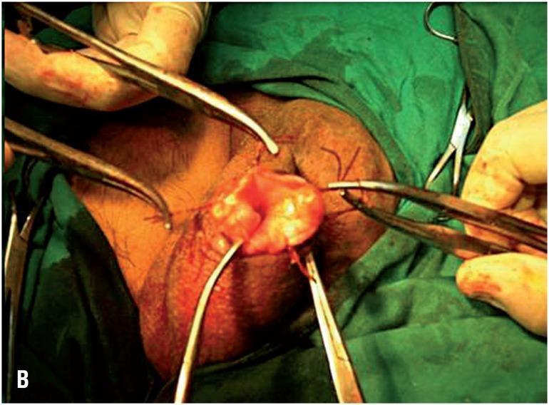
This clinical photograph captures an intraoperative view of a surgical procedure on the male scrotum, specifically a minimally invasive hydrocelectomy using the eversion technique (Lord's or Jaboulay-type modification). The surgical field shows the scrotum stabilized with surgical clamps and forceps. A small incision has been made, through which the parietal tunica vaginalis is everted. The image demonstrates the critical step of suturing the edge of the tunica vaginalis directly to the dartos fascia and scrotal skin. This maneuver aims to keep the visceral surface of the tunica vaginalis exposed to the lymph-rich subcutaneous tissues to facilitate fluid absorption and prevent hydrocele recurrence. Visible elements include surgical instrumentation (hemostats and forceps), interrupted suture lines securing the everted tissue, and the characteristic red, vascularized appearance of the visceral tunica. This content is intended for urological surgical training, illustrating anatomical landmarks and technical execution of scrotal sac eversion.
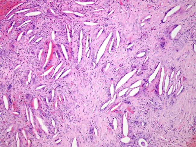
Light microscopy of a thickened hydrocele wall from tunica vaginalis demonstrates cross-sectional histology with dense fibrous stroma, scattered inflammatory cells, and cholesterol clefts. Acute neutrophilic bands and chronic lymphoplasmacytic infiltrates coexist, while multinucleated foreign body giant cells form around cholesterol clefts, reflecting a cholesterol granulomatous reaction. Fibrosis is prominent, with fascicular, collagen-rich architecture and minimal residual lining. The observed histology indicates chronic inflammatory and granulomatous response to leaked or degraded hydrocele contents, superimposed infection, and scarring. Diagnostic significance includes evidence of infectious complication and chronicity; findings support secondary infection in a long-standing hydrocele and help differentiate from neoplastic or purely serous processes. Clinically relevant search terms: hydrocele wall pathology, tunica vaginalis inflammation, cholesterol granuloma, foreign body giant cell reaction, chronic infection, fibrosis, scrotal hydrocele, hydrocelectomy planning, granulomatous scrotal disease.
TEP totally extraperitoneal hernia repair laparoscopic preperitoneal mesh placement
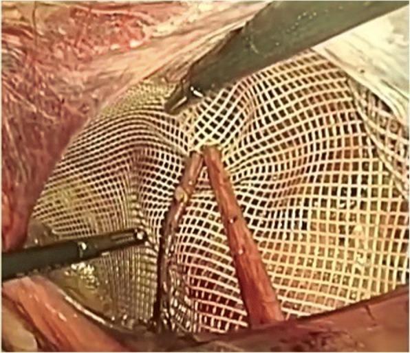
Laparoscopic intraoperative photograph demonstrating a 'key-holed' or slit mesh placement during a transabdominal preperitoneal (TAPP) or totally extraperitoneal (TEP) inguinal hernia repair. A white, rectangular polyester mesh with a macro-porous, grid-like structure is shown. The mesh has been vertically engraved to create a slit (keyhole), allowing it to be positioned around the spermatic cord structures, which are visible passing through the opening. Two laparoscopic graspers are seen: one superiorly manipulating the upper edge of the mesh and another inferiorly stabilizing the material. The background shows the dissected preperitoneal space, including the retroinguinal space of Bogros. This technique is designed to provide comprehensive coverage of the myopectineal orifice while accommodating the passage of the vas deferens and spermatic vessels to prevent entrapment or future recurrence. The surgical field displays typical anatomy of the posterior inguinal wall, used for teaching prosthetic reinforcement in hernia surgery.
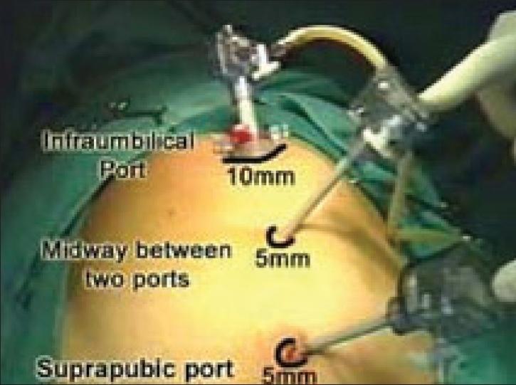
This clinical photograph illustrates the standard laparoscopic port placement for a Totally Extraperitoneal (TEP) inguinal hernia repair. The patient is positioned supine, with the abdomen draped in green surgical sheets. Three trocars are inserted along the midline into the preperitoneal space. The superior port is a 10-mm infraumbilical port, equipped with a valve mechanism and insufflation tubing, typically used for the 30-degree laparoscope. Positioned inferiorly in the midline are two working ports: a 5-mm port placed midway between the umbilical and pubic regions, and a 5-mm suprapubic port located approximately 2–3 cm above the pubic symphysis. The visual demonstrates the vertical triangulation required for instruments to reach the inguinal region. This configuration allows the surgeon to operate within the extraperitoneal plane without entering the peritoneal cavity, minimizing the risk of visceral injury and facilitating mesh placement over the direct, indirect, and femoral hernia sites.
Bassini hernia repair anatomy diagram conjoined tendon inguinal ligament
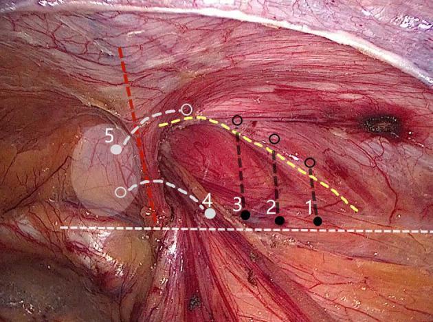
This clinical photograph shows a laparoscopic view of the inguinal region during an internal ring closure procedure, specifically demonstrating a 'five-stitches method' for hernia repair. The surgical field displays the reddish-pink fibrous texture of the conjoined tendon and the iliopubic tract, with prominent epigastric vessels visible. Overlaid on the anatomy are numbered markers (1-5) and colored dashed lines illustrating the suturing technique: black solid dots indicate needle entry points, while open circles represent extraction points. Points 1-3 follow a lateral-to-medial linear path along the iliopubic tract, connected by a dashed yellow line. Points 4 and 5 transition into a U-shaped configuration, marked by a red dashed line, to navigate around the epigastric vessels. A white dashed line indicates the alignment of tissues for approximation. The diagram highlights the clinical strategy of superficial, small-bite suturing intended to avoid underlying neurovascular structures, including the iliac artery and branches of the genitofemoral and ilioinguinal nerves.
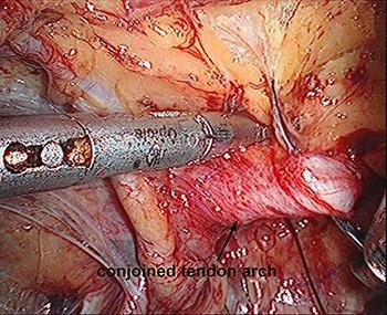
This endoscopic clinical photograph depicts an intraoperative view of the preperitoneal space during a Totally Extraperitoneal (TEP) inguinal hernia repair. The focal point is the conjoined tendon arch (indicated by a black arrow and text label), which appears as a reddish-pink, fibrous anatomical structure. A metallic laparoscopic instrument is visible, positioned to perform a puncture and suturing through the tendon arch using a suture needle. The surrounding surgical field shows thin, translucent connective tissues, small blood vessels, and adjacent musculature of the posterior abdominal wall. This image serves as an educational guide for identifying key landmarks in the inguinal region and demonstrates a specific technique for internal ring suturing to reduce the defect size before mesh placement. The educational focus is on laparoscopic anatomy and surgical maneuvers for direct or indirect hernia management.
Elaborate Surgical Procedures
SECTION A: HYDROCELE OPERATIONS - STEP BY STEP
1. LORD'S OPERATION (Plication)
- Incision: Transverse or longitudinal incision on the anterior scrotum over the most prominent part of the hydrocele.
- Layers dissected: Dartos, external spermatic fascia, cremasteric fascia, internal spermatic fascia are opened in sequence until the tunica vaginalis (hydrocele sac) is reached.
- Delivery: The hydrocele sac is grasped with tissue forceps and delivered through the wound into the operative field.
- Opening the sac: A small nick is made into the sac; fluid is aspirated or allowed to drain.
- Plication (key step): Multiple interrupted absorbable sutures (e.g., 2/0 Vicryl) are placed in a radial pattern around the sac, taking bites of the tunica vaginalis. When all sutures are tied simultaneously, the tunica vaginalis bunches up ("pleats") at its attachment to the testis and epididymis, obliterating the potential space.
- The testis is returned to the scrotum; no sac tissue is removed.
- Closure: Dartos closed with absorbable suture; skin closed.
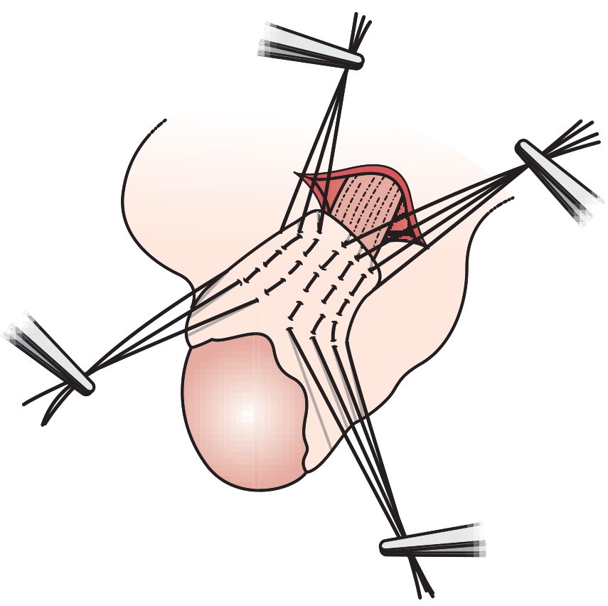
2. JABOULAY'S PROCEDURE (Eversion / Bottle Operation)
- Incision and delivery: Same as Lord's - scrotal incision, layers dissected, hydrocele sac delivered into wound.
- Opening the sac: The anterior wall of the tunica vaginalis is incised widely, exposing the testis and epididymis.
- Fluid evacuation: All fluid is drained.
- Inspection: Testis and epididymis are carefully inspected for secondary pathology (tumour, epididymitis).
- Eversion (key step): The cut edges of the tunica vaginalis are carried posteriorly behind the testis. The tunica is everted (turned inside-out) so its mesothelial surface now faces outward into the scrotal tissue. The two cut edges are sutured together behind the testis with interrupted or running absorbable sutures.
- Haemostasis: Critical - overrunning stitches at the cut edge are used to control bleeding, as the raw edges can bleed significantly into the scrotal tissue causing haematoma.
- The testis is returned to the scrotum.
- Closure: Layers closed in sequence.
3. ANDREWS' OPERATION / EXCISION OF THE SAC (Radical Cure)
- Incision and delivery: Same initial steps as above.
- Opening: The sac is opened and fluid drained.
- Excision (key step): The entire excess tunica vaginalis wall is excised, leaving only a small cuff of tissue around the testis and epididymis (to avoid injuring the blood supply and epididymis).
- Haemostasis: The cut edge is overrun with a continuous haemostatic suture - this is the most haematoma-prone operation of the three.
- The testis is replaced in the scrotum.
- Closure: Layers closed.
4. HERNIOTOMY (Children / Congenital Communicating Hydrocele)
- Incision: Small transverse incision in the skin crease just above and lateral to the pubic tubercle (inguinal crease).
- Layers: Scarpa's fascia opened, external oblique aponeurosis opened along the line of its fibers, inguinal canal entered.
- Cord identification: Spermatic cord identified, ilioinguinal nerve protected.
- Sac isolation: The processus vaginalis (indirect hernia sac) is identified anteriorly on the cord and carefully dissected away from cord structures (vas deferens, testicular vessels).
- High ligation (key step): The sac is opened, inspected, and ligated at the level of the deep inguinal ring (internal ring) with a transfixion suture. This obliterates the communication with the peritoneal cavity.
- Distal sac: The distal part of the sac (in the scrotum) is left open (not tied off) - this prevents formation of a new hydrocele.
- Closure: External oblique closed, skin closed.
SECTION B: HERNIA OPERATIONS - STEP BY STEP
INCISION AND INITIAL STEPS (Common to All Open Inguinal Repairs)
- Complete division of external oblique aponeurosis and transversalis fascia
- Differentiation between indirect and direct defects
- Isolation of spermatic cord or round ligament
- Ligation and removal of the sac at the deep inguinal ring flush with the peritoneum
- Oblique reconstruction of the inguinal canal with anterior and posterior wall plus internal and external rings
- Skin and subcutaneous tissue
- Scarpa's fascia
- External oblique aponeurosis (opened parallel to its fibers, from the external ring laterally)
- Nerves identified and protected: Ilioinguinal nerve (runs in the canal), iliohypogastric nerve (on internal oblique), genital branch of genitofemoral nerve (with posterior cremaster)
- Cremasteric fibers are divided or retracted; cord is slung with a soft rubber sling
- Indirect sac (if present) is found anteromedially on the cord, separated from cord structures, opened, reduced or excised, and ligated at the internal ring
- Direct hernia sac (if present) lies medially through the floor of the canal; contents reduced, sac inverted
5. BASSINI REPAIR (Tissue Repair)
- The transversalis fascia is divided from the internal ring to the pubic tubercle.
- The hernia sac is dealt with (ligated at internal ring for indirect; inverted for direct).
- The cord is retracted superiorly.
- Repair (key step): The "triple layer" - transversus abdominis aponeurosis + internal oblique + transversalis fascia together are sutured to the shelving edge of the inguinal ligament using interrupted non-absorbable sutures, from pubic tubercle to the internal ring.
- This reconstructs the posterior wall of the inguinal canal. The internal ring is tightened around the cord.
- The cord is replaced anterior to the repair; external oblique is closed over the cord.
6. SHOULDICE REPAIR (Multi-Layer Tissue Repair)
- After initial dissection and sac ligation, the cremaster muscle is excised (divided and ligated) to expose the internal ring fully.
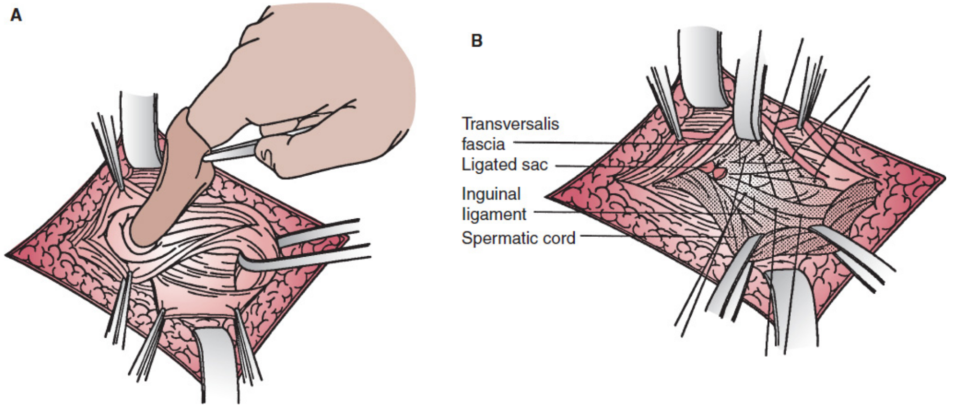
- Floor incision (critical step): Scissors are passed posterior to the transversalis fascia from the internal ring to the pubic tubercle. The transversalis fascia is incised (opened) along its entire length, creating an upper (medial) flap and a lower (lateral) flap.
- Preperitoneal fat is cleaned off the posterior surface of both flaps.
-
Layer 1 (of first suture): Starting at the pubic tubercle medially, the free edge of the lower (lateral/inferior) transversalis flap is sutured in a running, imbricated fashion to the posterior surface of the upper flap and posterior rectus sheath. Running 0.5-1 cm bites. This continues laterally to the internal ring, tightening the fascia around the cord ("recreating the internal ring"). Critically, the new ring must admit only the back of a DeBakey forceps - not a finger - to avoid ischaemia.
-
Layer 2 (same suture returning): The suture continues back (medially) as the second layer, suturing the upper transversalis flap to the base of the lower flap and the inguinal ligament (shelving edge). Tied to the tail of the first layer at the pubic tubercle.
-
Layer 3 (second suture, starting from internal ring): Internal oblique and transversus abdominis aponeurosis (conjoined tendon) are brought to the external oblique aponeurosis/inguinal ligament. Running continuous suture from lateral to medial.
-
Layer 4 (same suture returning): Second pass medially to laterally, creating the fourth layer. Tied at the internal ring.
- The cord is replaced in the canal; external oblique closed.
- Requires an experienced trained assistant (at least 3 people: surgeon + 2 assistants)
- The cremaster bundle ("scarf") is wrapped around the new internal ring to recreate it
- Not tying off the distal sac - opened widely to prevent post-op hydrocele
- Relaxing incision NOT needed in most cases (unlike McVay)
7. McVAY REPAIR (Cooper's Ligament Repair)
- Initial dissection same as above.
- The attenuated central portion of the inguinal floor (transversalis fascia) is excised.
- Cooper's ligament (pectineal ligament - on the superior surface of the superior pubic ramus) is clearly identified.
- Repair (key step): Transversus abdominis aponeurosis + transversalis fascia are sutured to Cooper's ligament from pubic tubercle medially to the femoral vein laterally.
- Transition stitch: A crucial stitch is placed between transversalis fascia, Cooper's ligament, and inguinal ligament to bridge the repair above the femoral vessels (prevents femoral hernia recurrence).
- The repair continues laterally along the inguinal ligament.
- Relaxing incision: MANDATORY - a vertical incision in the anterior rectus sheath (3-4 cm above pubis to level of internal ring) is required to reduce tension; the rectus muscle protects against herniation through this defect.
8. LICHTENSTEIN TENSION-FREE REPAIR (Open Mesh - Gold Standard)
- Initial dissection identical to tissue repairs - cord isolated, sac dealt with.
- The floor of the inguinal canal is assessed but NOT opened.
- Mesh preparation: A piece of polypropylene mesh (~15 x 8 cm) is fashioned. The mesh is trimmed to fit. A keyhole slit is made at the lateral end to accommodate the spermatic cord.
- Mesh placement (key step): The mesh is laid flat over the entire posterior wall of the inguinal canal, covering from 1.5 cm medial to the pubic tubercle to well lateral of the internal ring.
- Mesh fixation:
- Inferior edge: Sutured to the shelving edge of the inguinal ligament with a continuous non-absorbable suture (e.g., 2/0 Prolene) from the pubic tubercle to just lateral of the internal ring.
- Superior edge: Tacked to the internal oblique and conjoined tendon with interrupted sutures (being careful to avoid the iliohypogastric nerve).
- Medial end: Anchored to the pubic tubercle periosteum.
- Keyhole closure: The two tails of the keyhole are sutured together around the cord at the internal ring, snugly recreating the ring.
- Nerve management: Ilioinguinal nerve, iliohypogastric nerve, and genital branch of genitofemoral nerve are all identified and either preserved in situ or neurectomised (if entrapment risk is high).
- External oblique is closed over the mesh and cord.
9. TAPP REPAIR (Trans-Abdominal Pre-Peritoneal)
- Diagnostic laparoscopy: Both myopectineal orifices are inspected; unrelated pathology excluded.
- Peritoneal incision: A transverse incision is made in the peritoneum lateral to the medial umbilical ligament, extending to the anterior superior iliac spine. Must enter the TRUE preperitoneal plane (not retromuscular).
- Preperitoneal dissection: The preperitoneal space is developed, exposing all key landmarks:
- Cooper's ligament
- Inferior epigastric vessels
- Iliopubic tract
- Vas deferens and spermatic vessels (forming the "Triangle of Doom" - external iliac vessels between them - NO staples here)
- "Triangle of Pain" (lateral to the spermatic vessels - genitofemoral and lateral femoral cutaneous nerves - NO staples here)
- Hernia sac reduction: The indirect sac is reduced from the internal ring; large sacs may be divided.
- Mesh placement: A large mesh (at least 10 x 15 cm) is introduced and unrolled to cover all three hernia sites (indirect, direct, femoral) - the myopectineal orifice of Fruchaud.
- Mesh fixation: The mesh can be fixed with tacks (to Cooper's ligament, internal oblique - avoiding the triangles of doom and pain) or left unfixed ("self-gripping" mesh). Fixation below the iliopubic tract is AVOIDED to prevent nerve injury.
- Peritoneal closure: The peritoneal flap is re-closed with tacks or suture to exclude the mesh from the peritoneal cavity.
10. TEP REPAIR (Totally Extraperitoneal)
- Entry: Infraumbilical incision; anterior rectus sheath opened on one side; rectus muscle retracted laterally. Dissection begun between rectus and posterior rectus sheath (retrorectus space).
- Space creation: A balloon dissector or blunt dissection is used to develop the preperitoneal space. Two additional midline cannulas are inserted under direct vision.
- CO₂ insufflation: The preperitoneal space is insufflated with CO₂ to 8-12 mmHg.
- Preperitoneal dissection: Identical anatomy as TAPP is developed:
- Cooper's ligament, inferior epigastric vessels, iliopubic tract, vas deferens, spermatic vessels are all identified.
- The hernia sac is reduced.
- Triangle of Doom and Triangle of Pain are respected.
- Mesh placement: Large mesh (10 x 15 cm minimum) is inserted through the 10mm port and unrolled to cover the myopectineal orifice.
- No peritoneal closure needed - the peritoneum was never opened.
- Desufflation: The space is desufflated; the mesh is held in place by the intra-abdominal pressure (and optionally tacked).
11. STOPPA REPAIR (GPRVS - Giant Prosthetic Reinforcement of the Visceral Sac)
- Midline infraumbilical incision.
- The preperitoneal space is developed bilaterally by dissection behind the rectus and over the peritoneum ("visceral sac").
- A very large mesh (typically 24 x 16 cm or bigger) is placed in the preperitoneal space to cover the entire myopectineal orifice bilaterally.
- The mesh is spread out and held in place by the visceral weight (no fixation required).
- Wound closure in layers.
12. PROLENE HERNIA SYSTEM (PHS) - Bilayer Mesh
- Open inguinal approach (same initial steps).
- The deep component of PHS is inserted through the internal ring or defect into the preperitoneal space and unrolled.
- The connector sits in the ring.
- The superficial onlay component lies flat over the posterior wall like a Lichtenstein repair.
- Both layers are sutured in place.
CRITICAL ANATOMICAL TRIANGLES (for Laparoscopic Repairs)
| Structure | Boundaries | What to Avoid |
|---|---|---|
| Triangle of Doom | Medial: vas deferens; Lateral: spermatic vessels | External iliac artery and vein lie here - NO tacks/staples |
| Triangle of Pain | Lateral to spermatic vessels, below iliopubic tract | Genitofemoral nerve + lateral femoral cutaneous nerve - NO tacks/staples |
COMPARISON OF RECURRENCE RATES
| Operation | Type | Recurrence Rate |
|---|---|---|
| Bassini | Tissue | 10-15% (general practice) |
| Shouldice | Tissue | ~1-2% (Shouldice Hospital); up to 15% elsewhere |
| McVay | Tissue | ~5-10% |
| Lichtenstein | Open Mesh | 1-1.6% |
| TAPP / TEP | Laparoscopic Mesh | ~1-2% |
| Stoppa | Open Preperitoneal Mesh | ~1-2% |
POSTOPERATIVE CARE (All Hernia Repairs)
- Discharged same day (most cases); local anaesthesia + IV sedation for Shouldice/Lichtenstein.
- Multimodal analgesia: paracetamol + NSAIDs around the clock for 2-5 days; opioids as rescue only.
- Resume normal activities at approximately 4 weeks.
- Heavy lifting restriction for 4-6 weeks.
COMPLICATIONS OF HERNIA REPAIR
| Complication | Mechanism | Management |
|---|---|---|
| Recurrence | Technical failure, missed femoral hernia, inadequate mesh size | Posterior/laparoscopic approach if prior anterior repair |
| Hydrocele formation | Lymphatic obstruction post-repair | Lord's / Jaboulay's / excision |
| Cord ischaemia | Overtightened internal ring | Doppler U/S; usually self-limiting with atrophy |
| Haematoma | Scrotal/inguinal; vascular injury | Conservative; rarely surgical drainage |
| Nerve entrapment / chronic pain | Ilioinguinal, iliohypogastric, genitofemoral | Neurectomy if persistent |
| Dysejaculation | Suture repair related (Ben David syndrome) | Reassurance; self-limited, resolves over months-years |
| Mesh infection | Rare; ~0.1% | Antibiotics; mesh removal if required |