
Giant cells lesions of bone Pathology
Giant cells lesions of bone Pathology
"Giant Cell Tumor of Bone"[MeSH Terms] AND denosumab
giant cell tumor of bone histology multinucleated giant cells stromal cells
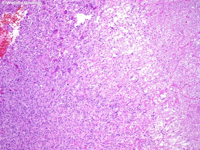
This is a histopathology slide of a giant cell tumor of bone, prepared from a skeletally located tumor and stained with hematoxylin and eosin (H&E). The preparation shows infarct-like necrosis within the neoplasm, a pattern frequently observed in GCTs. The neoplastic stroma is composed of monotony-rich mononuclear spindle-shaped cells with oval to round nuclei and inconspicuous atypia, interspersed by numerous osteoclast-like multinucleated giant cells. Ghost outlines of former giant cells persist as pale, hyalinized remnants amid necrotic debris, providing diagnostic clues even when tumor cells are extensively necrotic. There is typically little inflammatory infiltrate, and the background is variably fibrous and vascular. The overall architecture ranges from diffuse cellularity to focal, lace-like necrosis with preserved stromal landmarks, aiding recognition of GCT versus mimics. Clinically, identification of giant cells and stromal background supports a diagnosis of giant cell tumor of bone, a locally aggressive, typically epimetaphyseal lesion in adults. Diagnostic significance includes distinguishing GCT from aneurysmal bone cyst, chondroblastoma, and osteosarcoma with giant cells. The histology provides educational value for surgical oncology, pathology training, and radiologic-pathologic correlation; the pattern informs prognosis, surgical planning, and potential adjuvant therapy decisions. Keywords: giant cell tumor, GCT of bone, infarct-like necrosis, osteoclast-like giant cells, H&E, bone tumor, epiphysis.
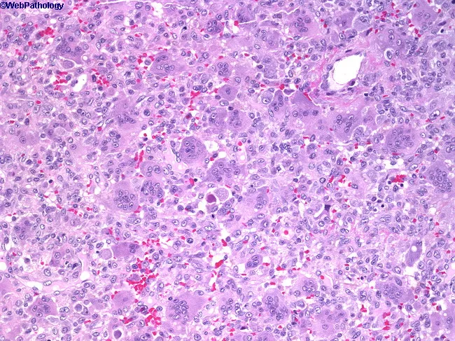
Giant cell tumor of bone (osteoclastoma) histopathology. This hematoxylin and eosin stained section shows numerous multinucleated osteoclast-like giant cells dispersed among a background of uniform, oval to spindle-shaped mononuclear stromal cells. The giant cells contain multiple nuclei with scant cytoplasm, while the mononuclear cells display vesicular nuclei, fine chromatin, and limited mitotic activity. The lesion often shows a vascular, hemorrhagic background and scattered hemosiderin deposits. There is typically minimal cytologic atypia; occasional atypical mitoses may be present in more aggressive variants. Architecturally, the tissue demonstrates a proliferative stromal framework with evenly distributed giant cells and occasional mitotic figures but not overt anaplasia. This histology correlates with radiologic eccentric, radiolucent, soap-bubble–appearing lesions that arise in the epiphysis of long bones. Clinically, patients present with pain and swelling; there is a female predominance in the first two decades, with peak incidence in the third decade. The diagnosis is important to distinguish from other giant cell–rich lesions, including aneurysmal bone cyst, brown tumor of hyperparathyroidism, fibrous dysplasia, chondroblastoma, and osteosarcoma with giant cells. Immunohistochemistry may help in uncertain cases, with stromal cells often expressing RANKL and osteoclast-like giant cells being TRAP-positive. Management relies on curettage with adjuvants or en bloc resection, with recurrence risk dependent on procedure.
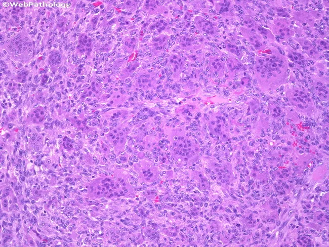
High-power light microscopic image of a hematoxylin and eosin (H&E) stained bone biopsy demonstrating a classical giant cell tumor of bone (GCTB). The sample comprises a vascular, myxoid to fibrous stromal background populated by numerous round to oval mononuclear cells and abundant multinucleated osteoclast-like giant cells. The nuclei of the giant cells resemble those of the surrounding mononuclear stromal cells, often with evenly distributed chromatin and small nucleoli. The cytoplasm of both cell types is eosinophilic and granular, giving a dense pink–purple matrix. Mitotic activity is limited, and atypia is minimal, consistent with a benign, though locally aggressive, neoplasm. In this image, the delicate interplay between mononuclear cells and giant cells can obscure boundary between populations, highlighting diagnostic challenges in GCTB. The overall cellularity is moderate to high, with occasional hemorrhagic foci and a storiform to lace-like stromal pattern. Clinically, GCTB presents in young to middle-aged adults and tends to arise in the epiphysis of long bones, near the adjacent articular surface. The histology is diagnostic when uniform nuclei and numerous osteoclast-like giant cells are present, aiding differential diagnosis from aneurysmal bone cyst and other giant cell-rich lesions. This depiction supports planning of curettage with adjuvant therapy and informs prognosis.
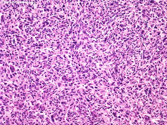
Histopathology of giant cell tumor of bone demonstrated on Hematoxylin and Eosin stained sections, visualized by bright-field light microscopy. The specimen represents a skeletally derived tumor bed with abundant spindle-shaped stromal cells interspersed with numerous multinucleated giant cells resembling osteoclasts. The mononuclear cells are typically evenly distributed, with minimal cytologic atypia and rare mitotic activity, providing a cellular yet non-atypical background. The giant cells are conspicuous but evenly dispersed, with numerous nuclei per cell in a peripherally oriented pattern. The matrix is typically collagenous with a vascular stroma, and hemorrhagic or foamy macrophages may be present. In this illustrative image, sheets of spindle cells exhibit mild pleomorphism but lack overt nuclear atypia, supporting a benign/low-grade neoplasm rather than high-grade sarcoma. Diagnostic significance lies in recognizing the characteristic osteoclast-like giant cells coexisting with a uniform mononuclear stromal component. Knowledge of this histology helps differentiate GCT from other sarcomas, guides management (curettage versus en bloc resection), and correlates with radiographic epiphyseal lesions in long bones. Additional differential considerations include fibrosarcoma and other spindle-cell sarcomas, but the uniform stromal population with evenly distributed osteoclast-like giant cells and absence of nuclear atypia aids accurate classification. This histologic profile informs surgical planning, biopsy interpretation, adjuvant decisions, and radiologic correlation in long-bone epiphyseal lesions.
Giant cell lesions of bone: pathology
1. Giant cell tumor (GCT) of bone, the prototype
- Most cells in the tumor are non-neoplastic osteoclasts and their precursors.
- The neoplastic cells are primitive osteoblast precursors with high RANKL expression. RANKL drives proliferation of osteoclast precursors and their differentiation into mature osteoclasts.
- Loss of normal osteoblast-osteoclast feedback causes localized, highly destructive bone resorption. - Robbins Basic Pathology, Giant Cell Tumor
- The neoplastic cells carry mutations in the H3-3A (H3F3A) gene, which encodes histone 3.3. How this leads to tumor formation is unknown. - Robbins, Cotran & Kumar
- The usual mutation is G34W in H3.3. Immunohistochemistry for H3.3 G34W is a useful diagnostic stain.
- The tumor usually sits in the epiphysis and may extend into the metaphysis. It abuts the subchondral bone.
- The most common sites are the distal femur and proximal tibia ("around the knee"), then the distal radius and sacrum.
- Pain and swelling are typical. Because it lies near a joint, it can mimic arthritis. Pathological fracture can be the presenting event.
- It is an eccentric, lytic, expansile lesion with a "soap-bubble" look.
- The cortex is thinned and there is no sclerotic rim.
- It reaches the subchondral bone, in a skeletally mature patient with a closed physis.
- The tumor is a red-brown, soft, hemorrhagic mass.
- Cystic degeneration is frequent.
- It destroys the overlying cortex and forms a bulging soft tissue mass bounded by a thin shell of reactive bone. - Robbins Basic Pathology
- Numerous osteoclast-type giant cells, with up to 100 or more nuclei, are evenly scattered among uniform oval mononuclear cells.
- The giant cell nuclei look identical to the nuclei of the stromal cells. This is a key feature.
- Mitoses may be present and necrosis can be prominent. Atypical mitoses and marked pleomorphism are not expected.
- Secondary changes include hemorrhage, hemosiderin, foamy macrophages, aneurysmal bone cyst-like change, and reactive woven bone.
- The neoplastic component is the mononuclear stromal cell, not the giant cell.
- Curettage is the standard treatment, but 40% to 60% recur locally. - Robbins, Cotran & Kumar
- A small proportion (a few percent) develop pulmonary metastases. These are histologically benign-looking and often indolent.
- Rarely, GCT undergoes malignant transformation. This happens mostly after radiotherapy or at sites of recurrence.
- Denosumab, a monoclonal antibody against RANKL, is used for unresectable or recurrent disease. It is also used as neoadjuvant therapy. - Rheumatology 2-Volume Set (2022), Giant Cell Tumor
- Denosumab's effect on recurrence is debated. A 2026 JBJS meta-analysis (PMID 41662446) examined perioperative denosumab and local recurrence after surgery. I haven't read the full paper, so check its findings before applying them clinically.
2. Other giant cell-containing lesions of bone
| Lesion | Key features |
|---|---|
| Aneurysmal bone cyst | Occurs in children and young adults, in the metaphysis. It shows blood-filled spaces without an endothelial lining, separated by fibrous septa with giant cells and reactive woven bone. It has a USP6 rearrangement. Fluid-fluid levels are seen on MRI. |
| Brown tumor of hyperparathyroidism | Osteoclast-rich reactive lesion with hemosiderin and hemorrhage. Serum calcium and PTH are high, and there is subperiosteal resorption. It is not a true neoplasm. |
| Chondroblastoma | Occurs in the epiphysis of adolescents. Chondroblasts have grooved nuclei and "chicken-wire" calcification. Giant cells are scattered. H3F3B K36M mutation. S100 positive. |
| Giant cell reparative granuloma | Occurs in the jaw and small bones of the hands and feet. Clusters of giant cells sit in a fibrous, hemorrhagic stroma. It is reactive. |
| Non-ossifying fibroma / fibrous cortical defect | Metaphyseal, eccentric, with a sclerotic rim. Spindle cells in a storiform pattern, with giant cells and foamy macrophages. |
| Osteosarcoma, giant cell-rich | The malignant cells make osteoid, and there is marked atypia. Giant cells are reactive. |
| Paget disease (and Paget sarcoma) | Giant cells can be abundant in giant cell-rich Paget sarcoma. |
| Cherubism | Jaw disease of children, caused by SH3BP2 mutation. Multilocular lesions with giant cells and perivascular cuffing. |
| Giant cell tumor of tendon sheath / PVNS | Soft tissue and synovial lesion, not bone. Related but distinct, with a CSF1 rearrangement. |
3. Quick points for exams
- GCT has two cell populations: neoplastic mononuclear stromal cells (RANKL+) and reactive osteoclast-type giant cells (RANK+, TRAP+).
- GCT is typically epiphyseal, with a "soap-bubble" lytic lesion near the knee, in adults aged 20 to 40.
- Marker: H3F3A (H3.3 G34W) mutation. Chondroblastoma has a related histone mutation.
- Treatment: curettage with adjuvant therapy, with denosumab for selected cases. Recurrence is common.
- If giant cells appear in a lesion of a child or in the metaphysis, think of aneurysmal bone cyst or non-ossifying fibroma first.
- If the patient has high calcium and PTH, think of brown tumor.
Create a differential diagnosis comparison table
Differential diagnosis of giant cell-rich lesions of bone
Table 1: Clinical and radiological comparison
| Feature | Giant cell tumor (GCT) | Aneurysmal bone cyst (ABC) | Brown tumor (hyperparathyroidism) | Chondroblastoma | Non-ossifying fibroma (NOF) | Giant cell-rich osteosarcoma | Giant cell reparative granuloma |
|---|---|---|---|---|---|---|---|
| Age | 20 to 40 years, with a closed physis | Under 20 years | Any adult, depending on the cause of the hyperparathyroidism | 10 to 25 years, with an open physis | 5 to 20 years | Usually 10 to 25 years, but older patients are possible | Under 30 years |
| Site | Epiphysis, extending to the metaphysis and subchondral bone. Distal femur, proximal tibia, distal radius, sacrum. | Metaphysis of long bones, and the posterior elements of the spine | Any bone, often multiple. Jaw, pelvis, ribs, femur. | Epiphysis or apophysis (proximal humerus, distal femur, proximal tibia) | Metaphysis of the distal femur or tibia, eccentric in the cortex | Metaphysis of long bones | Mandible and maxilla, small bones of the hands and feet |
| Radiology | Eccentric, lytic, expansile, "soap-bubble". Thin cortex, no sclerotic rim. | Eccentric, expansile, "blown-out" lesion. MRI shows fluid-fluid levels. | Lytic, well-defined lesion on a background of generalized osteopenia and subperiosteal resorption | Small, well-defined lytic lesion with a thin sclerotic rim. Calcifications are possible. | Eccentric, multiloculated lesion with a scalloped sclerotic border | Destructive, permeative lesion with periosteal reaction and matrix mineralization | Lytic, expansile, sometimes multilocular |
| Behavior | Benign but locally aggressive. 40 to 60% recur after curettage. Rare lung metastases. | Benign, locally aggressive. Can recur. | Resolves after correction of the parathyroid disease | Benign. Rare recurrence or lung metastasis. | Benign, often regresses spontaneously | Highly malignant | Benign. Recurrence is possible. |
Table 2: Pathological comparison
| Feature | GCT | ABC | Brown tumor | Chondroblastoma | NOF | Giant cell-rich osteosarcoma | Giant cell reparative granuloma |
|---|---|---|---|---|---|---|---|
| Giant cells | Large, evenly distributed, with many nuclei (up to 100 or more). Nuclei match those of the stromal cells. | Present in the septa, usually smaller and clustered | Clustered around areas of hemorrhage | Scattered, among chondroblasts | Scattered, among spindle cells | Reactive and scattered | Clustered around hemorrhage |
| Other cells | Uniform oval mononuclear stromal cells (the neoplastic component, RANKL+) | Fibroblasts and histiocytes | Fibroblasts, hemosiderin-laden macrophages | Chondroblasts with grooved nuclei, "chicken-wire" calcification | Bland spindle cells in a storiform pattern, foamy macrophages | Atypical, pleomorphic malignant cells | Fibroblasts and histiocytes |
| Distinguishing feature | Giant cells are uniformly distributed, with no matrix production | Blood-filled spaces without an endothelial lining, separated by septa | Indistinguishable from GCT histologically. Diagnosis needs biochemistry. | Chondroid matrix and pericellular calcification | Storiform spindle cell pattern, sclerotic border | Malignant osteoid. Marked atypia. | Fibrous stroma. Giant cells are clustered rather than evenly spread. |
| Genetics / markers | H3F3A (H3.3 G34W) mutation | USP6 rearrangement | None specific | H3F3B K36M mutation, S100 positive | None specific | Varies | None specific |
| Key lab or clue | Normal calcium | Normal calcium | High calcium and PTH, low phosphate | Normal calcium | Normal calcium | May have raised alkaline phosphatase | Normal calcium |
How to tell them apart
- Check age and growth plate. A lesion in an adult with a closed physis, reaching the subchondral bone, points to GCT. An epiphyseal lesion in an adolescent with an open physis points to chondroblastoma.
- Check location. An epiphyseal lesion is GCT or chondroblastoma. A metaphyseal lesion is more likely ABC, NOF or osteosarcoma.
- Check serum calcium and PTH. Campbell's notes that a brown tumor "resembles a giant cell tumor and is difficult to distinguish from one", and the diagnosis rests on serum calcium, phosphorus and alkaline phosphatase. - Campbell's Operative Orthopaedics 15th Ed, "Brown Tumor" of Hyperparathyroidism. Schwartz adds that hyperparathyroidism affects multiple areas and the serum calcium is abnormal, and that epiphyseal lesions such as chondroblastoma belong in the differential. - Schwartz's Principles of Surgery 11th Ed, Giant Cell Tumor of Bone
- Look for matrix. Osteoid with atypia means osteosarcoma. Chondroid matrix means chondroblastoma. Neither is present in GCT.
- Use molecular tests when they help. H3.3 G34W immunohistochemistry supports GCT. USP6 rearrangement supports primary ABC. This matters because ABC-like change can be a secondary feature of GCT, chondroblastoma and other lesions.
Pitfalls
- GCT with secondary ABC change can be mistaken for primary ABC. Look for the solid GCT component.
- Brown tumor needs a biochemical check before a GCT is treated with surgery.
- Biopsy of an osteosarcoma can sample only the giant cell-rich area. Radiology showing a destructive, mineralized, permeative lesion should raise suspicion.
- Do not treat any giant cell lesion as GCT without correlating the radiology and the patient's age and location.