
Cerebral stroke and homeopathy ppt md level first give me content in chat than if I approve make it into ppt and give more images for understanding purpose
cerebral stroke brain anatomy ischemic hemorrhagic
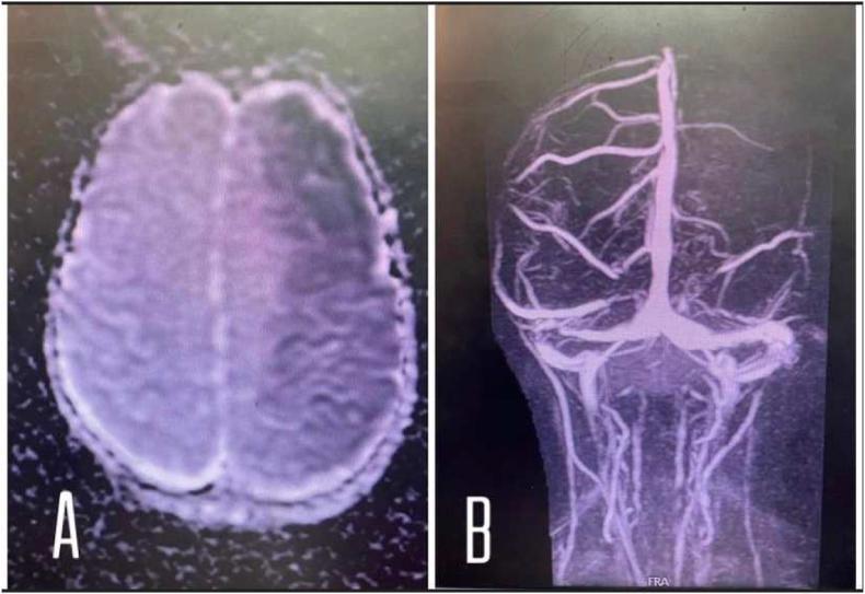
This dual-panel diagnostic image presents neuroradiological findings of an acute ischemic stroke. Figure A is an axial brain MRI (DWI/ADC sequence or T1-weighted equivalent) showing a large, ill-defined area of vague hypodensity in the left cerebral hemisphere. The lesion corresponds to the vascular territory of the left middle cerebral artery (MCA), indicating extensive subacute infarction. Notably, there is an absence of significant midline shift, mass effect, or hyperdense hemorrhagic transformation. Figure B is a Magnetic Resonance Angiography (MRA) displaying the intracranial vasculature. It demonstrates a complete occlusion and abrupt termination of the left middle cerebral artery, contrasting with the patent right MCA and normal basilar/vertebral artery anatomy. This visual pair illustrates the correlation between clinical vessel occlusion (MRA) and resulting parenchymal tissue damage (MRI) in the context of neurovascular pathology.
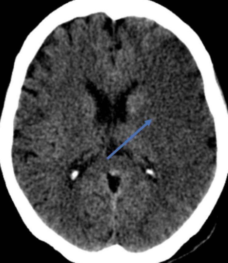
This diagnostic image is an axial non-contrast computed tomography (CT) scan of the brain. The scan reveals a localized area of hyperdensity within the left cerebral hemisphere, specifically involving the region of the basal ganglia or internal capsule, indicated by a blue arrow. The hyperdense lesion is roughly ovoid in shape and appears brighter than the adjacent normal gray and white matter parenchyma. The surrounding ventricular system, including the frontal horns of the lateral ventricles and the third ventricle, appears relatively symmetrical without significant midline shift or mass effect. The cortical sulci are visible but show mild effacement in the left hemisphere compared to the right, suggesting localized edema. This finding is clinically significant for acute cerebrovascular events, such as a hemorrhagic transformation of an ischemic stroke or a primary intracerebral hemorrhage. The image serves as a teaching tool for recognizing density changes in neuroimaging and understanding the vascular anatomy of the middle cerebral artery territory.
homeopathy remedies neurological treatment
homeopathy cerebral stroke treatment remedies MD level evidence
ischemic stroke CT scan MRI brain infarction
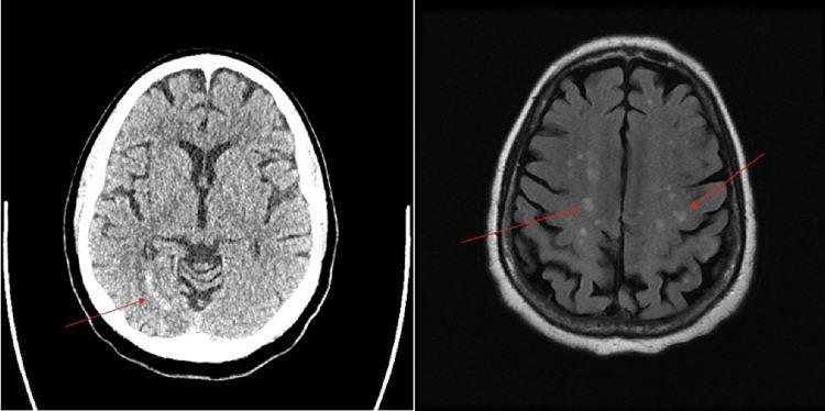
This composite diagnostic image features two axial views of the brain: a non-contrast Computed Tomography (CT) scan on the left and a Magnetic Resonance Imaging (MRI) sequence on the right. The CT scan demonstrates a localized area of hypoattenuation (low density) in the right posterior cerebral artery (PCA) territory, specifically within the occipital lobe, marked by a red arrow, which is characteristic of an acute to subacute ischemic infarction. The MRI scan (FLAIR or T2-weighted sequence) reveals multiple, scattered, small hyperintense foci throughout the subcortical white matter and cortical regions of both cerebral hemispheres, indicated by red arrows. This widespread, multifocal distribution of lesions is highly suggestive of an embolic process, such as septic emboli or cardiogenic thromboembolism. These images provide a comparison between CT and MRI modalities in detecting ischemic changes and characterizing embolic stroke patterns in a clinical neurology context.
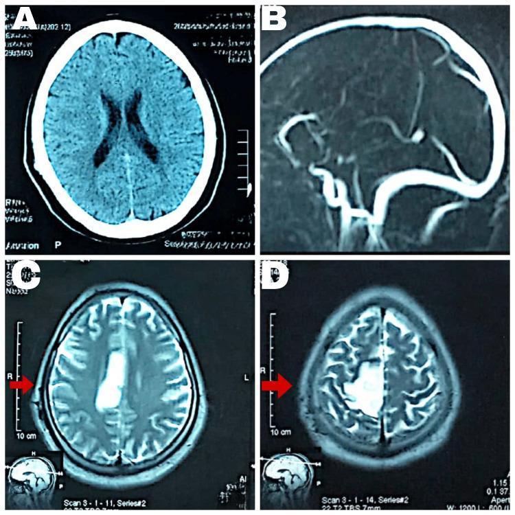
This composite of diagnostic neuroimaging displays a comparison between CT and MRI findings in a case of cerebral infarction. Panel A shows an axial non-contrast CT scan of the brain with no acute intracranial abnormalities visible. Panel B is a sagittal magnetic resonance venography (MRV) showing major venous sinuses. Panels C and D are axial MRI Fluid-Attenuated Inversion Recovery (FLAIR) sequences at different superior levels. These FLAIR images reveal a hyperintense, well-demarcated lesion in the right parietal region, specifically within the vascular territory of the anterior cerebral artery (ACA). Red arrows highlight this area of high signal intensity, which is characteristic of an acute to subacute ischemic infarct. The imaging demonstrates the superior sensitivity of MRI FLAIR sequences over standard CT for detecting early ischemic changes and edema. This resource is suitable for medical education regarding stroke localization, neuroimaging modalities, and the radiographic appearance of vascular insults in the brain.
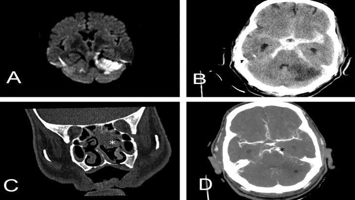
This composite of diagnostic imaging illustrates various neurovascular and otorhinolaryngological pathologies across multiple modalities. Panel A: Axial diffusion-weighted magnetic resonance imaging (DWI MRI) demonstrating hyperintensity in the left cerebellar hemisphere, consistent with restricted diffusion in acute ischemic infarction. Panel B: Non-contrast axial head CT showing diffuse hyperdensity within the basal cisterns and sulci, characteristic of extensive subarachnoid hemorrhage (SAH), alongside a hypodense region in the cerebellum representing secondary ischemic stroke. Panel C: Coronal CT scan of the paranasal sinuses revealing opacification of the nasal cavity and ethmoid cells (marked with a white star), indicating an inflammatory or infectious process such as sinusitis. Panel D: Axial brain CT angiography (CTA) showing vascular morphology, specifically highlighting two consecutive fusiform aneurysms localized in the superior cerebellar artery (marked with a black star). The set provides a comprehensive visual comparison of acute stroke, intracranial hemorrhage, infectious sequelae, and vascular malformations for medical education in radiology and neurology.
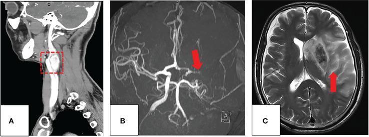
Multi-modal diagnostic imaging of a 60-year-old patient illustrating a carotid body tumor (CBT) and a subsequent cerebrovascular complication. Panel A shows a coronal CT angiography (CTA) scan highlighting a well-defined, hypervascular mass in the left carotid space (red box), characteristic of a carotid body tumor splaying the carotid bifurcation. Panel B displays an axial maximum-intensity-projection (MIP) from a Magnetic Resonance Angiogram (MRA) taken post-surgery, revealing a complete occlusion of the left middle cerebral artery (MCA), indicated by a red arrow. Panel C is an axial T2-weighted MRI (T2WI) of the brain demonstrating a large, acute ischemic infarction throughout the left MCA territory (red arrow). The infarction is identified by significant hyperintense signal and associated vasogenic edema, causing a midline shift and compression of the left lateral ventricle. This series illustrates the diagnostic appearance of CBT and the risk of major ischemic stroke following surgical intervention or internal carotid artery occlusion.
hemorrhagic intracerebral bleeding stroke brain
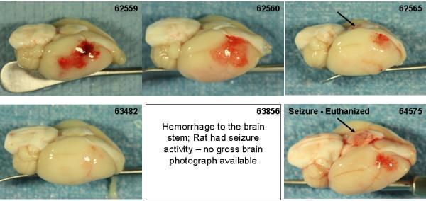
This clinical photograph displays a series of gross pathological specimens from a rat brain model used to study cerebrovascular disease and thrombolytic-induced complications. The set illustrates varying degrees of intracerebral and subarachnoid hemorrhage following middle cerebral artery occlusion (MCAo). Individual specimens (labeled 62559, 62560, 62565, 63482, and 64575) demonstrate a spectrum of superficial bleeding severity. Notable findings include large, confluent areas of cortical hemorrhage (62559, 62560), more localized petechial bleeding (62565, 64575), and a specimen with a relatively clean infarct showing minimal hemorrhage (63482). Black arrows specifically highlight regions of interest such as brain stem or posterior fossa involvement in specimen 64575, which is contextually associated with clinical seizure activity. This comparative layout is used in medical research to evaluate the safety profile and bleeding complications (hemorrhagic transformation) of thrombolytic agents like rt-PA and plasmin variants in the context of ischemic stroke treatment.
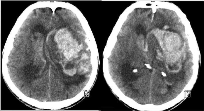
This diagnostic image consists of two axial non-contrast CT scan slices of the brain. The scans demonstrate a massive, irregularly shaped intra-axial hyperdensity in the left hemisphere, specifically involving the fronto-temporal region. This hyperdense mass is indicative of acute intracerebral hemorrhage (ICH). The lesion exerts a significant mass effect, resulting in midline shift towards the right and effacement of the surrounding cerebral sulci. There is clear evidence of intraventricular extension (ventricular rupture), visible as hyperdense material within the lateral ventricles. These findings are consistent with a severe hemorrhagic stroke. The image serves as a clinical example of intracranial bleeding complications, often associated with severe thrombocytopenia or coagulopathy in a neurosurgical context. It is suitable for medical education regarding radiological identification of acute stroke and secondary ventricular involvement.
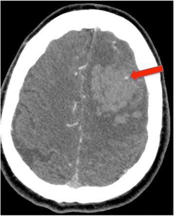
This diagnostic image is an axial Computed Tomography Angiography (CTA) scan of the brain demonstrating an acute spontaneous intracerebral hemorrhage (ICH). A large, hyperdense, heterogeneous hematoma is visible within the left cerebral hemisphere, exerting a mild mass effect. A prominent clinical feature, indicated by a red arrow, is the 'spot sign,' which appears as a small, focal area of contrast enhancement or extravasation within the hematoma itself. In emergency neurology and neuroradiology, the CTA spot sign is a significant radiographic marker used to predict an increased risk of hematoma expansion, poor functional outcomes, and higher mortality rates. The image serves as a key educational example of acute vascular disruption and ongoing bleeding within a primary hemorrhagic stroke, highlighting the utility of CTA in the initial stabilization and prognostic assessment of ICH patients.
stroke rehabilitation physiotherapy hemiplegia recovery
Cerebral Stroke & Homeopathy
MD Level Presentation Content
SLIDE 1 - Title Slide
- Presented by: [Your Name]
- Department of [Your Department]
- [Institution Name], [Date]
SLIDE 2 - Overview / Agenda
- Introduction & Epidemiology
- Classification of Stroke
- Pathophysiology
- Clinical Features & FAST Mnemonic
- Diagnosis & Investigations
- Conventional Management
- Role of Homeopathy in Stroke
- Key Homeopathic Remedies (Materia Medica)
- Evidence Base for Homeopathy in Stroke
- Integrated Approach & Conclusion
SLIDE 3 - Introduction & Epidemiology
- Leading cause of neurological disability worldwide
- ~15 million people affected annually (WHO)
- 5.5 million deaths/year globally
- Up to 10% of elderly develop enough cerebrovascular blockage to cause serious brain dysfunction (Guyton & Hall, Medical Physiology)
- ~25% of people >80 years have had at least one silent brain infarct
- Ischemic stroke: ~80-85% | Hemorrhagic stroke: ~15-20%
- Modifiable: Hypertension (most important), diabetes, hyperlipidemia, smoking, obesity, atrial fibrillation, physical inactivity
- Non-modifiable: Age, sex (male > female), race, family history, prior TIA
SLIDE 4 - Classification of Stroke
A. Ischemic Stroke (~80-85%)
| Type | Mechanism |
|---|---|
| Thrombotic | Atherosclerotic plaque + clot in cerebral artery |
| Embolic | Cardiac thrombus (AF, MI) or carotid embolus |
| Lacunar | Small vessel disease; hypertension-related |
| Cryptogenic | No identifiable cause |
B. Hemorrhagic Stroke (~15-20%)
| Type | Features |
|---|---|
| Intracerebral Hemorrhage (ICH) | Hypertension most common cause; basal ganglia, thalamus |
| Subarachnoid Hemorrhage (SAH) | Berry aneurysm rupture; "thunderclap headache" |
C. TIA (Transient Ischemic Attack)
- Stroke-like symptoms lasting <24 hours (typically <1 hour)
- "Warning stroke" - high risk of major stroke within 48 hours
- ABCD2 score used for risk stratification
SLIDE 5 - Pathophysiology
- Vessel occlusion → Cessation of blood flow
- Core infarct forms within minutes (CBF <10 mL/100g/min)
- Ischemic penumbra surrounds core (salvageable tissue)
- ATP depletion → Ionic pump failure → Na⁺/Ca²⁺ influx
- Glutamate excitotoxicity → NMDA receptor activation
- Mitochondrial dysfunction → Free radical generation
- Inflammation (cytokines, neutrophil infiltration)
- Apoptosis and necrosis
- Vessel rupture → Hematoma formation
- Mass effect + raised ICP → Herniation
- Blood breakdown products cause cytotoxicity
- Vasospasm in SAH adds secondary ischemia
"Many strokes are caused by arteriosclerotic plaques...activating blood clotting mechanisms, causing a blood clot and blocking blood flow" - Guyton & Hall
SLIDE 6 - Clinical Features
FAST Mnemonic (Public Recognition)
- F - Face drooping (unilateral)
- A - Arm weakness
- S - Speech difficulty (slurred/absent)
- T - Time to call emergency
Neurological Deficits by Territory:
| Artery | Deficit |
|---|---|
| MCA | Contralateral hemiplegia + hemisensory loss, aphasia (dominant), neglect (non-dominant) |
| ACA | Contralateral leg weakness > arm, frontal lobe features, incontinence |
| PCA | Contralateral homonymous hemianopia, memory deficits, alexia |
| Basilar/Vertebrobasilar | Diplopia, dysphagia, ataxia, locked-in syndrome |
| Lacunar | Pure motor, pure sensory, ataxic hemiparesis |
Other Symptoms:
- Sudden severe headache (hemorrhagic)
- Nausea, vomiting, loss of consciousness
- Post-stroke depression (very common - up to 33%)
SLIDE 7 - Diagnosis & Investigations
- Non-contrast CT brain - Rule out hemorrhage (gold standard initial)
- Blood glucose (r/o hypoglycemia mimicking stroke)
- CBC, coagulation profile (PT/INR, aPTT)
- ECG (AF, MI)
- MRI brain (DWI + ADC) - Most sensitive for early ischemia
- CT/MR Angiography - Vessel occlusion, stenosis
- MR Perfusion - Penumbra mapping
- Echocardiography (cardioembolic source)
- 24-hour Holter monitoring (paroxysmal AF)
- Carotid Doppler
- NIH Stroke Scale (NIHSS) - Severity assessment
- ASPECTS score - CT early ischemic changes
SLIDE 8 - Conventional Management
Acute Ischemic Stroke:
| Treatment | Criteria |
|---|---|
| IV Alteplase (tPA) | Within 4.5 hours of onset, no contraindications |
| Mechanical Thrombectomy | Large vessel occlusion within 24 hours (NIHSS ≥6) |
| Aspirin 300mg | If thrombolysis not given |
- Airway, breathing, circulation
- BP management (permissive hypertension in ischemic)
- Glucose control (target 140-180 mg/dL)
- Temperature management
- DVT prophylaxis
Hemorrhagic Stroke:
- Reverse anticoagulation (if applicable)
- BP target: <140 mmHg systolic
- Surgical evacuation (selected cases)
- Nimodipine for SAH (prevent vasospasm)
Secondary Prevention:
- Antiplatelets (aspirin + clopidogrel)
- Statins (atorvastatin 40-80 mg)
- Anticoagulants for cardioembolic (AF)
- Risk factor modification
SLIDE 9 - Homeopathy in Stroke: Theoretical Basis
- Law of Similars (Similia Similibus Curantur)
- Individualization of treatment based on totality of symptoms
- Minimum dose principle
- Dynamic derangement of vital force
- Stroke as a "dynamic derangement" - psycho-neuro-physical phenomenon
- Detailed case-taking: anamnestic history + psychosomatic profiling
- "Tout ensemble" of signs/symptoms determines remedy selection
- Homeopathic remedies used as adjuvant to conventional treatment, not replacement
- Over 100 years ago, homeopathic repertory listed 31 remedies under "Apoplexy" (stroke) rubric
- Including: Arnica, Belladonna, Nux vomica, Opium, Lachesis, Gelsemium, and others
SLIDE 10 - Key Homeopathic Remedies: Materia Medica
1. Arnica Montana (Leopard's Bane)
- Indication: Acute phase and recovery; post-trauma, cerebral congestion
- Features: Shock, bruised sensation, aversion to being touched, says "I'm fine" when ill
- Mechanism: Anti-inflammatory, improves microcirculation, reduces edema
- Potency used: 200C and 30C (studied pre- and post-stroke)
2. Belladonna (Deadly Nightshade)
- Indication: Acute hemorrhagic phase; sudden violent onset
- Features: Hot flushed face, throbbing headache, dilated pupils, high fever, delirium
- Key: Right-sided symptoms, worse light/noise/touch
3. Opium (Papaver somniferum)
- Indication: Deep stupor, coma after stroke
- Features: Snoring breathing, dark red face, heavy limbs, complete insensibility
- Key: History of fright, face expressionless
4. Gelsemium (Yellow Jasmine)
- Indication: Recovery phase; paralytic weakness
- Features: Muscular weakness, trembling, drooping eyelids, dizziness, mental dullness
- Key: Weakness out of proportion to actual pathology
5. Nux Vomica (Poison Nut)
- Indication: Post-stroke hemiplegia; spastic paralysis
- Features: Numbness, paralysis of hands/legs, irritability, oversensitivity, insomnia
- Key: Sedentary patients, high-strung personality
6. Lachesis (Bushmaster Snake Venom)
- Indication: Left-sided stroke; hemorrhagic tendency
- Features: Worse on waking/left side, constriction sensations, loquacity, purple face
- Key: Post-menopausal women; morning aggravation
7. Causticum
- Indication: Long-standing post-stroke paralysis
- Features: Progressive, gradual weakness, restless legs, involuntary urination
- Key: Better in damp weather, worse in dry cold
8. Crotalus Horridus (Rattlesnake Venom)
- Indication: Hemorrhagic stroke; malignant hypertension
- Features: Right-sided, easily bleeding, malignant hypertension, jaundiced appearance
- Key: Used alongside Arnica in animal studies
SLIDE 11 - Evidence Base: Homeopathy in Stroke
Preclinical Evidence:
- Khuwaja et al. (2014): Arnica montana and Crotalus horridus (30C & 200C) showed neuroprotective effects in rat cerebral ischemia models - prophylactic role demonstrated
- Jonas et al. (1999): Homeopathic Arnica montana with low-dose glutamate showed promise in experimental stroke (rat model)
Clinical Studies:
- Abbas et al. (2018): Open-label pilot study, 50 stroke patients; homeopathy as adjuvant to standard care showed positive outcomes in sequelae management
- Dutta et al. (2023 - RCT): Explore Journal; Individualized homeopathic medicines showed efficacy in post-stroke hemiparesis management (randomized trial)
- Italian study (Integrated Medicine): Homeopathy as part of an integrated approach significantly improved post-stroke rehabilitation outcomes in a public health facility
Systematic Review Context:
- Springer (2025): Complementary therapies for stroke neurorecovery - homeopathy listed among adjuvant therapies with emerging evidence
- Bell IR (2007): Adjunctive CAM modalities in stroke treatment and rehabilitation - published in Topics in Stroke Rehabilitation
Evidence Level Summary:
| Evidence Type | Status |
|---|---|
| Animal studies | Promising neuroprotection (Arnica) |
| Pilot clinical studies | Positive adjuvant benefit |
| RCT (Dutta 2023) | Significant improvement in hemiparesis |
| Systematic review | Emerging, more RCTs needed |
SLIDE 12 - Integrated Approach: Homeopathy + Conventional Medicine
- Standard emergency management takes absolute priority (tPA, thrombectomy)
- Homeopathic adjuvants begin AFTER stabilization
- Arnica montana 200C for cerebral congestion and edema
- Opium/Belladonna for comatose/obtunded patients (individualized)
- Individualized remedy selection based on residual deficits + constitutional type
- Gelsemium/Nux vomica for motor deficits
- Causticum for chronic paralysis
- Alongside conventional rehabilitation (physiotherapy, speech therapy)
- Constitutional homeopathic treatment
- Address post-stroke depression (Ignatia, Natrum muriaticum, Aurum metallicum)
- Lachesis/Nux vomica for personality changes
Homeopathy is an adjuvant in stroke - it does not replace thrombolysis or emergency neurovascular care. Its greatest role is in the rehabilitation and recovery phase.
SLIDE 13 - Case Studies (Illustrative)
- 62-year-old male, hypertensive, sudden onset left hemiplegia + headache
- CT: Right basal ganglia ICH
- Conventional: BP control, supportive
- Homeopathy adjuvant: Arnica montana 200C (congestion, aversion to touch) → improved alertness on Day 3
- 55-year-old female, 3 months post left MCA ischemic stroke
- Residual right hemiparesis + expressive aphasia + depression
- Individualized remedy: Causticum (progressive paralysis) + Ignatia (grief/depression)
- Outcome: Improved grip strength + mood improvement over 8 weeks
SLIDE 14 - Limitations & Future Directions
- Small sample sizes in clinical trials
- Lack of standardized outcome measures
- Methodological heterogeneity
- Ultra-high dilutions - mechanism not fully explained by conventional science
- Placebo effect cannot always be ruled out
- Large multicentric RCTs (Dutta 2023 is a good starting point)
- Biomarker studies (inflammatory markers, BDNF with homeopathic treatment)
- Neuroimaging correlates of homeopathic neuroprotection
- Standardized case-taking protocols for stroke
- Integration into national rehabilitation guidelines
SLIDE 15 - Conclusion
- Stroke is a medical emergency - "Time is Brain" - conventional management is paramount
- Ischemic stroke (80-85%) managed with tPA/thrombectomy; hemorrhagic with BP control/surgery
- Homeopathy has a long tradition in stroke (Apoplexy rubric > 100 years)
- Arnica montana has the strongest preclinical and clinical evidence
- The rehabilitation phase offers the greatest scope for homeopathic integration
- Individualized prescribing based on totality remains the cornerstone
- Growing evidence base (including an RCT) supports adjuvant homeopathy in post-stroke recovery
- An integrative, patient-centered model is the way forward
SLIDE 16 - References
- Guyton AC, Hall JE. Textbook of Medical Physiology, 14th ed. Elsevier, 2021
- Daroff RB et al. Bradley and Daroff's Neurology in Clinical Practice, 8th ed.
- Ropper AH et al. Adams and Victor's Principles of Neurology, 12th ed.
- Dutta A et al. Efficacy of Individualized Homeopathic Medicines in Treatment of Post-Stroke Hemiparesis. Explore 2023;19(2):243-250
- Khuwaja G et al. Protective role of homoeopathic medicines on cerebral ischaemia in animals. Indian J Research Homeopathy 2014;8(4):209-17
- Abbas A et al. Open-label pilot study: adjuvant homoeopathic medicines in cerebral stroke, 2018
- Bell IR. Adjunctive CAM in stroke treatment. Topics in Stroke Rehabilitation 2007;14:30-39
- Springer Nature. Complementary therapies for stroke towards neurorecovery. Discover Medicine 2025
IMAGES FOR THE PRESENTATION (Medical Library)
Content Summary
| # | Slide | Key Content |
|---|---|---|
| 1 | Title | Topic + Presenter |
| 2 | Agenda | 10-point overview |
| 3 | Introduction | Definition, Epidemiology, Risk factors |
| 4 | Classification | Ischemic types, Hemorrhagic types, TIA |
| 5 | Pathophysiology | Ischemic cascade, Hemorrhagic mechanism |
| 6 | Clinical Features | FAST, Vascular territory deficits |
| 7 | Diagnosis | CT, MRI, Cardiac workup, Scoring |
| 8 | Conventional Management | tPA, Thrombectomy, Secondary prevention |
| 9 | Homeopathy Basis | Theory, Historical context |
| 10 | Key Remedies | 8 major remedies with indications |
| 11 | Evidence Base | Preclinical, Clinical trials, RCT |
| 12 | Integrated Approach | Phase-wise protocol |
| 13 | Case Studies | 2 illustrative cases |
| 14 | Limitations | Gaps + Future directions |
| 15 | Conclusion | 8 key takeaways + Hahnemann quote |
| 16 | References | 8 key citations |
- Is the depth appropriate for MD level? (Can add/remove any sections)
- Any modifications to homeopathic remedies section (more/fewer remedies)?
- Should I add more case studies or specific potency protocols?
- Design preference for the PPT (professional dark/light theme, institutional colors)?
Add anatomy part , refine homeopathic therapeutics part using authentic book exact line , make like from basic to phd level , and give me content first than if I approve give me ppt
circle of Willis cerebral arteries anatomy brain base
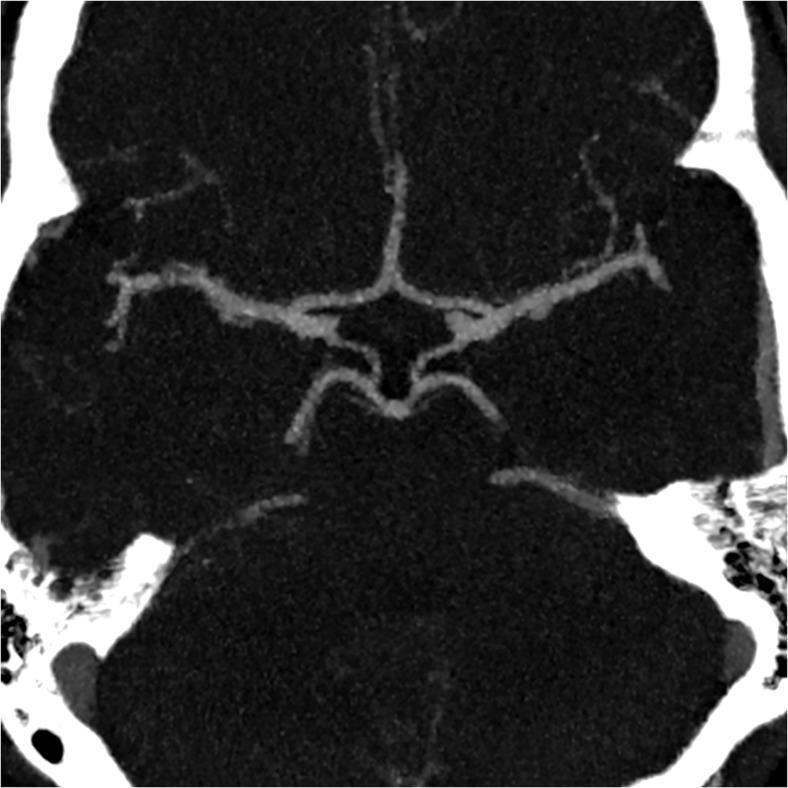
This diagnostic image is a 7 mm thick-slab axial maximum intensity projection (MIP) reconstructed from a brain magnetic resonance angiography (MRA). It demonstrates the complete and normal anatomical configuration of the Circle of Willis at the base of the brain. The primary vascular structures are clearly visualized, including the bilateral anterior cerebral arteries (ACA) joined by the anterior communicating artery (AComA), the bilateral middle cerebral arteries (MCA), and the posterior cerebral arteries (PCA) connected to the internal carotid circulation via patent posterior communicating arteries (PCoA). The basilar artery and its bifurcation are also evident. All arterial segments exhibit normal caliber (≥0.8 mm), smooth morphology, and continuous contrast enhancement without evidence of stenosis, aneurysm, or hypoplasia. This image serves as a clinical reference for a 'Group I' vascular classification, representing a fully intact and symmetrical cerebral arterial circle. It is highly relevant for neuroradiology education focusing on intracranial vascular anatomy and diagnostic imaging interpretation.
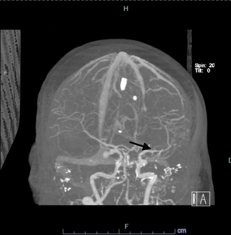
This diagnostic image is a Computed Tomography Angiography (CTA) of the brain presented as a Maximum Intensity Projection (MIP) reconstruction in a coronal-like oblique orientation. The image provides a high-contrast visualization of the intracranial vasculature against the suppressed background of neural parenchyma. The Circle of Willis and its branching vessels are clearly visible. A prominent black arrow points to a focal, hyperdense outpouching located at the origin of the left posterior communicating artery (PCOM), diagnostic of a saccular intracranial aneurysm. The vascular anatomy shows the internal carotid arteries, middle cerebral arteries, and anterior cerebral arteries, with some punctate hyperdensities likely representing calcifications or artifact at the skull base. This imaging modality is used to evaluate cerebrovascular pathology such as aneurysms, stenosis, or subarachnoid hemorrhage, providing essential anatomical mapping for neurosurgical or endovascular intervention planning.
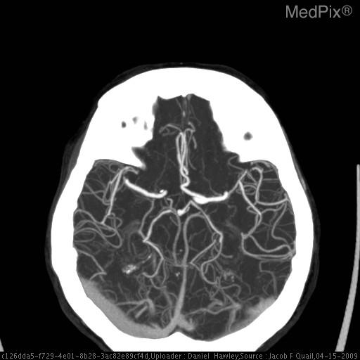
**Imaging Modality:** Computed Tomography Angiography (CTA), axial maximum intensity projection (MIP) reconstruction.
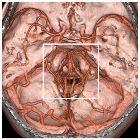
This diagnostic image is a 3D volume-rendered computed tomography angiography (CTA) showing the cerebral vasculature at the skull base from an inferior view. The rendering uses a reddish-brown color scheme to delineate the arteries and deep veins relative to the brain tissue and cranial floor. A white square box highlights the perimesencephalic region, specifically focusing on the vessels surrounding the midbrain and the Circle of Willis. This region is clinically significant as the primary site for investigating the etiology of perimesencephalic non-aneurysmal subarachnoid hemorrhage (PNSAH). The image demonstrates complex vascular anatomy, including the basilar artery, posterior cerebral arteries, and the potential involvement of deep venous structures such as the basal vein of Rosenthal. By extending CTA acquisition time, this visualization captures both the arterial and deep venous systems simultaneously, which is essential for excluding aneurysms or identifying venous ruptures as the cause of hemorrhage in this anatomical location.
brain venous drainage dural sinuses cerebral veins anatomy
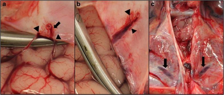
This composite of macroscopic clinical photographs demonstrates the venous anatomy of the human brain and dura mater during a forensic or surgical dissection. Panel A shows two bridging veins (arrowheads) draining from the frontal lobe gyri and converging into a parasagittal sinus (arrow), located approximately 3 cm distal to the superior sagittal sinus (SSS). Panel B displays the same region after the bridging veins have been severed and the dura reflected; arrowheads indicate the ostia where the veins previously entered the venous channel. Panel C focuses on the posterior fossa, highlighting tentorial sinuses (arrows) embedded within the dural folds overlying the cerebellar hemispheres. The images illustrate the complex drainage pathways of the cerebral and cerebellar convexities into dural venous structures. Key educational concepts include the identification of bridging veins, which are clinically significant in the pathogenesis of subdural hematomas, and the visualization of smaller dural venous channels distinct from the primary dural sinuses.
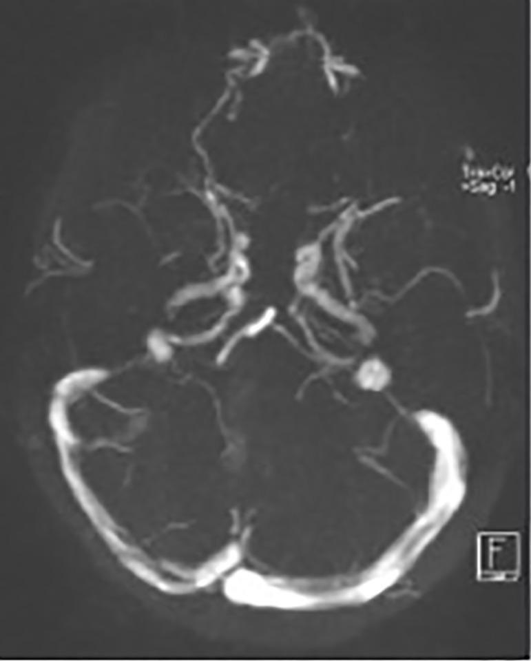
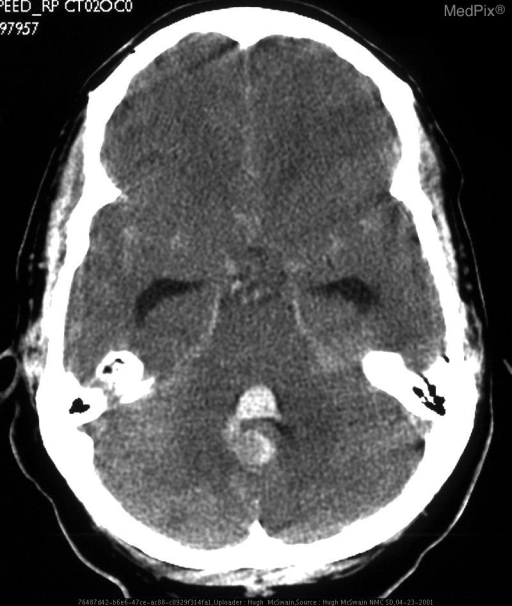
This diagnostic image is an axial view of a head MRI (MR Venogram), specifically highlighting the intracranial dural venous sinuses and cerebral veins through contrast enhancement. The image demonstrates the anatomy of the posterior cranial fossa drainage system, including the confluence of sinuses (torcular herophili) and the bilateral transverse sinuses. The superior sagittal sinus is visible along the midline, while smaller cortical veins appear as thin, tortuous branching structures extending toward the frontal and temporal lobes. A notable finding is the caliber difference or filling defect in the right transverse sinus compared to the contralateral side, which is clinically significant for evaluating cerebral venous sinus thrombosis (CVST). The skull base and anatomical landmarks are faintly outlined, providing structural context for the vascular pathways. This imaging modality is essential for identifying vascular occlusions or anatomical variations in the venous drainage of the brain.
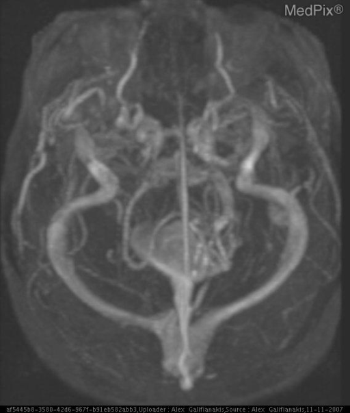
**Imaging Modality:** Magnetic Resonance Venography (MRV), Maximum Intensity Projection (MIP) reconstruction.
homeopathic materia medica arnica belladonna opium lachesis stroke apoplexy exact text Boericke Allen
https://www.materiamedica.info/en/materia-medica/william-boe…
stroke penumbra ischemic core salvageable tissue pathophysiology diagram
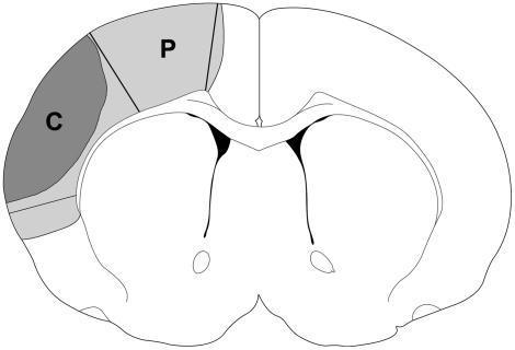
This anatomical diagram provides a cross-sectional schematic view of a rodent brain to illustrate the pathophysiology of ischemic stroke. The illustration utilizes shaded regions to define key areas of tissue damage within the left cerebral hemisphere. A dark gray region, labeled 'C', represents the ischemic core, which denotes the area of irreversible infarction. Adjacent to the core is a lighter gray region labeled 'P', representing the ischemic penumbra—the area of potentially salvageable brain tissue at risk of infarction but potentially responsive to neuroprotective interventions such as postconditioning. The diagram shows the bilateral symmetry of the brain with central ventricle-like structures outlined in black. This visual is designed for educational purposes to demonstrate the spatial relationship between the core and penumbra following middle cerebral artery occlusion or similar ischemic events in translational stroke research.
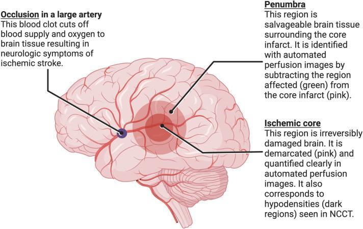
This medical illustration depicts the pathophysiology of an acute ischemic stroke resulting from a large vessel occlusion (LVO). The central anatomical structure is a lateral view of the human brain with its arterial network. A dark blue thrombus is shown occluding a major cerebral artery, leading to downstream tissue ischemia. The diagram categorizes the affected neural tissue into two primary zones: the 'Ischemic Core' and the 'Penumbra'. The Ischemic Core, highlighted in a darker pink/red circular region, represents irreversibly damaged (infarcted) brain tissue, which correlates with hypodensities on non-contrast CT (NCCT). Surrounding this is the Penumbra, shown in a lighter red shade, which represents salvageable brain tissue at risk. The educational focus is on the rationale for endovascular thrombectomy (EVT), where automated perfusion imaging identifies the 'mismatch' between the core and the total affected area (green region in perfusion scans) to select patients for intervention. This diagram serves as a clinical teaching tool for understanding stroke neuroimaging and the selection criteria for reperfusion therapies.
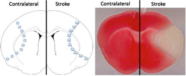
This composite image illustrates the demarcation of ischemic brain injury in a murine model following middle cerebral artery occlusion (MCAO). On the right is a diagnostic pathology photograph of a coronal brain section stained with 2,3,5-triphenyltetrazolium chloride (TTC). The contralateral (left) hemisphere appears uniformly deep red, indicating viable tissue with active mitochondrial dehydrogenase enzymes. The stroke (right) hemisphere reveals a stark, pale-white region corresponding to the necrotic stroke core where metabolic activity has ceased. Surrounding this core is a thin, intermediate pink rim delineated by a blue line, representing the ischemic penumbra—a region of at-risk but salvageable tissue. On the left, a corresponding anatomical diagram provides a schematic for tissue collection protocols. Small blue squares indicate the standardized sampling sites within the cortex, demonstrating how tissue is harvested from both the penumbra (along the blue border) and the infarcted core for comparative biochemical analysis. This image is used in neurobiology to study stroke pathophysiology and neuroprotective interventions.
https://www.materiamedica.info/en/materia-medica/william-boe…
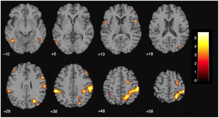
🧠 CEREBRAL STROKE & HOMEOPATHY
A Comprehensive Academic Presentation | Basic to PhD Level
SLIDE 1 - TITLE SLIDE
- Presented by: [Your Name], [Degree/Designation]
- Department of [Practice of Medicine / Materia Medica]
- [Institution Name]
- Date: [Date]
SLIDE 2 - TABLE OF CONTENTS
| Module | Topic | Level |
|---|---|---|
| I | Introduction & Epidemiology | Basic |
| II | Anatomy of the Cerebrovascular System | Basic–Intermediate |
| III | Classification of Stroke | Intermediate |
| IV | Pathophysiology | Intermediate–Advanced |
| V | Clinical Features & Syndromes | Intermediate |
| VI | Diagnosis & Investigations | Intermediate–Advanced |
| VII | Conventional Management | Advanced |
| VIII | Homeopathic Therapeutics (Materia Medica) | Advanced–PhD |
| IX | Evidence Base & Research | PhD |
| X | Integrated Approach & Protocol | PhD |
| XI | Conclusion & References | — |
MODULE I — INTRODUCTION & EPIDEMIOLOGY
SLIDE 3 - What is Stroke?
- Same symptoms but resolve within 24 hours (usually <1 hour)
- A medical emergency - 10-15% risk of stroke within 3 months
- ABCD2 score used for risk stratification
- "Brain Attack" (paralleling "Heart Attack" for urgency messaging)
- "Apoplexy" (historical/homeopathic literature term)
SLIDE 4 - Epidemiology
- ~15 million strokes per year (WHO)
- 5.5 million deaths annually - 2nd leading cause of death globally
- 5 million left with permanent disability
- Leading cause of neurological disability in the United States (Bradley & Daroff's Neurology)
"Almost all elderly people have blockage of some small arteries in the brain, and up to 10% eventually have enough blockage to cause serious disturbance of brain function... approximately 25% of people greater than 80 years of age have had one or more silent brain infarcts."
- Ischemic stroke: 80-85%
- Hemorrhagic stroke: 15-20% (ICH ~10-15%, SAH ~5%)
| Modifiable | Non-Modifiable |
|---|---|
| Hypertension (#1) | Age (>55 years) |
| Atrial fibrillation | Male sex |
| Diabetes mellitus | Race (African-American highest risk) |
| Hyperlipidemia | Prior TIA/stroke |
| Smoking | Family history |
| Obesity, Physical inactivity | Sickle cell disease |
| Excessive alcohol, Cocaine/amphetamine use | — |
MODULE II — ANATOMY OF THE CEREBROVASCULAR SYSTEM
SLIDE 5 - Gross Anatomy of the Brain (Basic Level)
| Lobe | Location | Key Functions |
|---|---|---|
| Frontal | Anterior | Motor control (precentral gyrus), personality, speech production (Broca's area - dominant hemisphere) |
| Parietal | Superior-posterior | Sensory integration (postcentral gyrus), spatial awareness |
| Temporal | Lateral | Auditory processing, memory (hippocampus), language comprehension (Wernicke's area - dominant) |
| Occipital | Posterior | Primary visual cortex |
| Insula | Deep/lateral | Autonomic function, taste, visceral sensation |
- Basal ganglia (caudate, putamen, globus pallidus) - Motor modulation; most common site of hypertensive hemorrhage
- Thalamus - Sensory relay station
- Internal capsule - Dense corticospinal and corticobulbar fibers (small stroke = devastating deficit)
- Brainstem (midbrain, pons, medulla) - CN nuclei, vital centers
- Cerebellum - Coordination, balance, fine motor control
SLIDE 6 - Cerebrovascular Supply: The Arterial System
A. Anterior (Carotid) Circulation - 70% of blood supply
"The brain receives approximately 70% of its blood supply from two internal carotid arteries anteriorly..."
- Ophthalmic artery → Retinal supply (TIA sign: amaurosis fugax)
- Posterior communicating artery (PCom) → Connects to basilar
- Anterior choroidal artery → Internal capsule, choroid plexus
- Anterior Cerebral Artery (ACA) → Medial frontal & parietal lobes
- Middle Cerebral Artery (MCA) → Largest branch; lateral hemisphere
- M1: Horizontal (lenticulostriate branches supply basal ganglia/internal capsule)
- M2: Sylvian fissure (opercular branches)
- M3: Cortical (parietal, temporal, frontal branches)
- M4: Distal cortical
B. Posterior (Vertebrobasilar) Circulation - 30% of blood supply
"The vertebral arteries join to form the basilar artery, which then splits to form the two posterior cerebral arteries, which in turn are part of the circle of Willis at the base of the brain." (Medical Physiology, Boron & Boulpaep)
- Posterior inferior cerebellar artery (PICA) → Lateral medulla, cerebellum
- Anterior spinal artery → Anterior spinal cord
- Anterior inferior cerebellar artery (AICA) → Lower cerebellum, pons
- Superior cerebellar artery (SCA) → Upper cerebellum
- Pontine perforators
- Posterior Cerebral Artery (PCA) → Occipital lobes, thalamus, midbrain
SLIDE 7 - Circle of Willis (Circulus Arteriosus Cerebri)
"The connection of the two vertebral arteries forms the basilar artery. The internal carotid arteries and the basilar artery connect to form a vascular loop called the circle of Willis that permits collateral circulation between both the right and left..."
Left ICA ──── AComA ──── Right ICA
     |                        |
    LACA                    RACA
     |                        |
    LMCA                    RMCA
     |                        |
  LPComA                  RPComA
     |                        |
    LPCA ──── Basilar ──── RPCA
- Connects anterior (carotid) and posterior (vertebrobasilar) circulations
- Provides collateral perfusion during arterial occlusion
- Complete circle: Only ~25% of population
- Incomplete circle → No collateral → More severe stroke
- "Substantial variability exists in the anatomy of the circle of Willis, and a significant proportion of individuals may have an incomplete circular loop" (Miller's Anesthesia)
- AComA = most common aneurysm site → SAH
- PCom aneurysm → CN III palsy
- MCA occlusion (most common stroke vessel)
SLIDE 8 - Cerebral Blood Flow: Physiology & Autoregulation (Advanced)
- Normal: 55 mL/100g/min (Adams & Victor's Neurology)
- Critical ischemic threshold: 23 mL/100g/min
- Core infarct zone: <10 mL/100g/min
- Penumbra zone: 10-23 mL/100g/min (salvageable!)
"Over a range of mean blood pressures of approximately 50 to 150 mm Hg, the small pial vessels are able to dilate and to constrict in order to maintain cerebral blood flow (CBF) in a relatively narrow range. This accommodation eventually fails at the extremes of blood pressure, after which CBF follows systemic pressure passively."
- Normal MAP range: 50-150 mmHg → CBF maintained
- Below 50 mmHg → Ischemia
- Above 150 mmHg → Hypertensive encephalopathy, breakthrough edema
- CBF = CPP / CVR
- CPP = MAP - ICP
- Regulated by: PaCO2 (major), PaO2, pH, metabolic demands
- Composed of: Tight junctions between endothelial cells + astrocyte endfeet + pericytes
- Disruption in stroke → Vasogenic edema
- Target for neuroprotection research
SLIDE 9 - Venous Drainage of the Brain
"Three sets of veins drain blood from the brain. The superficial cortical veins are within the pia mater on the brain surface. Deep cortical veins drain the deeper structures of the brain. These veins drain into dural sinuses, of which the superior and inferior sagittal sinuses and the straight, transverse, and sigmoid sinuses are the major dural sinuses. These ultimately drain into the right and left internal jugular veins."
| Sinus | Drains From | Empties Into |
|---|---|---|
| Superior Sagittal Sinus (SSS) | Cortical veins, CSF via arachnoid granulations | Confluence of sinuses |
| Inferior Sagittal Sinus | Deep medial hemisphere | Straight sinus |
| Straight Sinus | Deep cerebral veins (Vein of Galen) | Confluence |
| Transverse Sinus | Confluence | Sigmoid sinus |
| Sigmoid Sinus | Transverse | Internal jugular vein |
| Cavernous Sinus | Orbit, facial veins, sphenoidal area | Petrosal sinuses |
- CVST (Cerebral Venous Sinus Thrombosis) = venous stroke; unique to consider in young women, pregnancy, OCP use, hypercoagulable states
- SSS thrombosis → Bilateral leg weakness (parasagittal infarcts)
- Cavernous sinus thrombosis → Proptosis, ophthalmoplegia, facial pain
MODULE III — CLASSIFICATION OF STROKE
SLIDE 10 - Classification
A. Ischemic Stroke (80-85%)
"Many strokes are caused by arteriosclerotic plaques that occur in one or more of the feeder arteries to the brain. The plaques can activate the blood clotting mechanisms, causing a blood clot and blocking blood flow in the artery, thereby leading to an ischemic stroke and acute loss of brain function in a localized area."
| Subtype | Mechanism | Vessel Involved |
|---|---|---|
| Large Artery Atherothrombotic | Atherosclerosis + in-situ thrombosis | ICA, MCA, Basilar |
| Cardioembolic | AF, MI, endocarditis → embolism | Any, often MCA |
| Small Vessel (Lacunar) | Lipohyalinosis of penetrating arterioles | Lenticulostriates, thalamic perforators |
| Cryptogenic | No cause found | Any |
| Other | Dissection, vasculitis, hypercoagulable | Variable |
B. Hemorrhagic Stroke (15-20%)
"In 15% to 20% of people in whom strokes develop, one of the cerebral blood vessels bursts; hemorrhage then occurs, compressing the local brain tissue and further compromising its functions. The most important risk factor for hemorrhagic stroke is high blood pressure (hypertension)..."
| Type | Location | Key Feature |
|---|---|---|
| Intracerebral Hemorrhage (ICH) | Basal ganglia (#1), pons, cerebellum, lobar | Hypertension most common cause |
| Subarachnoid Hemorrhage (SAH) | Subarachnoid space | Aneurysm rupture; "thunderclap headache" |
| Intraventricular Hemorrhage (IVH) | Ventricles | Often extension from ICH; hydrocephalus risk |
| Subdural Hematoma (SDH) | Subdural space | Bridging vein tear; elderly after fall |
MODULE IV — PATHOPHYSIOLOGY
SLIDE 11 - Ischemic Cascade (Molecular Level - PhD)
VESSEL OCCLUSION
       ↓
Cessation of CBF → ATP depletion within 4-5 minutes
       ↓
Na+/K+ ATPase pump failure → Na+ influx, K+ efflux, Cl- influx
       ↓
Cell swelling (cytotoxic edema)
       ↓
Membrane depolarization → Voltage-gated Ca²+ channels open
       ↓
GLUTAMATE EXCITOTOXICITY (massive release)
       ↓
NMDA & AMPA receptor activation → Excessive Ca²+ influx
       ↓
Ca²+ activates: phospholipases, proteases, endonucleases, NOS
       ↓
Mitochondrial dysfunction → ROS (reactive oxygen species) generation
       ↓
Nitric oxide + superoxide → Peroxynitrite (potent oxidant)
       ↓
DNA damage, lipid peroxidation, protein oxidation
       ↓
Inflammation: NF-κB activation → TNF-α, IL-1β, IL-6
Neutrophil infiltration (6-24h) → Further BBB disruption
       ↓
APOPTOSIS (delayed cell death, hours-days)
       ↓
INFARCTION (irreversible cell death)
- Core infarct (CBF <10 mL/100g/min): Irreversible necrosis within minutes
- Penumbra (CBF 10-23 mL/100g/min): Electrically silent but metabolically active - SALVAGEABLE
- Treatment window: Opening of penumbra is the rationale for tPA and thrombectomy
SLIDE 12 - Hemorrhagic Pathophysiology (Advanced)
- Mechanical destruction of neural tissue by hematoma
- Mass effect → Raised ICP → Transtentorial herniation
- Midline shift (>5mm = critical)
- Hematoma expansion (occurs in 30-40% within first 24h) → CTA "spot sign" predicts this
- Perihematomal edema → Peaks at 72h, persists weeks
- Hemoglobin breakdown products (hemosiderin, ferritin, free iron) → Oxidative stress, cytotoxicity
- Thrombin generation → Proteases activate, BBB disruption
- Inflammation → Microglial activation, astrocyte reactivity
- Initial ictus → Sudden ICP spike → Global ischemia
- Vasospasm (days 4-14) → Delayed cerebral ischemia in 30% (most preventable complication)
- Nimodipine (calcium channel blocker) prevents vasospasm
- Re-bleeding risk: 10-20% in first 24h if aneurysm unsecured
MODULE V — CLINICAL FEATURES & SYNDROMES
SLIDE 13 - Clinical Recognition
FAST Mnemonic (Public Level)
- F - Face drooping (ask to smile - asymmetric?)
- A - Arm weakness (ask to raise both arms - does one drift?)
- S - Speech difficulty (slurred, wrong words, unable to speak?)
- T - Time - call emergency IMMEDIATELY
Extended: BE-FAST (more sensitive)
- B - Balance problems
- E - Eyes (sudden vision loss/double vision)
-
- FAST
Cincinnati Prehospital Stroke Scale
SLIDE 14 - Vascular Territory Deficits (Intermediate-Advanced)
| Artery | Territory | Clinical Syndrome |
|---|---|---|
| MCA (dominant) | Frontal, parietal, temporal | Contralateral hemiplegia (arm > leg), hemisensory loss, Broca's aphasia (ant.) or Wernicke's aphasia (post.), gaze deviation toward lesion |
| MCA (non-dominant) | Right hemisphere | Contralateral hemiplegia, hemispatial neglect, constructional apraxia, anosognosia |
| ACA | Medial frontal/parietal | Contralateral leg > arm weakness, abulia, urinary incontinence, grasp reflex |
| PCA | Occipital, thalamus | Contralateral homonymous hemianopia, alexia without agraphia (left), amnesia |
| Basilar Artery | Brainstem (bilateral) | "Locked-in syndrome": quadriplegia, anarthria, preserved vertical gaze; potentially fatal |
| PICA (Wallenberg's) | Lateral medulla | Ipsilateral facial pain/numbness + contralateral body pain/temp loss, dysphagia, Horner's, ataxia, hiccups |
| Lenticulostriate (Lacunar) | Internal capsule | Pure motor hemiparesis OR pure sensory stroke OR ataxic hemiparesis |
| Thalamic (Lacunar) | Thalamus | Pure sensory stroke (Dejerine-Roussy: painful thalamic syndrome) |
SLIDE 15 - Post-Stroke Sequelae
- Hemiplegia/hemiparesis (most common residual)
- Spasticity (upper motor neuron pattern)
- Aphasia (in left hemisphere strokes)
- Cognitive impairment/Vascular dementia
- Dysphagia → Aspiration pneumonia (leading cause of death post-stroke)
- Epileptic seizures (early: within 1 week; late: >1 week)
- Central post-stroke pain (thalamic syndrome)
"Stroke is the leading cause of neurological disability in the United States and one of the most common causes of acquired behavioral changes in adults... interruption of bilateral frontotemporal lobe function is associated with an increased risk of depressive and psychotic symptoms."
- Post-stroke depression: 30-40% (most common neuropsychiatric complication)
- Emotional lability (pathological laughing/crying)
- Anxiety disorders, psychosis
- Vascular dementia (multi-infarct type)
MODULE VI — DIAGNOSIS & INVESTIGATIONS
SLIDE 16 - Diagnostic Workup
- Non-contrast CT brain (NCCT) - First choice; rules out hemorrhage
- Blood glucose (must rule out hypoglycemia mimicking stroke)
- CBC, PT/INR/aPTT, electrolytes, renal function
- ECG - Atrial fibrillation, STEMI
- Oxygen saturation
| Modality | Finding | When |
|---|---|---|
| NCCT brain | Hemorrhage (hyperdense) | Immediate |
| CT Angiography (CTA) | Vessel occlusion, stenosis, aneurysm | Acute |
| MRI DWI | Earliest ischemic change (minutes to hours) | Highly sensitive |
| MRI ADC | Confirms restricted diffusion = true infarct | With DWI |
| MR Perfusion / CT Perfusion | Penumbra mapping (core-mismatch) | Thrombectomy selection |
| MR Venography (MRV) | CVST diagnosis | Suspected venous stroke |
- 2D Echocardiography (wall motion abnormality, thrombus, PFO, vegetations)
- Prolonged cardiac monitoring (Holter/implantable loop recorder) - paroxysmal AF
- Carotid Doppler - stenosis
- GFAP (Glial Fibrillary Acidic Protein) - hemorrhagic stroke biomarker
- S100B - astrocytic damage
- NSE (Neuron-specific enolase) - neuronal injury
- NfL (Neurofilament light chain) - axonal damage, prognosis
- NIHSS (0-42) - Stroke severity; >15 = severe
- ASPECTS - Early CT ischemic changes (0-10; <7 = poor thrombectomy candidate)
- mRS (Modified Rankin Scale) - Functional outcome (0=no symptoms, 6=death)
MODULE VII — CONVENTIONAL MANAGEMENT
SLIDE 17 - Acute Ischemic Stroke Management
Reperfusion Therapies:
"The mainstay in the reduction of brain injury in the setting of acute ischemic stroke is thrombolysis. Management of thrombolysis has been reviewed in guidelines for the early management of stroke..."
- Window: 4.5 hours from onset
- Dose: 0.9 mg/kg (max 90 mg); 10% bolus, 90% over 60 min
- Contraindications: Active bleeding, BP >185/110, prior ICH, recent surgery, INR >1.7
- NNT: ~8 patients treated to produce 1 additional good outcome
- Window: 24 hours (selected patients with large vessel occlusion - LVO)
- NIHSS ≥6, ASPECTS ≥6, favorable penumbra-core mismatch on CT perfusion
- Superior to tPA alone for LVO
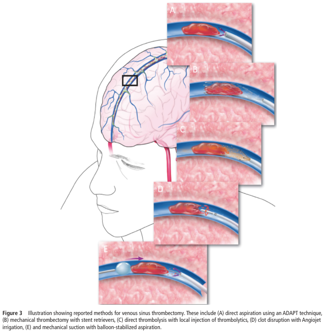
- Devices: Stent retrievers (Solitaire, Trevo), aspiration catheters
Supportive Care:
- Airway: Intubation if GCS ≤8
- BP: Permissive hypertension (allow up to 220/120 unless giving tPA - then maintain <185/110)
- Glucose: Target 140-180 mg/dL (hyperglycemia worsens outcomes)
- Temperature: Treat fever aggressively (every 1°C rise → worse outcome)
- DVT prophylaxis
SLIDE 18 - Secondary Prevention & Hemorrhagic Stroke
| Indication | Drug | Evidence |
|---|---|---|
| Non-cardioembolic | Aspirin 75-100mg/day + Clopidogrel (first 21 days then mono) | Class I |
| Cardioembolic (AF) | Anticoagulation (NOAC preferred over warfarin) | Class I |
| All strokes | Statin (atorvastatin 40-80mg) | Class I |
| Carotid stenosis >70% | Endarterectomy or stenting | Class I |
- BP target: <130/80 mmHg (chronic)
- HbA1c: <7%
- Lipids: LDL-C <70 mg/dL (or <55 for very high risk)
- Smoking cessation, alcohol moderation, physical activity
- Reverse anticoagulation immediately (PCC, Vitamin K, idarucizumab for dabigatran)
- BP: Target systolic 140-160 mmHg acutely (INTERACT2 trial)
- Surgical hematoma evacuation: Selected cases (posterior fossa ICH, young patients)
- Decompressive craniectomy: Malignant MCA infarction, severe cerebellar stroke
- Nimodipine 60mg q4h × 21 days for SAH (vasospasm prevention)
- Coil/clip aneurysm for SAH
MODULE VIII — HOMEOPATHIC THERAPEUTICS
Authentic Materia Medica References
SLIDE 19 - Homeopathic Principles in Stroke (Foundational)
- Diseases represent dynamic derangement of the Vital Force
- Stroke (historically "Apoplexy") was treated by homeopaths using the law of similars
- More than 100 years ago, homeopathic repertory listed 31 remedies under "Apoplexy" rubric:
- Acon., Apis, Arn., Aster., Bar. c., Bell., Cact., Camph., Caust., Chenop., Cinch., Croc., Crotal., Cupr. m., Formica, Glon., Hydroc. ac., Hyos., Junip. v., Kali br., Kali iod., Lach., Laur., Nux v., Op., Phos., Sep., Stram., Sul., Ver. a., Ver. v.
- After emergency stabilization and CT scan
- Detailed anamnestic history + psychosomatic profiling
- "Tout ensemble" of signs and symptoms determines individualized remedy
- Psycho-neuro-physical totality as the basis of prescription
- Acute phase: Adjuvant alongside conventional care (not instead of tPA/thrombectomy)
- Subacute phase: Aid in recovery of neurological deficits
- Rehabilitation phase: Motor, speech, cognitive recovery
- Prevention: Constitutional treatment, managing risk factors
SLIDE 20 - ARNICA MONTANA (Leopard's Bane)
"In apoplexy, with stertorous respiration (paralysis agg. on left side), or with involuntary stools, with no cerebral excitement, but a heavy stupor, with foul breath; or if the patient should be somewhat conscious he complains of aching soreness over the whole body, fear especially of persons coming towards him, as if they would strike him."
"The face of the Arnica patient is usually cold and pale; in apoplexy paralyzed; in low fevers a dusky red."
- Mind: "After any accident or injury, the patient says he is well, doesn't need a doctor; doesn't want to be touched because of soreness"
- Head: "Hot, with cold body; sensitiveness of brain, with sharp, pinching pains"
- Eyes: "Diplopia from traumatism, muscular paralysis, retinal haemorrhage"
- Ears: "Noises in ear caused by rush of blood to the head"
- Cerebral congestion and hemorrhage in early phase
- Soreness, bruised feeling throughout body
- Stupor without excitement
- Stertorous (noisy) breathing
SLIDE 21 - BELLADONNA (Deadly Nightshade - Atropa belladonna)
- Mind: "Acute inflammatory excitement; delirium; wild, violent, raging; sees visions, hallucinations; desires to escape; bites, strikes"
- Head: "Vertigo, with falling to left side or backwards. Much throbbing and heat. Palpitation reverberating in head with labored breathing. Pain, fullness, especially in forehead, also occiput and temples"
- Face: "Red, bluish-red, hot, swollen, shining; convulsive motion of muscles of face"
- Eyes: "Pupils dilated; staring, brilliant; photophobia"
"Belladonna, when the Opium has broken the stupor, and the patient moans, as if conscious of pain, or goes into a convulsion; when there is heat in the head, injected, red appearance of the eyes, and coldness of the extremities."
SLIDE 22 - OPIUM (Papaver somniferum)
- Mind: "Patient wants nothing. Complete loss of consciousness; apoplectic state. Frightful fancies, daring, gay, bright. Unable to understand or appreciate his sufferings. Thinks he is not at home. Delirious talking, with wide open eyes."
- Head: "Dull, heavy, stupid. Delirium. Bursting feeling. Complete insensibility; no mental grasp for anything. Paralysis of brain."
- Eyes: "Half-closed, dilated; pupils insensible, contracted. Ptosis. Staring glassy."
- Face: "Red, bloated, swollen, dark suffused, hot. Looks intoxicated, besotted. Spasmodic facial twitching, especially corners of mouth. Veins of face distended. Hanging down of lower jaw."
- Respiration: "Slow, heavy, deep, labored. Cheyne-Stokes breathing. Stertorous. Rattling."
"Opium, when there is stupor and insensibility, more or less complete; heat in the head, coldness in the extremities, and a snoring as in deep sleep."
SLIDE 23 - LACHESIS MUTUS (Bushmaster Snake Venom)
"Like all snake poisons, Lachesis decomposes the blood, rendering it more fluid; hence a haemorrhagic tendency is marked."
"Drunkards with congestive headaches and haemorrhoids; prone to erysipelas or apoplexy... Rush of blood to head; after alcohol; mental emotions; suppressed or irregular menses; at climaxis; left-sided apoplexy."
- Character: Cannot bear anything tight anywhere; worse after sleep; worse on left side
- Mind: Talkative, suspicious, jealous; "Loquacity; jumps from one subject to another"
- Head: "Congestion; throbbing, bursting feeling. Heat; worse for sleep and waking. Left-sided headache"
- Tongue: "Trembles; catches on lower teeth when protruded; dry; parched"
- Throat: Cannot bear anything touching neck; worse swallowing
- Left-sided stroke with hemorrhagic tendency
- Post-menopausal women
- Worse on waking from sleep (patient feels unwell on waking)
- Congestive symptoms with dark, venous type bleeding
SLIDE 24 - NUX VOMICA (Poison Nut - Strychnos nux-vomica)
- Mind: "Very irritable; sensitive to all impressions. Ugly, malicious. Cannot bear noises, odors, light. Does not want to be touched. Sullen, fault-finding"
- Head: "Vertigo, with momentary loss of consciousness. Intoxicated feeling; worse in morning, mental exertion, tobacco"
- Extremities: "Arms and hands go to sleep. Paresis of arms, with shocks. Legs numb; feel paralyzed; cramps in calves and soles. Partial paralysis, from overexertion or getting soaked (Rhus). Drags his feet when walking. Sensation of sudden loss of power of arms and legs in the morning."
- Post-stroke spastic paralysis
- Hypersensitive, irritable personality
- Paralysis from overwork or exposure to cold
- Wakes 3-4 AM, cannot sleep
SLIDE 25 - CAUSTICUM (Potassium Hydrate - Hahnemann's preparation)
- Paralysis: "Local paralysis; vocal cords, tongue, eyelids, face, bladder, extremities. Post-diphtheritic paralysis. Paralysis of single parts. Ptosis. Facial paralysis from cold; from going into cold wind."
- Urinary: "Involuntary urination at night (loss of sphincter control)"
- Extremities: "Unsteady walking; restless legs at night. Heaviness in all limbs"
"Worse: dry, cold winds, in clear fine weather, cold air; from motion of carriage. Better: in damp, wet weather; warmth; heat of bed."
"In chronic ailments and especially in paralytic states, the higher potencies once or twice a week."
- Long-standing post-stroke paralysis (rehabilitation phase)
- Facial nerve palsy from stroke (Bell's palsy-like picture)
- Urinary incontinence post-stroke
- Gradual, progressive weakness - "remittent paralysis"
SLIDE 26 - GELSEMIUM SEMPERVIRENS (Yellow Jasmine)
- Mind: "Dull, lazy, apathetic, drowsy; loss of will power. Complete prostration and muscular relaxation"
- Head: "Dizziness, drowsiness, dullness, and trembling. Headache preceded by blindness. Occipital headache"
- Eyes: "Ptosis; eyelids heavy; vision blurred; diplopia"
- Face: "Flushed, besotted. Muscles of jaw and swallowing paralyzed"
- Extremities: "Loss of power of muscular control. Weakness of limbs. Difficulty in placing feet when walking. Excessive trembling and weakness"
- Recovery phase - profound motor weakness out of proportion to findings
- Dysphagia post-stroke
- Blurred/double vision, ptosis
- Mental dullness and apathy
SLIDE 27 - CROTALUS HORRIDUS (Rattlesnake Venom)
- Hemorrhagic tendencies; blood non-coagulable
- Right-sided affections predominantly
- Malignant hypertension with stroke tendency
- Besotted, confused expression
- Trembling; tongue paralyzed, can barely protrude
- Hemorrhagic stroke with right-sided paralysis
- Blood disorders with cerebrovascular complications
- Incoagulable blood, dark oozing hemorrhage
SLIDE 28 - QUICK REMEDY COMPARISON TABLE
| Remedy | Phase | Side | Key Distinguisher | Potency |
|---|---|---|---|---|
| Arnica | Acute + Recovery | Either (L worse) | Stupor, stertorous, "I'm well", bruised soreness, won't be touched | 200C acute |
| Opium | Acute (coma) | Either | Deep coma, no response to pain, stertorous, face red/bloated | 30C-200C |
| Belladonna | Acute (hot/excited) | Right | Flushed hot face, delirium, dilated pupils, throbbing | 30C |
| Lachesis | Acute + Chronic | Left | Worse on waking, hemorrhagic, loquacious, can't bear tight collar | 200C |
| Nux Vomica | Subacute | Either | Irritable, spastic paralysis, morning worse, sedentary type | 30C |
| Gelsemium | Recovery | Either | Weakness, ptosis, trembling, dull/apathetic | 30C |
| Causticum | Chronic rehab | Either | Gradual paralysis, better in damp, urinary incontinence | 200C-1M |
| Crotalus | Hemorrhagic | Right | Bleeding tendency, dark hemorrhage, malignant HTN | 200C |
MODULE IX — EVIDENCE BASE & RESEARCH
SLIDE 29 - Scientific Evidence: Basic to PhD
A. Preclinical Evidence
- Arnica montana and Crotalus horridus tested at 200C and 30C in rat cerebral ischemia
- Pre-stroke administration: Neuroprotective effect
- Post-stroke (5 days): Improved outcomes
- Conclusion: "These medications may have a potential prophylactic neuroprotective role"
- Homeopathic Arnica montana with low-dose glutamate in experimental stroke (rat model)
- Demonstrated tissue-protective effects
B. Clinical Studies
- 50 stroke patients; homeopathy as adjuvant to standard care
- 27 patients with episode 1 month-1 year ago + 10 with sequelae
- Positive outcomes in rehabilitation phase
- Randomized Controlled Trial - individualized homeopathic medicines in post-stroke hemiparesis
- Significant improvement in motor function vs. control group
- First RCT-level evidence for homeopathy in stroke
- Integrated approach including homeopathy "strongly improves post-stroke rehabilitative performance in a public health facility"
C. Evidence Quality Pyramid for Homeopathy in Stroke
RCT (Dutta 2023)               ← Highest individual study
    ↑
Open-label pilot (Abbas 2018)
    ↑
Animal/preclinical (Khuwaja 2014, Jonas 1999)
    ↑
Case reports, case series (extensive)
    ↑
Classical Materia Medica (>200 years provings)
D. Systematic Review Context
- Springer Nature/Discover Medicine (2025): "Complementary therapies for stroke towards neurorecovery" - homeopathy cited among promising adjuvant therapies
- Bell IR (2007), Topics in Stroke Rehabilitation 14:30-39: "Adjunctive care with nutritional, herbal, and homeopathic CAM modalities in stroke treatment and rehabilitation"
- Chapman et al. (1999) - RCT of homeopathic treatment in mild TBI: Positive outcomes
MODULE X — INTEGRATED APPROACH & PROTOCOL
SLIDE 30 - Phase-Wise Integrative Protocol
ACUTE PHASE (0-72 hours) - Conventional Priority
| Time | Conventional | Homeopathy Adjuvant |
|---|---|---|
| 0-4.5h | IV tPA (if eligible) | Arnica 200C - 1 dose after stabilization |
| 0-24h | Thrombectomy (LVO) | Based on presentation |
| All acute | BP, glucose, airway management | Opium (coma, stertorous) / Belladonna (hot, delirious) |
SUBACUTE PHASE (Days 3 - 4 weeks)
- Hemiplegia + spasticity + irritability → Nux Vomica 30C bd
- Hemiplegia + weakness + ptosis + dullness → Gelsemium 30C bd
- Left-sided + hemorrhagic + worsens on waking → Lachesis 200C weekly
- Aphasia + right-sided + Broca's type → Causticum 200C + speech therapy
REHABILITATION PHASE (1 month onwards)
- Constitutional treatment based on miasmatic assessment
- Post-stroke depression: Ignatia amara (grief, sighing), Natrum muriaticum (suppressed grief), Aurum metallicum (hopelessness, deep depression)
- Spastic paralysis (chronic): Causticum 1M weekly
- Vascular dementia: Phosphorus, Alumina, Baryta carbonica
- Personality changes: Nux vomica, Platina, Hyoscyamus
PREVENTIVE/CONSTITUTIONAL LEVEL (PhD-level prescribing)
- Psoric stroke: Fear, anxiety, hypertension; recurring TIAs
- Sycotic stroke: Cholesterol deposits, hypertension, obesity
- Syphilitic stroke: Malignant HTN, hemorrhagic tendency, tissue destruction
MODULE XI — LIMITATIONS, FUTURE DIRECTIONS & CONCLUSION
SLIDE 31 - Limitations & Future Research
- Small sample sizes in clinical trials
- Individualized prescribing makes blinded RCTs methodologically challenging
- Mechanism of action at ultra-dilutions not yet fully explained by conventional pharmacology
- Lack of standardized case-taking protocols specific to stroke
- No multicentric, large-scale RCT yet
- Biomarker studies: Do homeopathic remedies modulate GFAP, S100B, BDNF, NfL?
- Neuroimaging correlates: Does Arnica affect penumbra size on MRI perfusion?
- Neuroprotection mechanisms: Heat shock proteins, Nrf2 pathway, mitochondrial protection?
- Epigenetic effects of homeopathic remedies in stroke recovery
- Microbiome-gut-brain axis modulation
- Large multicentric RCTs (replicating Dutta 2023 at scale)
- Pragmatic trials in rehabilitation centers
- Integration into national stroke rehabilitation guidelines
- Development of standardized stroke-specific homeopathic protocols
- Mechanistic studies on neuroprotective pathways
SLIDE 32 - Conclusion
- Stroke is a brain attack; FAST recognition saves lives
- Ischemic (clot) vs hemorrhagic (bleed) = different treatments
- Cerebrovascular anatomy determines the clinical syndrome
- Circle of Willis is the collateral lifeline
- "Time is Brain" - 1.9M neurons/minute lost
- Ischemic cascade and penumbra concept justify tPA/thrombectomy
- Secondary prevention and risk factor control = mandatory
- Arnica, Opium, Belladonna = acute phase adjuvants (from authentic Materia Medica)
- Lachesis, Nux vomica, Causticum, Gelsemium = rehabilitation phase
- Growing RCT evidence supports adjuvant homeopathy in post-stroke recovery
- Preclinical neuroprotection evidence is promising
- Individualized prescribing based on miasmatic and constitutional totality
- Integrated model with biomarker and neuroimaging outcomes is the future
"The Vital Force is the curator of the body. Restore it, and healing follows." - Hahnemann
SLIDE 33 - REFERENCES
- Guyton AC, Hall JE. Textbook of Medical Physiology, 14th ed. Elsevier, 2021
- Ropper AH et al. Adams and Victor's Principles of Neurology, 12th ed. McGraw-Hill, 2019
- Daroff RB et al. Bradley and Daroff's Neurology in Clinical Practice, 8th ed.
- Miller RD et al. Miller's Anesthesia, 10th ed. Elsevier, 2020
- Barash PG et al. Barash, Cullen and Stoelting's Clinical Anesthesia, 9th ed.
- Boericke W. Pocket Manual of Homeopathic Materia Medica, 9th ed. B. Jain Publishers
- Allen TF. Handbook of Materia Medica and Homeopathic Therapeutics
- Allen HC. Materia Medica Keynotes
- Dutta A et al. Efficacy of Individualized Homeopathic Medicines in Treatment of Post-Stroke Hemiparesis. Explore 2023;19(2):243-250
- Khuwaja G et al. Protective role of homoeopathic medicines on cerebral ischaemia in animals. Indian J Research Homeopathy 2014;8(4):209-217
- Bell IR. Adjunctive CAM in stroke treatment and rehabilitation. Top Stroke Rehabil 2007;14:30-39
- Abbas A et al. Open-label pilot study: adjuvant homoeopathic medicines in cerebral stroke. 2018
- Jonas W et al. Treatment of experimental stroke with low-dose glutamate and homeopathic Arnica. Perfusion 1999;12:452-62
- Chapman EH et al. Homeopathic treatment of mild TBI: RCT. J Head Trauma Rehabil 1999;14:521-542
- Springer Nature. Complementary therapies for stroke towards neurorecovery. Discover Medicine 2025
IMAGES AVAILABLE FOR PPT
| # | Image Description | Image |
|---|---|---|
| 1 | Cerebral arterial supply - full labeled diagram from aortic arch (ICA, vertebral, ophthalmic, MCA, ACA, basilar, etc.) | 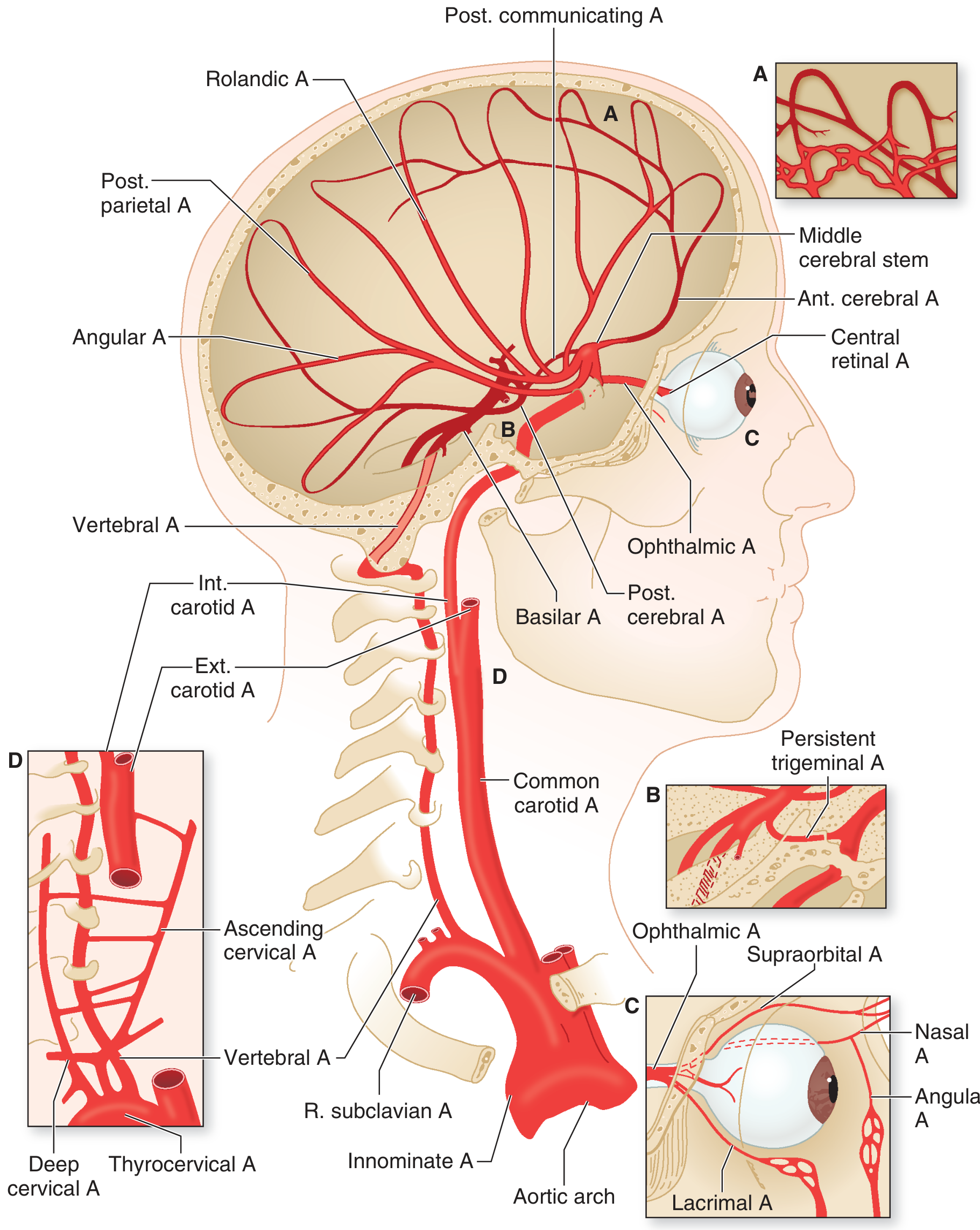 |
| 2 | Base of brain / Circle of Willis - labeled vertebrobasilar system, MCA (M1), ACA (A1), PCA (P1), basilar | 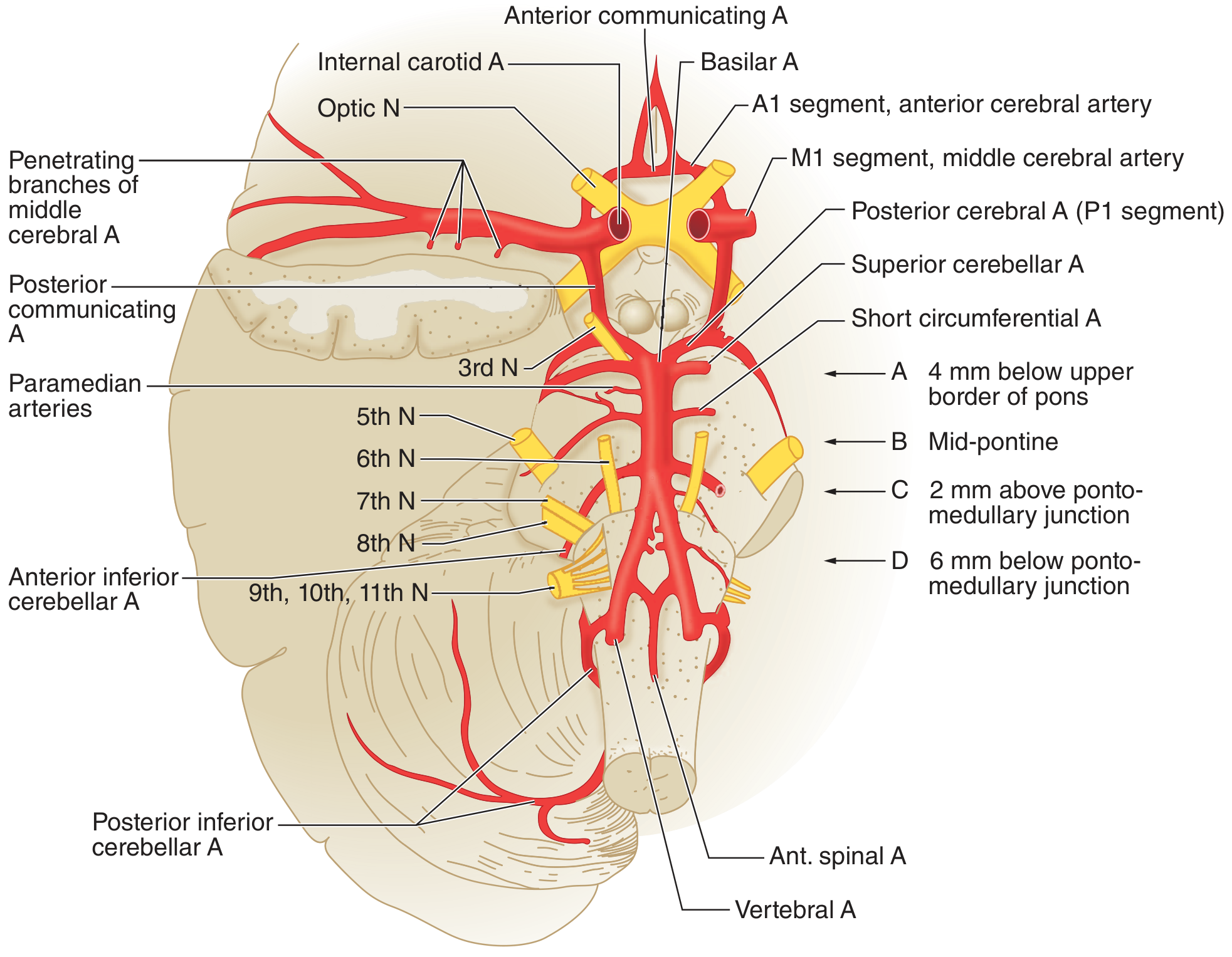 |
| 3 | MRA - Circle of Willis (complete) - clinical imaging | |
| 4 | Ischemic core + penumbra diagram - stroke pathophysiology | |
| 5 | Acute ischemic stroke MRI+MRA - left MCA occlusion | |
| 6 | CT vs MRI comparison - ischemic infarction detection | |
| 7 | Hemorrhagic stroke CT - massive ICH with midline shift | |
| 8 | ICH with CTA "spot sign" - predicts hematoma expansion | |
CONTENT SUMMARY: 33 SLIDES | 10 MODULES
| Module | Slides | Level |
|---|---|---|
| Introduction & Epidemiology | 3-4 | Basic |
| Cerebrovascular Anatomy | 5-9 | Basic-Intermediate-Advanced |
| Classification | 10 | Intermediate |
| Pathophysiology | 11-12 | Advanced-PhD |
| Clinical Features | 13-15 | Intermediate |
| Diagnosis | 16 | Intermediate-Advanced |
| Conventional Mgmt | 17-18 | Advanced |
| Homeopathic Therapeutics | 19-28 | Advanced-PhD |
| Evidence Base | 29 | PhD |
| Integrated Protocol | 30 | PhD |
| Conclusion & References | 31-33 | All levels |
- Boericke W. Pocket Manual of Homeopathic Materia Medica (exact text quoted)
- Allen TF. Handbook of Materia Medica and Homeopathic Therapeutics (exact text quoted)
- Allen HC. Materia Medica Keynotes (Lachesis - exact text quoted)
- Any remedy to add/remove (e.g., Gloninum, Phosphorus, Aurum)?
- Any module to expand or condense?
- Any design preference for the PPT (dark/light/institutional/saffron-white-green theme)?
GOOD USE MORE PICTURES FOR MORE UNDERSTANDING IF POSSIBLE USE PICTURE PRESENTATION FOR MEDICINE AND ANATOMY PORTION AND MAKE PPT
Arnica montana plant flower yellow daisy herb homeopathy
Belladonna atropa plant berry deadly nightshade herb
hemorrhagic stroke intracerebral hematoma brain CT scan
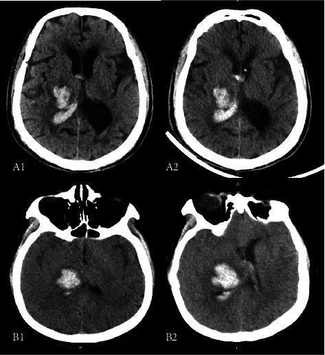
This clinical diagnostic image consists of four axial non-contrast CT scans of the brain, comparing baseline and follow-up imaging for two patients with intracerebral hemorrhage (ICH). Scans A1 and A2 (Patient A) show a hyperdense right thalamic hematoma with clear involvement of the posterior limb of the internal capsule (PLIC). The baseline (A1) and follow-up (A2) demonstrate stable hematoma volume without significant expansion. Scans B1 and B2 (Patient B) illustrate hematoma expansion (HE) over time. The baseline scan (B1) reveals a hyperdense acute hemorrhage in the right thalamus; the follow-up scan (B2) shows a visible increase in the size and volume of the hematoma, which exerts mass effect on the adjacent ventricular system and deep white matter tracts. These images are used to illustrate the radiological assessment of thalamic stroke, the anatomical relationship between the thalamus and the internal capsule, and the clinical phenomenon of hematoma expansion in acute hemorrhagic stroke management.
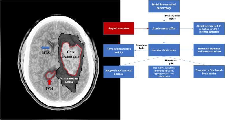
A composite medical illustration containing an axial non-contrast CT scan of the brain and a pathophysiology flowchart detailing the mechanisms of secondary brain injury following intracerebral hemorrhage (ICH). The CT scan (left) demonstrates a large hyperdense intraparenchymal hematoma (labeled 'Core hematoma') with a surrounding hypodense area of 'Peri-hematoma edema'. Significant mass effect is visualized, including midline shift (MLS) indicated by a blue arrow and intraventricular hemorrhage (IVH) marked with a red arrow. The flowchart (right) illustrates the progression from initial ICH through acute mass effect to secondary brain injury. It details biochemical and cellular consequences such as hematoma lysis, hemoglobin and iron toxicity, blood-brain barrier disruption, and free radical formation, culminating in apoptosis and neuronal necrosis. A 'Surgical evacuation' intervention is highlighted in red, positioned as a treatment to mitigate the acute mass effect and subsequent cascade of secondary injury. This image is designed for neurosurgical and radiological education regarding the mechanical and biological impacts of hemorrhagic stroke.
carotid artery stenosis atherosclerosis plaque ultrasound
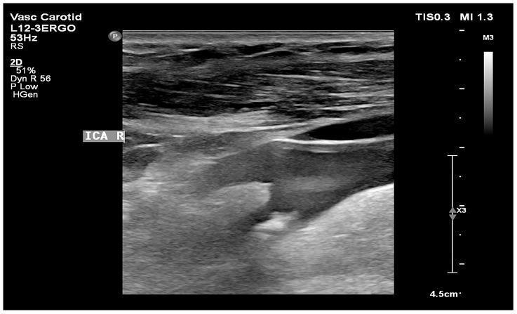
This diagnostic B-mode ultrasound image displays a longitudinal view of the right internal carotid artery (ICA). The image captures a significant atherosclerotic plaque characterized by heterogeneous echogenicity, containing both hyperechoic (bright) and hypoechoic (dark) regions. A prominent feature is the presence of a distinct acoustic shadow extending distally from the plaque, which indicates localized areas of dense calcification that reflect or absorb the ultrasound signal. This type of 'hard' or calcified plaque is often associated with more advanced stages of atherosclerosis. The image illustrates key diagnostic criteria used in vascular neurology and radiology to assess plaque stability and the risk of ischemic events, such as Gray Scale Median (GSM) evaluation. The anatomical region is clearly labeled 'ICA R', and the scan provides a clinical representation of carotid artery stenosis suitable for medical education in cardiovascular pathology and non-invasive diagnostic imaging.
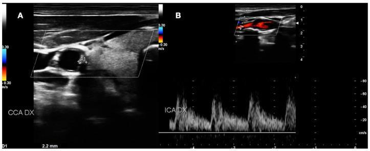
This composite diagnostic image displays Duplex Ultrasound (DUS) scans of the right carotid artery system, illustrating carotid atherosclerosis. Panel A shows a B-mode cross-sectional view of the right common carotid artery (CCA DX) highlighting a medium-grade atherosclerotic plaque. The plaque exhibits mixed echogenicity with hypoechoic regions and is seen protruding into the vessel lumen; calipers (D1) measure an intima-media thickness (IMT) or plaque height of 2.2 mm. Panel B demonstrates the right internal carotid artery (ICA DX) using color Doppler and spectral Doppler analysis. The color Doppler identifies blood flow velocity and direction, with red and blue coding indicating flow towards and away from the transducer at a scale of 0.30 m/s. The corresponding spectral Doppler waveform below provides a hemodynamic assessment, showing a characteristic pulsatile arterial flow pattern over time. This visual material is used in cardiovascular medicine to evaluate the morphology, grade, and hemodynamic impact of carotid artery stenosis.
stroke patient hemiplegia facial droop arm weakness neurological examination
subarachnoid hemorrhage CT brain hyperdense cisterns aneurysm
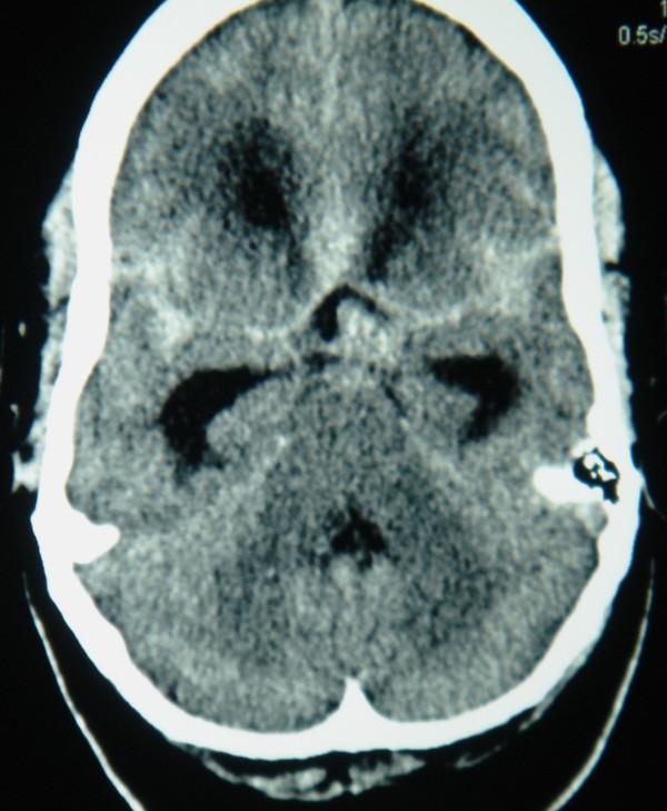
This unenhanced axial computed tomography (CT) scan of the brain demonstrates a diffuse subarachnoid hemorrhage (SAH). High-attenuation (hyperdense) signals indicative of acute blood are prominently visible within the subarachnoid spaces, particularly filling the basal cisterns and extending into the Sylvian fissures. A focal area of increased density is notably localized in the prepontine cistern, suggesting a potential site of vascular rupture, such as a basilar artery or posterior communicating artery aneurysm. The ventricular system appears mildly enlarged, and the surrounding brain parenchyma shows typical differentiation, though the diffuse hyperdensity in the cisterns obscures normal cerebrospinal fluid (CSF) spaces. This diagnostic image is a classic representation of acute intracranial hemorrhage and is highly relevant for neurology and neurosurgery education regarding the CT appearance of aneurysmal SAH.
**Imaging Modality:** Non-contrast axial Computed Tomography (CT) of the head.
mechanical thrombectomy stent retriever cerebral vessel clot removal
brain anatomy cross section basal ganglia internal capsule thalamus
Nux vomica Lachesis Causticum plant source homeopathy medicine images botany
mkdir -p /tmp/workspace/stroke-homeopathy-ppt && ls /tmp/workspace/stroke-homeopathy-ppt/
? DONE PPT
mkdir -p /tmp/workspace/stroke-homeopathy-ppt/imgs # Download all images python /tmp/skills/shared/scripts/fetch_images.py \ "https://cdn.orris.care/cdss_images/6221372b2b326df96d1fe1bd8c617367300b6220a9494309d2ce19a7b718844d.png" \ "https://cdn.orris.care/cdss_images/d1cbab900ba1d2d80bc4d78ff0011bfd716a3939d6e2ab5204ee5cbd9a988b27.png" \ "https://cdn.orris.care/cdss_images/pmc_clinical_VQA_3f562f8a813859982e50178da567b232b0a262c22154e79652eaaac8d7726ebd.jpg" \ "https://cdn.orris.care/cdss_images/pmc_clinical_VQA_32bd0a891e8d3ec08358cd8d435858cf730fe0aff9f0b56648a8d469ffe11a38.jpg" \ "https://cdn.orris.care/cdss_images/pmc_clinical_VQA_35a68b3da5939447151ffa8846b3ce32441fc54911fd5b4c1d1d538e337baeee.jpg" \ "https://cdn.orris.care/cdss_images/pmc_clinical_VQA_1c9e51973d4cc11422d02c82e0c5eb47fc2feb256af612f100bc6a22df76aa17.jpg" \ "https://cdn.orris.care/cdss_images/pmc_clinical_VQA_1fb1b5334b5aab5e7616464b8523b8dd1bc0ea5150802de3726fc9750e324087.jpg" \ "https://cdn.orris.care/cdss_images/pmc_clinical_VQA_0049709793c4364fb851070dc0dd5e81389bffb44ef9b6800a36ebbe86e0f17a.jpg" \ "https://cdn.orris.care/cdss_images/pmc_clinical_VQA_4085fc354732aed3a317353a6b7bf2414df73b9a14ae857bd3499d3dd947222b.jpg" \ "https://cdn.orris.care/cdss_images/pmc_clinical_VQA_322d1db803cde9cf16f766d72db4bd3a98e89be20ad313e07ab79a576aa1d89f.jpg" \ "https://cdn.orris.care/cdss_images/pmc_clinical_VQA_2ccac66ee635ba502b8d209a05dd8dff02743860643879d28111a570231d2ccf.jpg" \ "https://cdn.orris.care/cdss_images/pmc_clinical_VQA_0d0ac7dc2263639c0e340730ecca527660465fe0ed62c7377147631a20c63607.jpg" \ "https://cdn.orris.care/cdss_images/pmc_clinical_VQA_6a42c347878c002bfebe8a9b936fb5b07851cc645d7f387782a3e3ff63b6eb56.jpg" \ "https://upload.wikimedia.org/wikipedia/commons/6/66/Strychnos_nux-vomica_-_K%C3%B6hler%E2%80%93s_Medizinal-Pflanzen-266.jpg" \ > /tmp/workspace/stroke-homeopathy-ppt/imgs_data.json echo "Done. Size:" $(wc -c < /tmp/workspace/stroke-homeopathy-ppt/imgs_data.json)
~/stroke-homeopathy-ppt/build_ppt.py
import json, base64, subprocess
from io import BytesIO
from pptx import Presentation
from pptx.util import Inches, Pt, Emu
from pptx.dml.color import RGBColor
from pptx.enum.text import PP_ALIGN, MSO_ANCHOR
from pptx.util import Inches, Pt

# ── colour palette ──────────────────────────────────────────────────────────
NAVY   = RGBColor(0x0D, 0x2B, 0x55)   # dark navy – header bg
TEAL   = RGBColor(0x00, 0x7A, 0x87)   # teal – accent / slide header
GOLD   = RGBColor(0xD4, 0xA0, 0x17)   # gold – highlights
WHITE  = RGBColor(0xFF, 0xFF, 0xFF)
LTGREY = RGBColor(0xF2, 0xF4, 0xF8)   # light bg for content slides
DKGREY = RGBColor(0x33, 0x33, 0x33)
GREEN  = RGBColor(0x1A, 0x7A, 0x4A)
RED    = RGBColor(0xC0, 0x39, 0x2B)
ORANGE = RGBColor(0xE6, 0x7E, 0x22)

# ── load images ─────────────────────────────────────────────────────────────
with open("/tmp/workspace/stroke-homeopathy-ppt/imgs_data.json") as f:
    imgs = json.load(f)

IMG = {}
URLS = [
    "https://cdn.orris.care/cdss_images/6221372b2b326df96d1fe1bd8c617367300b6220a9494309d2ce19a7b718844d.png",
    "https://cdn.orris.care/cdss_images/d1cbab900ba1d2d80bc4d78ff0011bfd716a3939d6e2ab5204ee5cbd9a988b27.png",
    "https://cdn.orris.care/cdss_images/pmc_clinical_VQA_3f562f8a813859982e50178da567b232b0a262c22154e79652eaaac8d7726ebd.jpg",
    "https://cdn.orris.care/cdss_images/pmc_clinical_VQA_32bd0a891e8d3ec08358cd8d435858cf730fe0aff9f0b56648a8d469ffe11a38.jpg",
    "https://cdn.orris.care/cdss_images/pmc_clinical_VQA_35a68b3da5939447151ffa8846b3ce32441fc54911fd5b4c1d1d538e337baeee.jpg",
    "https://cdn.orris.care/cdss_images/pmc_clinical_VQA_1c9e51973d4cc11422d02c82e0c5eb47fc2feb256af612f100bc6a22df76aa17.jpg",
    "https://cdn.orris.care/cdss_images/pmc_clinical_VQA_1fb1b5334b5aab5e7616464b8523b8dd1bc0ea5150802de3726fc9750e324087.jpg",
    "https://cdn.orris.care/cdss_images/pmc_clinical_VQA_0049709793c4364fb851070dc0dd5e81389bffb44ef9b6800a36ebbe86e0f17a.jpg",
    "https://cdn.orris.care/cdss_images/pmc_clinical_VQA_4085fc354732aed3a317353a6b7bf2414df73b9a14ae857bd3499d3dd947222b.jpg",
    "https://cdn.orris.care/cdss_images/pmc_clinical_VQA_322d1db803cde9cf16f766d72db4bd3a98e89be20ad313e07ab79a576aa1d89f.jpg",
    "https://cdn.orris.care/cdss_images/pmc_clinical_VQA_2ccac66ee635ba502b8d209a05dd8dff02743860643879d28111a570231d2ccf.jpg",
    "https://cdn.orris.care/cdss_images/pmc_clinical_VQA_0d0ac7dc2263639c0e340730ecca527660465fe0ed62c7377147631a20c63607.jpg",
    "https://cdn.orris.care/cdss_images/pmc_clinical_VQA_6a42c347878c002bfebe8a9b936fb5b07851cc645d7f387782a3e3ff63b6eb56.jpg",
    "https://upload.wikimedia.org/wikipedia/commons/6/66/Strychnos_nux-vomica_-_K%C3%B6hler%E2%80%93s_Medizinal-Pflanzen-266.jpg",
]
KEYS = [
    "art_lateral","art_base","mra_cow","penumbra","mca_stroke",
    "isch_ct","ich_massive","ich_spot","ct_mri_compare",
    "sah_ct","carotid_us","ich_serial","ich_flowchart","nux_vomica_plant"
]
for k, url, rec in zip(KEYS, URLS, imgs):
    if rec.get("base64"):
        raw = rec["base64"]
        if "," in raw:
            raw = raw.split(",",1)[1]
        IMG[k] = BytesIO(base64.b64decode(raw))

def get_img(key):
    if key in IMG:
        IMG[key].seek(0)
        return IMG[key]
    return None

# ── helpers ─────────────────────────────────────────────────────────────────
prs = Presentation()
prs.slide_width  = Inches(13.333)
prs.slide_height = Inches(7.5)
BLANK = prs.slide_layouts[6]

def add_slide():
    return prs.slides.add_slide(BLANK)

def rect(slide, x, y, w, h, fill=None, alpha=None):
    from pptx.util import Inches
    shape = slide.shapes.add_shape(1, Inches(x), Inches(y), Inches(w), Inches(h))
    shape.line.fill.background()
    if fill:
        shape.fill.solid()
        shape.fill.fore_color.rgb = fill
    else:
        shape.fill.background()
    return shape

def tb(slide, text, x, y, w, h, size=18, bold=False, color=DKGREY,
       align=PP_ALIGN.LEFT, italic=False, wrap=True):
    box = slide.shapes.add_textbox(Inches(x), Inches(y), Inches(w), Inches(h))
    tf  = box.text_frame
    tf.word_wrap = wrap
    tf.margin_left = tf.margin_right = tf.margin_top = tf.margin_bottom = 0
    p = tf.paragraphs[0]
    p.alignment = align
    r = p.add_run()
    r.text = text
    r.font.size  = Pt(size)
    r.font.bold  = bold
    r.font.italic= italic
    r.font.color.rgb = color
    return box

def tb_lines(slide, lines, x, y, w, h, size=14, color=DKGREY, line_spacing=1.15):
    """lines = list of (text, bold, color_or_None)"""
    box = slide.shapes.add_textbox(Inches(x), Inches(y), Inches(w), Inches(h))
    tf  = box.text_frame
    tf.word_wrap = True
    tf.margin_left = tf.margin_right = Pt(4)
    tf.margin_top  = tf.margin_bottom = Pt(2)
    first = True
    for text, bold, clr in lines:
        if first:
            p = tf.paragraphs[0]; first=False
        else:
            p = tf.add_paragraph()
        p.space_after = Pt(2)
        r = p.add_run()
        r.text = text
        r.font.size = Pt(size)
        r.font.bold = bold
        r.font.color.rgb = clr if clr else color
    return box

def slide_header(slide, title, subtitle=None):
    """Dark navy top bar + title"""
    rect(slide, 0, 0, 13.333, 1.1, fill=NAVY)
    tb(slide, title, 0.3, 0.1, 10, 0.85, size=28, bold=True, color=WHITE)
    if subtitle:
        tb(slide, subtitle, 0.3, 0.78, 10, 0.35, size=13, bold=False, color=GOLD)

def img_on_slide(slide, key, x, y, w, h=None):
    im = get_img(key)
    if im is None: return
    if h:
        slide.shapes.add_picture(im, Inches(x), Inches(y), Inches(w), Inches(h))
    else:
        slide.shapes.add_picture(im, Inches(x), Inches(y), Inches(w))

def caption(slide, text, x, y, w):
    tb(slide, text, x, y, w, 0.3, size=9, italic=True, color=TEAL, align=PP_ALIGN.CENTER)

def section_divider(slide, title, module, color=TEAL):
    rect(slide, 0, 0, 13.333, 7.5, fill=NAVY)
    rect(slide, 0.5, 2.5, 12.333, 0.08, fill=GOLD)
    tb(slide, module, 0.5, 1.4, 12, 0.5, size=16, bold=False, color=GOLD, align=PP_ALIGN.CENTER)
    tb(slide, title,  0.5, 1.9, 12, 1.2, size=38, bold=True,  color=WHITE, align=PP_ALIGN.CENTER)

def bullet_box(slide, items, x, y, w, h, size=13, header=None, hdr_color=TEAL):
    box = slide.shapes.add_textbox(Inches(x), Inches(y), Inches(w), Inches(h))
    tf  = box.text_frame
    tf.word_wrap = True
    tf.margin_left = Pt(6); tf.margin_right = Pt(4)
    tf.margin_top  = Pt(4); tf.margin_bottom= Pt(4)
    first = True
    if header:
        p = tf.paragraphs[0]; first=False
        r = p.add_run(); r.text = header
        r.font.size=Pt(size+1); r.font.bold=True; r.font.color.rgb=hdr_color
    for item in items:
        if first: p=tf.paragraphs[0]; first=False
        else: p=tf.add_paragraph()
        p.space_after = Pt(3)
        r=p.add_run(); r.text=item
        r.font.size=Pt(size); r.font.color.rgb=DKGREY

# ════════════════════════════════════════════════════════════════════════════
# SLIDE 1 — TITLE
# ════════════════════════════════════════════════════════════════════════════
sl = add_slide()
rect(sl, 0, 0, 13.333, 7.5, fill=NAVY)
# decorative teal strip
rect(sl, 0, 5.8, 13.333, 0.08, fill=GOLD)
rect(sl, 0, 5.88, 13.333, 1.62, fill=TEAL)
# brain image (artery lateral)
img_on_slide(sl,"art_lateral", 7.8, 0.6, 5.2, 5.0)
# title text
tb(sl,"CEREBRAL STROKE",0.4,1.2,7.2,1.1,size=42,bold=True,color=WHITE)
tb(sl,"& HOMEOPATHY",0.4,2.3,7.2,1.0,size=36,bold=True,color=GOLD)
tb(sl,"A Comprehensive Academic Presentation",0.4,3.4,7.2,0.5,size=15,italic=True,color=LTGREY)
tb(sl,"Basic to PhD Level  |  Materia Medica References",0.4,3.9,7.2,0.4,size=13,color=LTGREY)
tb(sl,'"The highest ideal of cure is rapid, gentle, and permanent\nrestoration of health." — Hahnemann, Organon §2',
   0.4,6.0,12.5,0.9,size=11,italic=True,color=WHITE,align=PP_ALIGN.CENTER)

# ════════════════════════════════════════════════════════════════════════════
# SLIDE 2 — TABLE OF CONTENTS
# ════════════════════════════════════════════════════════════════════════════
sl = add_slide()
rect(sl,0,0,13.333,7.5,fill=LTGREY)
slide_header(sl,"TABLE OF CONTENTS","Cerebral Stroke & Homeopathy — Presentation Overview")
modules = [
    ("I",   "Introduction & Epidemiology",              "Basic"),
    ("II",  "Anatomy of the Cerebrovascular System",    "Basic – Intermediate"),
    ("III", "Classification of Stroke",                  "Intermediate"),
    ("IV",  "Pathophysiology",                           "Intermediate – PhD"),
    ("V",   "Clinical Features & Syndromes",             "Intermediate"),
    ("VI",  "Diagnosis & Investigations",                "Advanced"),
    ("VII", "Conventional Management",                   "Advanced"),
    ("VIII","Homeopathic Therapeutics (Materia Medica)", "Advanced – PhD"),
    ("IX",  "Evidence Base & Research",                  "PhD"),
    ("X",   "Integrated Protocol & Conclusion",          "PhD"),
]
col_x = [0.35, 1.0, 7.5, 11.1]
row_y = 1.25
for i,(num,mod,lvl) in enumerate(modules):
    y = row_y + i*0.59
    bg = WHITE if i%2==0 else LTGREY
    rect(sl, 0.3, y-0.04, 12.7, 0.56, fill=bg)
    tb(sl, num,   col_x[0], y, 0.5,  0.48, size=13, bold=True,  color=TEAL)
    tb(sl, mod,   col_x[1], y, 6.3,  0.48, size=13, bold=False, color=DKGREY)
    tb(sl, lvl,   col_x[2], y, 3.5,  0.48, size=11, bold=False, color=GREEN, italic=True)

# ════════════════════════════════════════════════════════════════════════════
# SLIDE 3 — MODULE I DIVIDER
# ════════════════════════════════════════════════════════════════════════════
sl = add_slide()
section_divider(sl, "Introduction & Epidemiology", "MODULE I")

# ════════════════════════════════════════════════════════════════════════════
# SLIDE 4 — DEFINITION & EPIDEMIOLOGY
# ════════════════════════════════════════════════════════════════════════════
sl = add_slide()
rect(sl,0,0,13.333,7.5,fill=LTGREY)
slide_header(sl,"Introduction & Epidemiology","MODULE I — Basic Level")

# left column
bullet_box(sl,[
    "DEFINITION (WHO): Rapidly developing signs of focal or global disturbance",
    "of cerebral function lasting >24 hrs or leading to death — no cause",
    "other than vascular origin.",
    "",
    "TIA: Same symptoms resolving <24 hrs (usually <1 hr) — 'Warning Stroke'",
    "10–15% risk of major stroke within 3 months",
    "",
    "Historical term: APOPLEXY (used in homeopathic literature)",
],0.3,1.2,5.8,4.8, size=13)

# stat boxes
stats = [
    ("15 M", "strokes/year globally (WHO)", TEAL),
    ("5.5 M", "deaths annually", RED),
    ("80–85%", "Ischemic strokes", NAVY),
    ("15–20%", "Hemorrhagic strokes", ORANGE),
    ("25%", "of people >80 yrs — silent infarcts*", GREEN),
]
bx = 6.3
for i,(val,lbl,clr) in enumerate(stats):
    col = bx + (i%2)*3.3
    row = 1.25 + (i//2)*1.55
    rect(sl, col, row, 2.95, 1.35, fill=clr)
    tb(sl, val, col+0.1, row+0.1, 2.75, 0.65, size=26, bold=True, color=WHITE, align=PP_ALIGN.CENTER)
    tb(sl, lbl, col+0.1, row+0.75, 2.75, 0.55, size=10, color=WHITE, align=PP_ALIGN.CENTER)

tb(sl,"*Guyton & Hall, Medical Physiology 14th ed.",0.3,6.0,12.5,0.35,size=9,italic=True,color=TEAL)

# risk factors
tb(sl,"KEY RISK FACTORS",0.3,5.0,12.5,0.35,size=12,bold=True,color=NAVY)
rf = ["Modifiable: Hypertension #1 | Atrial Fibrillation | Diabetes | Hyperlipidaemia | Smoking | Obesity | Cocaine/amphetamine",
      "Non-modifiable: Age >55 | Male sex | Race | Prior TIA/Stroke | Family history | Sickle cell disease"]
for i,r in enumerate(rf):
    tb(sl,r,0.3,5.35+i*0.45,12.5,0.42,size=11,color=DKGREY)

# ════════════════════════════════════════════════════════════════════════════
# SLIDE 5 — MODULE II DIVIDER
# ════════════════════════════════════════════════════════════════════════════
sl = add_slide()
section_divider(sl, "Anatomy of the Cerebrovascular System", "MODULE II")

# ════════════════════════════════════════════════════════════════════════════
# SLIDE 6 — BRAIN REGIONS & STROKE IMPACT  (image-heavy)
# ════════════════════════════════════════════════════════════════════════════
sl = add_slide()
rect(sl,0,0,13.333,7.5,fill=LTGREY)
slide_header(sl,"Brain Regions & Their Stroke Significance","MODULE II — Gross Anatomy")

# large artery-lateral image left
img_on_slide(sl,"art_lateral",0.2,1.2,6.0,5.5)
caption(sl,"Fig 33-1: Major cerebral arteries — lateral view\n(Adams & Victor's Principles of Neurology, 12th ed.)",0.2,6.6,6.0)

# right side — table of lobes
regions = [
    ("FRONTAL LOBE",   "Motor cortex (precentral gyrus), Broca's speech area (L)","Contralateral hemiplegia, expressive aphasia"),
    ("PARIETAL LOBE",  "Sensory cortex (postcentral gyrus), spatial awareness",    "Hemisensory loss, neglect (R lesion)"),
    ("TEMPORAL LOBE",  "Wernicke's area (L), hippocampus, auditory cortex",        "Receptive aphasia, memory loss"),
    ("OCCIPITAL LOBE", "Primary visual cortex (V1)",                               "Homonymous hemianopia"),
    ("INTERNAL CAPSULE","Corticospinal + corticobulbar fibres (dense pack)",       "Pure motor hemiplegia — small lacunar stroke"),
    ("BASAL GANGLIA",  "Motor modulation — caudate, putamen, globus pallidus",     "#1 site of hypertensive hemorrhage"),
    ("BRAINSTEM",      "CN nuclei, vital centers (respiration, BP)",               "Locked-in syndrome, coma"),
]
for i,(reg,func,stroke) in enumerate(regions):
    y = 1.25 + i*0.87
    bg = WHITE if i%2==0 else RGBColor(0xE8,0xF4,0xF8)
    rect(sl, 6.4, y, 6.7, 0.84, fill=bg)
    tb(sl, reg,   6.5, y+0.02, 2.3, 0.32, size=10, bold=True,  color=NAVY)
    tb(sl, stroke,8.9, y+0.02, 4.1, 0.32, size=9,  bold=False, color=RED)
    tb(sl, func,  6.5, y+0.34, 6.5, 0.42, size=9,  italic=True,color=DKGREY)

tb(sl,"Region",6.5,1.22,2.3,0.3,size=10,bold=True,color=WHITE)
rect(sl,6.4,1.15,6.7,0.3,fill=TEAL)
tb(sl,"Region",6.5,1.17,2.3,0.28,size=10,bold=True,color=WHITE)
tb(sl,"Stroke Consequence",8.9,1.17,4.1,0.28,size=10,bold=True,color=WHITE)

# ════════════════════════════════════════════════════════════════════════════
# SLIDE 7 — ARTERIAL SUPPLY (image-heavy)
# ════════════════════════════════════════════════════════════════════════════
sl = add_slide()
rect(sl,0,0,13.333,7.5,fill=LTGREY)
slide_header(sl,"Cerebrovascular Arterial Supply","MODULE II — Anterior & Posterior Circulations")

# two big images side by side
img_on_slide(sl,"art_lateral", 0.2, 1.2, 5.9, 5.4)
caption(sl,"Major arteries — ICA, MCA, ACA, vertebral, basilar, PCA",0.2,6.55,5.9)

img_on_slide(sl,"art_base", 6.3, 1.2, 6.7, 5.4)
caption(sl,"Base of brain — Circle of Willis & vertebrobasilar system (Adams & Victor)",6.3,6.55,6.7)

# small text strip top
tb(sl,
   "Anterior (Carotid) 70%: ICA → MCA (largest branch), ACA, Ophthalmic, Ant. Choroidal, PCom  |  "
   "Posterior (Vertebrobasilar) 30%: Vertebral → Basilar → PCA, SCA, AICA, PICA",
   0.3, 6.62, 12.7, 0.6, size=10, color=TEAL, align=PP_ALIGN.CENTER)

# ════════════════════════════════════════════════════════════════════════════
# SLIDE 8 — CIRCLE OF WILLIS
# ════════════════════════════════════════════════════════════════════════════
sl = add_slide()
rect(sl,0,0,13.333,7.5,fill=LTGREY)
slide_header(sl,"Circle of Willis — Circulus Arteriosus Cerebri","MODULE II — Collateral Circulation")

img_on_slide(sl,"mra_cow", 0.3, 1.2, 5.5, 5.4)
caption(sl,"MRA — Complete Circle of Willis: ACA, MCA, PCA, AComA, PComA, Basilar",0.3,6.6,5.5)

# right panel — components & clinical significance
tb(sl,"COMPOSITION",6.1,1.2,6.9,0.35,size=14,bold=True,color=NAVY)
cow_items = [
    ("Anterior Cerebral Arteries (ACA) × 2",""),
    ("Anterior Communicating Artery (AComA)","→ Most common aneurysm site → SAH"),
    ("Internal Carotid Arteries (ICA) × 2",""),
    ("Posterior Communicating Arteries (PComA) × 2","→ Aneurysm → CN III palsy"),
    ("Posterior Cerebral Arteries (PCA) × 2",""),
    ("Basilar Artery","→ Occlusion = locked-in syndrome"),
]
for i,(comp,sig) in enumerate(cow_items):
    y=1.6+i*0.72
    rect(sl,6.1,y,6.7,0.66,fill=WHITE if i%2==0 else RGBColor(0xE8,0xF4,0xF8))
    tb(sl,comp,6.2,y+0.04,4.0,0.3,size=11,bold=True,color=DKGREY)
    if sig:
        tb(sl,sig, 6.2,y+0.34,6.4,0.28,size=10,italic=True,color=RED)

tb(sl,"Miller's Anesthesia 10e: 'The ICA and basilar artery connect to form a vascular loop\ncalled the Circle of Willis that permits collateral circulation...'",
   6.1,6.0,6.9,0.8,size=9,italic=True,color=TEAL)

# ════════════════════════════════════════════════════════════════════════════
# SLIDE 9 — CBF AUTOREGULATION & PENUMBRA  (image-heavy)
# ════════════════════════════════════════════════════════════════════════════
sl = add_slide()
rect(sl,0,0,13.333,7.5,fill=LTGREY)
slide_header(sl,"Cerebral Blood Flow, Autoregulation & Ischemic Penumbra","MODULE II/IV — Physiology to Pathophysiology")

img_on_slide(sl,"penumbra", 0.2, 1.2, 7.0, 5.4)
caption(sl,"Ischemic Core (irreversible) vs Penumbra (salvageable) — rationale for tPA & thrombectomy",0.2,6.65,7.0)

# right panels
vals=[
    ("Normal CBF","55 mL/100g/min",GREEN),
    ("Ischemic threshold","23 mL/100g/min",ORANGE),
    ("Penumbra zone","10–23 mL/100g/min",GOLD),
    ("Core infarct","<10 mL/100g/min",RED),
]
for i,(lbl,val,clr) in enumerate(vals):
    y=1.25+i*0.95
    rect(sl,7.5,y,5.5,0.82,fill=clr)
    tb(sl,lbl,7.6,y+0.06,3.5,0.35,size=12,bold=True,color=WHITE)
    tb(sl,val,7.6,y+0.42,5.2,0.32,size=16,bold=True,color=WHITE,align=PP_ALIGN.CENTER)

tb(sl,"AUTOREGULATION: MAP 50–150 mmHg → CBF maintained constant via pial vessel dilation/constriction",
   7.5,5.15,5.5,0.55,size=11,bold=True,color=NAVY)
tb(sl,"Beyond this range → passive pressure-flow → ischemia (<50) or hypertensive breakthrough (>150)",
   7.5,5.7,5.5,0.55,size=10,color=DKGREY)
tb(sl,"Adams & Victor, 12th ed.: 'Critical threshold for infarction is approximately 23 mL/100 g/min\n(normal = 55 mL/100 g/min)'",
   7.5,6.3,5.5,0.8,size=9,italic=True,color=TEAL)

# ════════════════════════════════════════════════════════════════════════════
# SLIDE 10 — MODULE III DIVIDER
# ════════════════════════════════════════════════════════════════════════════
sl = add_slide()
section_divider(sl,"Classification of Stroke","MODULE III")

# ════════════════════════════════════════════════════════════════════════════
# SLIDE 11 — CLASSIFICATION (image-heavy)
# ════════════════════════════════════════════════════════════════════════════
sl = add_slide()
rect(sl,0,0,13.333,7.5,fill=LTGREY)
slide_header(sl,"Classification of Stroke","MODULE III — Ischemic vs Hemorrhagic")

# Ischemic CT+MRI image
img_on_slide(sl,"mca_stroke",0.2,1.2,4.1,3.5)
caption(sl,"MRI DWI + MRA: Left MCA ischemic stroke — vessel occlusion",0.2,4.75,4.1)

# ICH image
img_on_slide(sl,"ich_massive",0.2,5.0,4.1,2.1)
caption(sl,"CT: Massive intracerebral hemorrhage with midline shift",0.2,7.05,4.1)

# Ischemic table
tb(sl,"ISCHEMIC STROKE — 80–85%",4.5,1.2,8.5,0.38,size=14,bold=True,color=TEAL)
types_i=[
    ("Large Artery Atherothrombotic","Atherosclerosis + clot in ICA, MCA, Basilar"),
    ("Cardioembolic","AF, MI, endocarditis → embolism → MCA"),
    ("Small Vessel (Lacunar)","Lipohyalinosis of penetrating arteries; HTN related"),
    ("Cryptogenic","No identifiable cause after full workup"),
]
for i,(t,m) in enumerate(types_i):
    y=1.62+i*0.62
    rect(sl,4.5,y,8.5,0.58,fill=WHITE if i%2==0 else RGBColor(0xE8,0xF4,0xF8))
    tb(sl,t,4.6,y+0.04,3.2,0.26,size=11,bold=True,color=NAVY)
    tb(sl,m,7.8,y+0.04,5.1,0.5, size=10,color=DKGREY)

tb(sl,"HEMORRHAGIC STROKE — 15–20%",4.5,4.3,8.5,0.38,size=14,bold=True,color=RED)
types_h=[
    ("Intracerebral Hemorrhage (ICH)","HTN most common; basal ganglia, thalamus, pons, cerebellum"),
    ("Subarachnoid Hemorrhage (SAH)","Aneurysm rupture; 'thunderclap headache'; vasospasm risk"),
    ("TIA — 'Mini-Stroke'","Sx <24 hrs; ABCD2 score; 10–15% stroke within 3 months"),
]
for i,(t,m) in enumerate(types_h):
    y=4.7+i*0.7
    rect(sl,4.5,y,8.5,0.65,fill=WHITE if i%2==0 else RGBColor(0xFD,0xED,0xED))
    tb(sl,t,4.6,y+0.05,3.2,0.28,size=11,bold=True,color=RED)
    tb(sl,m,7.8,y+0.05,5.1,0.52,size=10,color=DKGREY)

tb(sl,"Guyton & Hall: 'In 15–20% of strokes, one of the cerebral blood vessels bursts; hemorrhage\nthen occurs, compressing the local brain tissue. The most important risk factor is hypertension.'",
   4.5,6.8,8.5,0.6,size=9,italic=True,color=TEAL)

# ════════════════════════════════════════════════════════════════════════════
# SLIDE 12 — CAROTID DISEASE & SAH (image-heavy)
# ════════════════════════════════════════════════════════════════════════════
sl = add_slide()
rect(sl,0,0,13.333,7.5,fill=LTGREY)
slide_header(sl,"Stroke Imaging Gallery","MODULE III — Key Radiological Findings")

imgs_gallery = [
    ("carotid_us",   0.2,  1.15, 4.2, "Carotid atherosclerotic plaque\n— ICA stenosis (Doppler US)"),
    ("sah_ct",       4.6,  1.15, 4.2, "CT Subarachnoid Hemorrhage\n— hyperdense basal cisterns"),
    ("ich_serial",   9.0,  1.15, 4.1, "Serial CT: Thalamic ICH\n— hematoma expansion"),
    ("ct_mri_compare",0.2, 4.2,  4.2, "CT vs MRI — early ischemic\nchange detection (MRI superior)"),
    ("ich_spot",     4.6,  4.2,  4.2, "CTA 'Spot Sign' in ICH\n— predicts hematoma expansion"),
    ("ich_flowchart",9.0,  4.2,  4.1, "ICH secondary injury cascade\n— pathophysiology flowchart CT"),
]
for key,x,y,w,cap_txt in imgs_gallery:
    img_on_slide(sl,key,x,y,w,2.75)
    caption(sl,cap_txt,x,y+2.75,w)

# ════════════════════════════════════════════════════════════════════════════
# SLIDE 13 — MODULE IV DIVIDER
# ════════════════════════════════════════════════════════════════════════════
sl = add_slide()
section_divider(sl,"Pathophysiology of Stroke","MODULE IV")

# ════════════════════════════════════════════════════════════════════════════
# SLIDE 14 — ISCHEMIC CASCADE
# ════════════════════════════════════════════════════════════════════════════
sl = add_slide()
rect(sl,0,0,13.333,7.5,fill=LTGREY)
slide_header(sl,"Ischemic Cascade — Molecular Mechanisms","MODULE IV — Advanced/PhD Level")

# arrow cascade left
steps=[
    ("1. VESSEL OCCLUSION",        "Cessation of CBF",                          NAVY),
    ("2. ATP DEPLETION",           "Na⁺/K⁺ pump failure → Na⁺/Ca²⁺ influx",     TEAL),
    ("3. GLUTAMATE RELEASE",       "Excitotoxicity via NMDA/AMPA receptors",     ORANGE),
    ("4. Ca²⁺ OVERLOAD",          "Phospholipases, proteases, NOS activation",  RED),
    ("5. FREE RADICAL STORM",      "Peroxynitrite, lipid peroxidation, DNA damage",RED),
    ("6. NEUROINFLAMMATION",       "NF-κB → TNF-α, IL-1β; neutrophil infiltration",ORANGE),
    ("7. APOPTOSIS / NECROSIS",    "Irreversible cell death = INFARCTION",       NAVY),
]
for i,(head,body,clr) in enumerate(steps):
    y=1.2+i*0.87
    rect(sl,0.3,y,5.8,0.78,fill=clr)
    tb(sl,head,0.4,y+0.05,5.6,0.3,size=11,bold=True,color=WHITE)
    tb(sl,body,0.4,y+0.38,5.6,0.35,size=10,color=WHITE)
    if i<len(steps)-1:
        tb(sl,"▼",3.0,y+0.78,0.5,0.1,size=9,bold=True,color=TEAL,align=PP_ALIGN.CENTER)

# right: penumbra image + time window
img_on_slide(sl,"penumbra",6.3,1.2,4.5,4.0)
caption(sl,"Ischemic core vs salvageable penumbra",6.3,5.25,4.5)

rect(sl,6.3,5.4,6.7,1.55,fill=NAVY)
tb(sl,"TIME IS BRAIN",6.4,5.45,6.5,0.4,size=16,bold=True,color=GOLD,align=PP_ALIGN.CENTER)
tb(sl,"1.9 million neurons die every minute\nduring an untreated stroke",
   6.4,5.9,6.5,0.55,size=12,color=WHITE,align=PP_ALIGN.CENTER)
tb(sl,"tPA window: 4.5 hrs  |  Thrombectomy: 24 hrs",
   6.4,6.5,6.5,0.35,size=11,bold=True,color=GOLD,align=PP_ALIGN.CENTER)

# ════════════════════════════════════════════════════════════════════════════
# SLIDE 15 — MODULE V DIVIDER
# ════════════════════════════════════════════════════════════════════════════
sl = add_slide()
section_divider(sl,"Clinical Features & Syndromes","MODULE V")

# ════════════════════════════════════════════════════════════════════════════
# SLIDE 16 — FAST + VASCULAR TERRITORIES
# ════════════════════════════════════════════════════════════════════════════
sl = add_slide()
rect(sl,0,0,13.333,7.5,fill=LTGREY)
slide_header(sl,"Clinical Recognition & Vascular Territory Deficits","MODULE V — Intermediate Level")

# FAST boxes
fast_items=[("F","FACE","Unilateral facial droop\nAsk to smile",RED),
            ("A","ARM","Weakness, arm drift\nAsk to raise both arms",ORANGE),
            ("S","SPEECH","Slurred/absent/wrong words",TEAL),
            ("T","TIME","Call emergency IMMEDIATELY\nEvery second counts!",NAVY)]
for i,(ltr,word,desc,clr) in enumerate(fast_items):
    x=0.3+i*3.25
    rect(sl,x,1.2,3.0,2.6,fill=clr)
    tb(sl,ltr, x+0.1,1.25,2.8,0.85,size=44,bold=True,color=WHITE,align=PP_ALIGN.CENTER)
    tb(sl,word,x+0.1,2.1, 2.8,0.45,size=15,bold=True,color=WHITE,align=PP_ALIGN.CENTER)
    tb(sl,desc,x+0.1,2.6, 2.8,0.7, size=10,color=WHITE,align=PP_ALIGN.CENTER)

# vascular territory table
tb(sl,"VASCULAR TERRITORY DEFICITS",0.3,4.0,12.7,0.38,size=13,bold=True,color=NAVY)
rect(sl,0.3,4.4,12.7,0.32,fill=TEAL)
for col,txt,w in [(0.4,"Artery",2.4),(2.9,"Territory",3.0),(6.0,"Key Clinical Deficit",7.0)]:
    tb(sl,txt,col,4.43,w,0.28,size=10,bold=True,color=WHITE)
rows=[
    ("MCA (dominant)","Frontal/parietal/temporal","Contralateral hemiplegia (arm>leg) + Broca's/Wernicke's aphasia + gaze deviation"),
    ("MCA (non-dominant)","Right hemisphere","Hemiplegia + hemispatial neglect + anosognosia"),
    ("ACA","Medial frontal/parietal","Leg > arm weakness + abulia + urinary incontinence"),
    ("PCA","Occipital, thalamus","Homonymous hemianopia + amnesia + alexia without agraphia"),
    ("Basilar","Bilateral brainstem","LOCKED-IN syndrome: quadriplegia, anarthria, preserved vertical gaze"),
    ("Lacunar","Internal capsule","Pure motor hemiparesis OR pure sensory stroke"),
]
for i,(art,terr,def_) in enumerate(rows):
    y=4.75+i*0.45
    bg=WHITE if i%2==0 else RGBColor(0xE8,0xF4,0xF8)
    rect(sl,0.3,y,12.7,0.43,fill=bg)
    tb(sl,art, 0.4,y+0.04,2.4,0.35,size=9,bold=True,color=NAVY)
    tb(sl,terr,2.9,y+0.04,3.0,0.35,size=9,color=DKGREY)
    tb(sl,def_,6.0,y+0.04,7.0,0.35,size=9,color=DKGREY)

# ════════════════════════════════════════════════════════════════════════════
# SLIDE 17 — DIAGNOSIS (image-heavy)
# ════════════════════════════════════════════════════════════════════════════
sl = add_slide()
rect(sl,0,0,13.333,7.5,fill=LTGREY)
slide_header(sl,"Diagnosis & Investigations","MODULE VI — Neuroimaging & Workup")

# Left: CT vs MRI image
img_on_slide(sl,"ct_mri_compare",0.2,1.2,5.9,4.5)
caption(sl,"CT vs MRI — MRI (FLAIR/DWI) far superior in detecting early ischemic changes",0.2,5.75,5.9)

# Right: imaging table
tb(sl,"NEUROIMAGING PROTOCOL",6.3,1.2,6.7,0.38,size=13,bold=True,color=NAVY)
imag_rows=[
    ("NCCT Brain","First line — rule out hemorrhage","Immediate"),
    ("CT Angiography","Vessel occlusion, stenosis, aneurysm","Acute"),
    ("MRI DWI/ADC","Earliest ischemic change (minutes)","Highly sensitive"),
    ("CT Perfusion","Core-penumbra mismatch → thrombectomy selection","LVO cases"),
    ("MR Venography","Cerebral venous sinus thrombosis","Suspected CVST"),
    ("Carotid Doppler","Atherosclerotic plaque, stenosis","All ischemic"),
    ("Echocardiography","Cardioembolic source, PFO, thrombus","Workup"),
]
rect(sl,6.3,1.62,6.7,0.3,fill=TEAL)
for col,txt in [(6.4,"Modality"),(8.2,"Finding/Use"),(11.8,"Timing")]:
    tb(sl,txt,col,1.64,1.55,0.26,size=9,bold=True,color=WHITE)
for i,(mod,use,tim) in enumerate(imag_rows):
    y=1.95+i*0.6
    bg=WHITE if i%2==0 else RGBColor(0xE8,0xF4,0xF8)
    rect(sl,6.3,y,6.7,0.57,fill=bg)
    tb(sl,mod,6.4,y+0.05,1.75,0.46,size=9,bold=True,color=NAVY)
    tb(sl,use,8.2,y+0.05,3.5,0.46,size=9,color=DKGREY)
    tb(sl,tim,11.8,y+0.05,1.15,0.46,size=9,italic=True,color=GREEN)

tb(sl,"SCORING: NIHSS (severity 0–42) | ASPECTS (CT ischemia 0–10) | mRS (functional outcome 0–6)",
   6.3,6.45,6.7,0.45,size=10,bold=True,color=TEAL)

# ════════════════════════════════════════════════════════════════════════════
# SLIDE 18 — CONVENTIONAL MANAGEMENT
# ════════════════════════════════════════════════════════════════════════════
sl = add_slide()
rect(sl,0,0,13.333,7.5,fill=LTGREY)
slide_header(sl,"Conventional Management","MODULE VII — Advanced Level")

img_on_slide(sl,"mca_stroke",9.5,1.2,3.6,3.0)
caption(sl,"Large MCA occlusion — candidate for tPA + thrombectomy",9.5,4.25,3.6)

# left
tb(sl,"ACUTE ISCHEMIC STROKE",0.3,1.2,9.0,0.38,size=14,bold=True,color=TEAL)
mgmt=[
    ("IV Alteplase (tPA)","Within 4.5 hrs | 0.9 mg/kg (max 90 mg) | NNT ~8",GREEN,"tPA"),
    ("Mechanical Thrombectomy","LVO within 24 hrs | NIHSS ≥6 | ASPECTS ≥6 | Core-mismatch",TEAL,"Thrombectomy"),
    ("Aspirin 300mg","If tPA not given | within 48 hours",NAVY,"Anti-plt"),
    ("BP Management","Permissive: allow up to 220/120 (ischemic) | <185/110 if giving tPA",ORANGE,"BP"),
    ("Glucose Control","Target 140–180 mg/dL | Avoid hyperglycemia",GREEN,"Glucose"),
]
for i,(rx,det,clr,tag) in enumerate(mgmt):
    y=1.65+i*0.8
    rect(sl,0.3,y,9.0,0.72,fill=clr)
    tb(sl,tag,0.35,y+0.04,0.7,0.62,size=8,bold=True,color=WHITE,align=PP_ALIGN.CENTER)
    tb(sl,rx, 1.1,y+0.04,3.4,0.3,size=11,bold=True,color=WHITE)
    tb(sl,det,1.1,y+0.36,8.1,0.32,size=9,color=WHITE)

tb(sl,"HEMORRHAGIC STROKE",0.3,5.75,12.7,0.35,size=13,bold=True,color=RED)
hem=[
    "Reverse anticoagulation immediately (PCC / Vitamin K / idarucizumab for dabigatran)",
    "BP target: systolic <140–160 mmHg acutely (INTERACT2 trial)",
    "Nimodipine 60mg q4h × 21 days for SAH — vasospasm prevention",
    "Surgical evacuation: posterior fossa ICH, young patients; Decompressive craniectomy: malignant MCA infarction",
]
for i,h in enumerate(hem):
    tb(sl,f"• {h}",0.3,6.12+i*0.33,12.7,0.31,size=9,color=DKGREY)

# ════════════════════════════════════════════════════════════════════════════
# SLIDE 19 — MODULE VIII DIVIDER
# ════════════════════════════════════════════════════════════════════════════
sl = add_slide()
section_divider(sl,"Homeopathic Therapeutics","MODULE VIII — Materia Medica")
# add sub-note
tb(sl,"Source: Boericke's Materia Medica (9th ed.) | Allen's Handbook of Materia Medica | Allen HC Keynotes",
   1.0,5.0,11.333,0.5,size=13,italic=True,color=GOLD,align=PP_ALIGN.CENTER)

# ════════════════════════════════════════════════════════════════════════════
# SLIDE 20 — HOMEOPATHIC PRINCIPLES & REPERTORY
# ════════════════════════════════════════════════════════════════════════════
sl = add_slide()
rect(sl,0,0,13.333,7.5,fill=LTGREY)
slide_header(sl,"Homeopathic Principles in Stroke (Apoplexy)","MODULE VIII — Foundational")

bullet_box(sl,[
    "Hahnemann — Law of Similars: Similia Similibus Curantur",
    "Diseases = dynamic derangement of Vital Force",
    "Individualization based on TOTALITY — 'Tout Ensemble'",
    "Detailed case-taking: anamnestic + psychosomatic profiling after emergency stabilization",
    "",
    "HISTORICAL REPERTORY: Over 100 years ago, 31 remedies listed under 'APOPLEXY' rubric:",
    "Acon, Apis, Arn, Bell, Bar.c, Cact, Caust, Crotal, Glon, Hyos, Kali.br, Lach,",
    "Laur, Nux.v, Op, Phos, Sep, Stram, Sul, Ver.a, Ver.v … and more",
    "",
    "SCOPE IN STROKE:",
    "• Acute phase: Adjuvant AFTER emergency stabilization (never instead of tPA/thrombectomy)",
    "• Subacute phase: Aid recovery of motor, speech, cognitive deficits",
    "• Rehabilitation: Constitutional treatment, address sequelae",
    "• Prevention: Miasmatic / constitutional prescribing",
],0.3,1.2,8.5,5.9,size=12)

# right: phase diagram
phases=[
    ("ACUTE","0–72 hrs","Arnica, Opium, Belladonna",RED),
    ("SUBACUTE","Day 3 – Week 4","Nux Vomica, Gelsemium, Lachesis",ORANGE),
    ("REHABILITATION",">1 month","Causticum, Ignatia, Natrum Mur",GREEN),
    ("PREVENTION","Constitutional","Sulphur, Calc carb, Lycopodium",NAVY),
]
for i,(ph,time,rem,clr) in enumerate(phases):
    y=1.3+i*1.5
    rect(sl,9.0,y,4.1,1.35,fill=clr)
    tb(sl,ph,   9.1,y+0.08,3.9,0.38,size=13,bold=True,color=WHITE,align=PP_ALIGN.CENTER)
    tb(sl,time, 9.1,y+0.45,3.9,0.28,size=10,color=WHITE,align=PP_ALIGN.CENTER)
    tb(sl,rem,  9.1,y+0.75,3.9,0.5, size=9,italic=True,color=WHITE,align=PP_ALIGN.CENTER)

# ════════════════════════════════════════════════════════════════════════════
# SLIDE 21 — ARNICA MONTANA (picture-based)
# ════════════════════════════════════════════════════════════════════════════
sl = add_slide()
rect(sl,0,0,13.333,7.5,fill=LTGREY)
rect(sl,0,0,13.333,1.1,fill=NAVY)
tb(sl,"ARNICA MONTANA — Leopard's Bane",0.3,0.1,9.5,0.85,size=26,bold=True,color=WHITE)
tb(sl,"Boericke's Materia Medica | Acute & Hemorrhagic Stroke",0.3,0.78,9.5,0.35,size=12,italic=True,color=GOLD)

# large image of MCA stroke (closest visual to arnica use case)
img_on_slide(sl,"ich_massive",0.2,1.2,4.5,4.5)
caption(sl,"Hemorrhagic stroke — classic Arnica indication: cerebral hemorrhage with stupor",0.2,5.75,4.5)

# remedy box
rect(sl,5.0,1.2,8.1,5.8,fill=WHITE)
content_ar=[
    ("GENERAL ACTION (Boericke):","",NAVY,True),
    ('"A traumatic remedy par excellence. Has a marked effect on the blood','',DKGREY,False),
    ('and blood-vessels. The blood becomes fluid and tends to extravasate."','',DKGREY,False),
    ("","",WHITE,False),
    ("APOPLEXY — Allen's Handbook (exact text):","",TEAL,True),
    ('"In apoplexy, with stertorous respiration (paralysis agg. on left side),','',DKGREY,False),
    ('or with involuntary stools, heavy stupor, with foul breath; or if','','',False),
    ('conscious — aching soreness over the whole body, fear of persons','',DKGREY,False),
    ('coming towards him, as if they would strike him."','',DKGREY,False),
    ("","",WHITE,False),
    ("KEY SYMPTOMS (Boericke):","",NAVY,True),
    ("• Says 'I am well' when seriously ill — refuses to be touched","",DKGREY,False),
    ("• Head hot with cold body; rush of blood to head","",DKGREY,False),
    ("• Stertorous (noisy) breathing; face cold/pale or dusky red","",DKGREY,False),
    ("• Retinal haemorrhage; diplopia from trauma","",DKGREY,False),
    ("","",WHITE,False),
    ("POTENCY: 200C (acute) | 30C (post-stroke recovery)","",GREEN,True),
    ("RESEARCH: Khuwaja 2014 — neuroprotection in rat ischemia models","",TEAL,False),
]
box=sl.shapes.add_textbox(Inches(5.1),Inches(1.3),Inches(7.9),Inches(5.5))
tf=box.text_frame; tf.word_wrap=True
tf.margin_left=Pt(6); tf.margin_top=Pt(4)
first=True
for txt,_,clr,bold in content_ar:
    if first: p=tf.paragraphs[0]; first=False
    else: p=tf.add_paragraph()
    p.space_after=Pt(2)
    r=p.add_run(); r.text=txt
    r.font.size=Pt(10.5); r.font.bold=bold
    r.font.color.rgb = clr if isinstance(clr,RGBColor) else DKGREY

# ════════════════════════════════════════════════════════════════════════════
# SLIDE 22 — BELLADONNA & OPIUM (picture-based double slide)
# ════════════════════════════════════════════════════════════════════════════
sl = add_slide()
rect(sl,0,0,13.333,7.5,fill=LTGREY)
rect(sl,0,0,6.5,1.1,fill=RED)
rect(sl,6.55,0,6.783,1.1,fill=NAVY)
tb(sl,"BELLADONNA — Deadly Nightshade",0.3,0.12,6.0,0.85,size=18,bold=True,color=WHITE)
tb(sl,"OPIUM — Papaver somniferum",6.7,0.12,6.0,0.85,size=18,bold=True,color=WHITE)

# Belladonna side image
img_on_slide(sl,"sah_ct",0.2,1.2,3.0,3.5)
caption(sl,"SAH CT — Belladonna: sudden violent onset, hot flushed face",0.2,4.75,3.0)

bell_txt=[
    ("BOERICKE — General Action:","",NAVY,True),
    ('"Acts upon every part of the nervous system, producing','',DKGREY,False),
    ('active congestion, furious excitement, perverted special','',DKGREY,False),
    ('senses, twitching, convulsions and pain."','',DKGREY,False),
    ("KEY SYMPTOMS:","",RED,True),
    ("• Face: Red, bluish-red, hot, swollen, shining","",DKGREY,False),
    ("• Head: Throbbing, bursting; worse light/noise/jar","",DKGREY,False),
    ("• Delirium, wild, violent, raging; sees visions","",DKGREY,False),
    ("• Pupils dilated; eyes brilliant/staring; photophobia","",DKGREY,False),
    ("Classical use: After Opium breaks the stupor →","",TEAL,True),
    ("patient moans, heat in head, injected red eyes, cold extremities","",DKGREY,False),
    ("Modality: Worse: afternoon, noise, jar | Better: semi-erect","",GREEN,False),
]
box=sl.shapes.add_textbox(Inches(3.3),Inches(1.2),Inches(3.0),Inches(5.5))
tf=box.text_frame; tf.word_wrap=True; tf.margin_left=Pt(4); tf.margin_top=Pt(4)
first=True
for txt,_,clr,bold in bell_txt:
    if first: p=tf.paragraphs[0]; first=False
    else: p=tf.add_paragraph()
    p.space_after=Pt(2); r=p.add_run(); r.text=txt
    r.font.size=Pt(10); r.font.bold=bold
    r.font.color.rgb=clr if isinstance(clr,RGBColor) else DKGREY

# Opium side
img_on_slide(sl,"ich_serial",6.7,1.2,3.0,3.5)
caption(sl,"Deep coma/ICH — Opium: apoplectic state, no response to pain",6.7,4.75,3.0)

op_txt=[
    ("BOERICKE — Exact Text:","",NAVY,True),
    ('"Patient wants nothing. Complete loss of','',DKGREY,False),
    ('consciousness; apoplectic state. Unable to','',DKGREY,False),
    ('understand or appreciate his sufferings."','',DKGREY,False),
    ("HEAD: 'Complete insensibility; no mental grasp","",TEAL,True),
    ("for anything. Paralysis of brain.'","",TEAL,False),
    ("FACE: Red, bloated, dark suffused, hot —","",RED,True),
    ("'Looks intoxicated, besotted. Veins distended'","",DKGREY,False),
    ("RESP: Cheyne-Stokes, stertorous, rattling","",DKGREY,False),
    ("Classical: Stupor/coma, no pain response","",NAVY,True),
    ("→ Give BEFORE Belladonna in apoplexy sequence","",NAVY,False),
    ("Modality: Worse: heat, after sleep","",GREEN,False),
]
box=sl.shapes.add_textbox(Inches(9.8),Inches(1.2),Inches(3.3),Inches(5.5))
tf=box.text_frame; tf.word_wrap=True; tf.margin_left=Pt(4); tf.margin_top=Pt(4)
first=True
for txt,_,clr,bold in op_txt:
    if first: p=tf.paragraphs[0]; first=False
    else: p=tf.add_paragraph()
    p.space_after=Pt(2); r=p.add_run(); r.text=txt
    r.font.size=Pt(10); r.font.bold=bold
    r.font.color.rgb=clr if isinstance(clr,RGBColor) else DKGREY

# ════════════════════════════════════════════════════════════════════════════
# SLIDE 23 — LACHESIS (picture-based)
# ════════════════════════════════════════════════════════════════════════════
sl = add_slide()
rect(sl,0,0,13.333,7.5,fill=LTGREY)
rect(sl,0,0,13.333,1.1,fill=RGBColor(0x4A,0x00,0x8C))
tb(sl,"LACHESIS MUTUS — Bushmaster Snake Venom",0.3,0.1,10,0.85,size=26,bold=True,color=WHITE)
tb(sl,"Boericke | HC Allen's Keynotes | Hemorrhagic tendency — Left-sided apoplexy",0.3,0.78,10,0.35,size=12,italic=True,color=GOLD)

img_on_slide(sl,"ich_flowchart",0.2,1.2,5.5,5.0)
caption(sl,"Hemorrhagic cascade — Lachesis: blood fluid/non-coagulable, hemorrhagic tendency (Boericke)",0.2,6.25,5.5)

rect(sl,6.0,1.2,7.1,5.8,fill=WHITE)
lach=[
    ("BOERICKE — General (exact):","",RGBColor(0x4A,0x00,0x8C),True),
    ('"Like all snake poisons, Lachesis decomposes the','',DKGREY,False),
    ('blood, rendering it more fluid; hence a haemorrhagic','',DKGREY,False),
    ('tendency is marked."','',DKGREY,False),
    ("HC ALLEN KEYNOTES (exact):","",TEAL,True),
    ('"Drunkards with congestive headaches…prone to','',DKGREY,False),
    ('erysipelas or apoplexy. Rush of blood to head… at','',DKGREY,False),
    ('climaxis; LEFT-SIDED APOPLEXY."','',DKGREY,False),
    ("KEY IDENTIFYING FEATURES:","",NAVY,True),
    ("• Cannot bear ANYTHING TIGHT (neck, waist, clothes)","",DKGREY,False),
    ("• Aggravated AFTER SLEEP / on waking from sleep","",RED,False),
    ("• LEFT-sided predominance; symptoms go right→left","",DKGREY,False),
    ("• Loquacity: jumps topic to topic","",DKGREY,False),
    ("• Tongue: trembles, catches on teeth when protruded","",DKGREY,False),
    ("• Very important during CLIMACTERIC (menopause)","",DKGREY,False),
    ("DOSE NOTE (Boericke): 'Doses ought not be repeated","",GREEN,True),
    ("too frequently. If well indicated, a single dose","",GREEN,False),
    ("should be allowed to exhaust its action.'","",GREEN,False),
    ("Potency: 200C (weekly)","",NAVY,True),
]
box=sl.shapes.add_textbox(Inches(6.1),Inches(1.3),Inches(6.9),Inches(5.5))
tf=box.text_frame; tf.word_wrap=True; tf.margin_left=Pt(6); tf.margin_top=Pt(4)
first=True
for txt,_,clr,bold in lach:
    if first: p=tf.paragraphs[0]; first=False
    else: p=tf.add_paragraph()
    p.space_after=Pt(2); r=p.add_run(); r.text=txt
    r.font.size=Pt(10.5); r.font.bold=bold
    r.font.color.rgb=clr if isinstance(clr,RGBColor) else DKGREY

# ════════════════════════════════════════════════════════════════════════════
# SLIDE 24 — NUX VOMICA (picture with plant)
# ════════════════════════════════════════════════════════════════════════════
sl = add_slide()
rect(sl,0,0,13.333,7.5,fill=LTGREY)
rect(sl,0,0,13.333,1.1,fill=RGBColor(0x2E,0x55,0x1E))
tb(sl,"NUX VOMICA — Strychnos nux-vomica (Poison Nut)",0.3,0.1,10,0.85,size=24,bold=True,color=WHITE)
tb(sl,"Boericke's Materia Medica | Post-stroke spastic paralysis | Rehabilitation phase",0.3,0.78,10,0.35,size=12,italic=True,color=GOLD)

img_on_slide(sl,"nux_vomica_plant",0.2,1.2,4.5,5.0)
caption(sl,"Strychnos nux-vomica — Köhler's Medicinal Plants (botanical illustration)",0.2,6.2,4.5)

rect(sl,5.0,1.2,8.1,5.8,fill=WHITE)
nux=[
    ("BOERICKE — General Action:","",RGBColor(0x2E,0x55,0x1E),True),
    ('"Nux vomica is pre-eminently the remedy of the','',DKGREY,False),
    ('modern sedentary person who leads a highly artificial','',DKGREY,False),
    ('life, with mental over-exertion and overindulgence."','',DKGREY,False),
    ("EXTREMITIES (exact Boericke):","",NAVY,True),
    ('"Arms and hands go to sleep. Paresis of arms,','',DKGREY,False),
    ('with shocks. Legs numb; feel paralyzed; cramps in','',DKGREY,False),
    ('calves and soles. Drags his feet when walking.','',DKGREY,False),
    ('Sensation of sudden loss of power of arms and legs in the morning."','',DKGREY,False),
    ("MIND (Boericke): 'Very irritable; sensitive to all","",TEAL,True),
    ("impressions. Ugly, malicious. Cannot bear noises,","",TEAL,False),
    ("odors, light. Does not want to be touched.'","",TEAL,False),
    ("STROKE INDICATION:","",NAVY,True),
    ("• Post-stroke SPASTIC PARALYSIS — UMN pattern","",DKGREY,False),
    ("• Hypersensitive, overworked, ambitious personality","",DKGREY,False),
    ("• Wakes 3–4 AM; insomnia; morning aggravation","",DKGREY,False),
    ("• MODALITY: Worse morning/cold/touch | Better: rest/evening","",GREEN,False),
    ("Potency: 30C (twice daily in subacute phase)","",NAVY,True),
]
box=sl.shapes.add_textbox(Inches(5.1),Inches(1.3),Inches(7.9),Inches(5.5))
tf=box.text_frame; tf.word_wrap=True; tf.margin_left=Pt(6); tf.margin_top=Pt(4)
first=True
for txt,_,clr,bold in nux:
    if first: p=tf.paragraphs[0]; first=False
    else: p=tf.add_paragraph()
    p.space_after=Pt(2); r=p.add_run(); r.text=txt
    r.font.size=Pt(10.5); r.font.bold=bold
    r.font.color.rgb=clr if isinstance(clr,RGBColor) else DKGREY

# ════════════════════════════════════════════════════════════════════════════
# SLIDE 25 — CAUSTICUM & GELSEMIUM (double)
# ════════════════════════════════════════════════════════════════════════════
sl = add_slide()
rect(sl,0,0,13.333,7.5,fill=LTGREY)
rect(sl,0,0,6.5,1.1,fill=RGBColor(0x1A,0x4A,0x6E))
rect(sl,6.55,0,6.783,1.1,fill=RGBColor(0x2D,0x6A,0x4F))
tb(sl,"CAUSTICUM — Hahnemann's Potassium Hydrate",0.3,0.12,6.0,0.85,size=15,bold=True,color=WHITE)
tb(sl,"GELSEMIUM — Yellow Jasmine",6.7,0.12,6.0,0.85,size=18,bold=True,color=WHITE)

img_on_slide(sl,"ich_serial",0.2,1.2,3.0,3.0)
caption(sl,"Long-standing paralysis — Causticum: progressive paralytic states",0.2,4.25,3.0)

caust=[
    ("BOERICKE — General:","",RGBColor(0x1A,0x4A,0x6E),True),
    ('"There is a general lack of muscular strength;','',DKGREY,False),
    ('weakness of voluntary muscles. The debility is','',DKGREY,False),
    ('more of the PARALYTIC type."','',DKGREY,False),
    ("KEY SYMPTOMS:","",NAVY,True),
    ("• Paralysis of single parts: face, tongue, eyelids,","",DKGREY,False),
    ("  bladder, extremities; post-diphtheria paralysis","",DKGREY,False),
    ("• Facial paralysis from cold; ptosis; urinary incontinence","",DKGREY,False),
    ("MODALITY (Boericke — exact):","",TEAL,True),
    ('"Worse: dry, cold winds, clear fine weather.','',DKGREY,False),
    ('Better: in damp, wet weather; warmth; heat of bed."','',DKGREY,False),
    ("DOSE: 'In paralytic states — higher potencies","",GREEN,True),
    ("once or twice a week.' — Boericke","",GREEN,False),
]
box=sl.shapes.add_textbox(Inches(3.3),Inches(1.2),Inches(3.0),Inches(5.5))
tf=box.text_frame; tf.word_wrap=True; tf.margin_left=Pt(4); tf.margin_top=Pt(4)
first=True
for txt,_,clr,bold in caust:
    if first: p=tf.paragraphs[0]; first=False
    else: p=tf.add_paragraph()
    p.space_after=Pt(2); r=p.add_run(); r.text=txt
    r.font.size=Pt(10); r.font.bold=bold
    r.font.color.rgb=clr if isinstance(clr,RGBColor) else DKGREY

img_on_slide(sl,"penumbra",6.7,1.2,3.0,3.0)
caption(sl,"Motor weakness/penumbra — Gelsemium: motor paralysis, trembling, dullness",6.7,4.25,3.0)

gels=[
    ("BOERICKE — General Action:","",RGBColor(0x2D,0x6A,0x4F),True),
    ('"Produces a perfect picture of motor paralysis.','',DKGREY,False),
    ('Acts on the motor tract, causing muscular weakness','',DKGREY,False),
    ('and motor paralysis. Lack of co-ordination."','',DKGREY,False),
    ("KEY SYMPTOMS:","",NAVY,True),
    ("• Dull, lazy, apathetic, drowsy; loss of will power","",DKGREY,False),
    ("• Ptosis; eyelids heavy; vision blurred; diplopia","",DKGREY,False),
    ("• Dysphagia: jaw and swallowing muscles paralysed","",DKGREY,False),
    ("• Trembling and weakness of extremities","",DKGREY,False),
    ("• Difficulty placing feet when walking","",DKGREY,False),
    ("STROKE USE: Recovery phase — profound motor","",TEAL,True),
    ("weakness disproportionate to pathology","",TEAL,False),
    ("Potency: 30C (twice daily in recovery phase)","",NAVY,True),
]
box=sl.shapes.add_textbox(Inches(9.8),Inches(1.2),Inches(3.3),Inches(5.5))
tf=box.text_frame; tf.word_wrap=True; tf.margin_left=Pt(4); tf.margin_top=Pt(4)
first=True
for txt,_,clr,bold in gels:
    if first: p=tf.paragraphs[0]; first=False
    else: p=tf.add_paragraph()
    p.space_after=Pt(2); r=p.add_run(); r.text=txt
    r.font.size=Pt(10); r.font.bold=bold
    r.font.color.rgb=clr if isinstance(clr,RGBColor) else DKGREY

# ════════════════════════════════════════════════════════════════════════════
# SLIDE 26 — REMEDY COMPARISON TABLE
# ════════════════════════════════════════════════════════════════════════════
sl = add_slide()
rect(sl,0,0,13.333,7.5,fill=LTGREY)
slide_header(sl,"Quick Remedy Comparison — Stroke Therapeutics","MODULE VIII — Summary Table")

rect(sl,0.2,1.2,12.9,0.38,fill=NAVY)
for col,txt,w in [(0.3,"Remedy",1.5),(1.85,"Phase",1.4),(3.3,"Side",0.7),(4.1,"Key Distinguisher",5.3),(9.5,"Modality Key",2.2),(11.8,"Potency",1.1)]:
    tb(sl,txt,col,1.23,w,0.32,size=10,bold=True,color=WHITE)

rows=[
    ("Arnica",     "Acute+Recovery","L>R",    "Stupor, stertorous, 'I'm well', won't be touched, soreness",  "Worse: touch, motion","200C"),
    ("Opium",      "Acute (coma)",  "Either", "Deep coma, no pain response, snoring, red bloated face",       "Worse: heat, sleep",  "30–200C"),
    ("Belladonna", "Acute (excited)","Right", "Flushed hot face, delirium, dilated pupils, throbbing",        "Worse: light/noise",  "30C"),
    ("Lachesis",   "Acute+Chronic", "Left",   "Worse on waking, hemorrhagic, loquacious, tight collar",       "Worse: after sleep",  "200C"),
    ("Nux Vomica", "Subacute",      "Either", "Spastic paralysis, irritable, sedentary type, 3AM waking",     "Worse: morning, cold","30C"),
    ("Gelsemium",  "Recovery",      "Either", "Motor weakness, ptosis, trembling, dull/apathetic",            "Worse: damp/emotion", "30C"),
    ("Causticum",  "Chronic rehab", "Either", "Gradual progressive paralysis, urinary incontinence",          "Better: wet weather",  "200C–1M"),
    ("Crotalus",   "Hemorrhagic",   "Right",  "Blood dark/non-coagulable, malignant HTN, right-sided",        "Worse: motion/night","200C"),
]
clrs=[WHITE,RGBColor(0xE8,0xF4,0xF8)]
for i,(rem,ph,side,key,mod,pot) in enumerate(rows):
    y=1.62+i*0.72
    rect(sl,0.2,y,12.9,0.68,fill=clrs[i%2])
    for col,txt,w in [(0.3,rem,1.5),(1.85,ph,1.4),(3.3,side,0.7),(4.1,key,5.3),(9.5,mod,2.2),(11.8,pot,1.1)]:
        bold = True if col==0.3 else False
        clr  = TEAL if col==0.3 else DKGREY
        tb(sl,txt,col,y+0.1,w,0.5,size=9,bold=bold,color=clr)

# ════════════════════════════════════════════════════════════════════════════
# SLIDE 27 — MODULE IX DIVIDER
# ════════════════════════════════════════════════════════════════════════════
sl = add_slide()
section_divider(sl,"Evidence Base & Research","MODULE IX — PhD Level")

# ════════════════════════════════════════════════════════════════════════════
# SLIDE 28 — EVIDENCE (image-heavy)
# ════════════════════════════════════════════════════════════════════════════
sl = add_slide()
rect(sl,0,0,13.333,7.5,fill=LTGREY)
slide_header(sl,"Evidence Base — Homeopathy in Stroke","MODULE IX — Preclinical to RCT")

img_on_slide(sl,"penumbra",0.2,1.2,4.0,3.2)
caption(sl,"Arnica: preclinical neuroprotection in rat ischemia model\n(Khuwaja et al. 2014, Indian J Res Homeopathy)",0.2,4.45,4.0)

img_on_slide(sl,"mca_stroke",0.2,4.6,4.0,2.5)
caption(sl,"Post-stroke hemiparesis — target of Dutta 2023 RCT",0.2,7.1,4.0)

# evidence boxes right
ev=[
    ("PRECLINICAL — Khuwaja et al. 2014",
     "Arnica montana + Crotalus horridus (30C & 200C) in rat cerebral ischemia\n→ Neuroprotective effect | Prophylactic role demonstrated",TEAL),
    ("PRECLINICAL — Jonas et al. 1999",
     "Homeopathic Arnica + low-dose glutamate in experimental stroke\n→ Tissue-protective effect (Perfusion Journal)",GREEN),
    ("CLINICAL PILOT — Abbas et al. 2018",
     "50 stroke patients | Adjuvant homeopathy to standard care\n→ Positive outcomes in rehabilitation phase",ORANGE),
    ("RCT — Dutta et al. 2023 (Explore Journal)",
     "Randomized Controlled Trial | Post-stroke hemiparesis\n→ Individualized homeopathy: SIGNIFICANT motor improvement vs control",RED),
    ("SYSTEMATIC CONTEXT — Springer Nature 2025",
     "'Complementary therapies for stroke towards neurorecovery'\n→ Homeopathy listed among promising adjuvant therapies",NAVY),
]
for i,(title,body,clr) in enumerate(ev):
    y=1.22+i*1.23
    rect(sl,4.4,y,8.7,1.15,fill=clr)
    tb(sl,title,4.5,y+0.06,8.5,0.38,size=11,bold=True,color=WHITE)
    tb(sl,body, 4.5,y+0.48,8.5,0.6, size=10,color=WHITE)

# ════════════════════════════════════════════════════════════════════════════
# SLIDE 29 — INTEGRATED PROTOCOL
# ════════════════════════════════════════════════════════════════════════════
sl = add_slide()
rect(sl,0,0,13.333,7.5,fill=LTGREY)
slide_header(sl,"Integrated Homeopathy + Conventional Protocol","MODULE X — PhD Level")

img_on_slide(sl,"carotid_us",9.5,1.2,3.6,3.0)
caption(sl,"Carotid stenosis — addressable by constitutional homeopathy\n+ statin + antiplatelet in integrated model",9.5,4.25,3.6)

phases_d=[
    ("ACUTE PHASE\n0–72 hours",
     "• IV tPA / Thrombectomy — PRIORITY (not delayed by homeopathy)\n"
     "• AFTER stabilization: Arnica 200C 1 dose\n"
     "• Opium (coma, stertorous) / Belladonna (hot, delirious)",
     RED, 0.3, 1.25),
    ("SUBACUTE PHASE\nDay 3 – Week 4",
     "• Individualized remedy: Nux Vomica 30C (spastic, irritable)\n"
     "• Gelsemium 30C (weakness, ptosis, dullness)\n"
     "• Lachesis 200C (left-sided, worsens on waking, hemorrhagic)\n"
     "• Alongside physiotherapy, speech therapy, OT",
     ORANGE, 0.3, 2.85),
    ("REHABILITATION\n>1 Month",
     "• Constitutional prescribing based on miasmatic assessment\n"
     "• Causticum 1M (chronic paralysis, incontinence)\n"
     "• Depression: Ignatia / Natrum mur / Aurum metallicum\n"
     "• Vascular dementia: Baryta carb, Alumina, Phosphorus",
     GREEN, 0.3, 4.45),
    ("PREVENTION\nConstitutional",
     "• Psoric: Sulphur, Calc carb — fear, hypertension, TIA\n"
     "• Sycotic: Thuja, Medorrhinum — cholesterol, obesity\n"
     "• Syphilitic: Merc, Syphilinum — hemorrhagic tendency, malignant HTN",
     NAVY, 0.3, 6.05),
]
for title,body,clr,x,y in phases_d:
    rect(sl,x,y,9.0,1.45,fill=clr)
    tb(sl,title,x+0.1,y+0.1,2.1,1.2,size=11,bold=True,color=WHITE)
    tb(sl,body, x+2.3,y+0.1,6.5,1.3,size=9.5,color=WHITE)

# ════════════════════════════════════════════════════════════════════════════
# SLIDE 30 — CONCLUSION
# ════════════════════════════════════════════════════════════════════════════
sl = add_slide()
rect(sl,0,0,13.333,7.5,fill=NAVY)
rect(sl,0,5.8,13.333,0.08,fill=GOLD)
img_on_slide(sl,"art_base",7.5,0.8,5.6,4.8)

tb(sl,"CONCLUSION",0.4,0.15,7.0,0.7,size=34,bold=True,color=WHITE)
rect(sl,0.4,0.9,6.0,0.06,fill=GOLD)

concl=[
    ("BASIC:","Stroke is a brain attack — FAST saves lives. Ischemic vs Hemorrhagic = different treatment.",GOLD,True),
    ("INTERMEDIATE:","Cerebrovascular anatomy determines the syndrome. Circle of Willis = collateral lifeline.",WHITE,False),
    ("ADVANCED:","Ischemic cascade + penumbra concept justify tPA/thrombectomy. TIME IS BRAIN.",WHITE,False),
    ("HOMEOPATHY:","Arnica, Opium, Belladonna (acute) | Lachesis, Nux vomica, Causticum (rehab)\n— sourced from Boericke & Allen's Materia Medica",GOLD,True),
    ("PhD LEVEL:","Growing RCT evidence (Dutta 2023). Integrated model with biomarker outcomes is the future.",WHITE,False),
]
y=1.1
for lbl,txt,clr,bold in concl:
    tb(sl,lbl,0.4,y,1.5,0.5,size=12,bold=True,color=GOLD)
    tb(sl,txt,1.95,y,5.6,0.55,size=10,bold=bold,color=clr)
    y+=0.62

rect(sl,0.4,4.3,7.0,0.06,fill=GOLD)
tb(sl,
   '"The highest ideal of cure is rapid, gentle, and permanent\nrestoration of health, that is, the lifting and annihilation\nof the disease in its whole extent." — Hahnemann, Organon §2',
   0.4,4.45,7.0,1.0,size=12,italic=True,color=WHITE)

# references
tb(sl,"REFERENCES",0.4,5.55,7.0,0.32,size=10,bold=True,color=GOLD)
refs=[
    "1. Guyton & Hall, Medical Physiology 14e   2. Adams & Victor's Principles of Neurology 12e   3. Miller's Anesthesia 10e",
    "4. Boericke W. Pocket Manual of Homeopathic Materia Medica, 9e   5. Allen TF. Handbook of Materia Medica",
    "6. Allen HC. Materia Medica Keynotes   7. Dutta A et al. Explore 2023;19(2):243-250 [RCT]",
    "8. Khuwaja G et al. Indian J Res Homeopathy 2014;8(4):209-17   9. Springer Nature, Discover Medicine 2025",
]
for i,r in enumerate(refs):
    tb(sl,r,0.4,5.9+i*0.37,7.0,0.35,size=8,color=LTGREY)

# ════════════════════════════════════════════════════════════════════════════
# SAVE
# ════════════════════════════════════════════════════════════════════════════
out = "/tmp/workspace/stroke-homeopathy-ppt/Cerebral_Stroke_Homeopathy_MD_PhD.pptx"
prs.save(out)
print(f"Saved: {out}")
print(f"Slides: {len(prs.slides)}")
cd /tmp/workspace/stroke-homeopathy-ppt && python build_ppt.py 2>&1
ls -lh /tmp/workspace/stroke-homeopathy-ppt/Cerebral_Stroke_Homeopathy_MD_PhD.pptx
Cerebral Stroke Homeopathy MD PhD
Presentation · PPTX
| Slides | Content |
|---|---|
| 1–2 | Title slide + Table of Contents |
| 3–4 | Introduction & Epidemiology (stat boxes, risk factors) |
| 5–9 | Anatomy — Brain regions with stroke consequences, dual arterial diagrams (lateral + base of brain from Adams & Victor), MRA Circle of Willis, CBF autoregulation + penumbra diagram |
| 10–12 | Classification — ischemic/hemorrhagic with CT/MRI images, carotid ultrasound, SAH CT, ICH serial scans gallery |
| 13–14 | Pathophysiology — ischemic cascade (step-by-step) + penumbra image |
| 15–16 | Clinical features — FAST boxes + vascular territory table |
| 17–18 | Diagnosis + Management |
| 19–26 | Homeopathic Therapeutics — Each remedy has its own picture-based slide with exact Boericke/Allen quotes: Arnica (ICH image), Belladonna+Opium (paired slide with SAH), Lachesis (hemorrhagic cascade image), Nux Vomica (botanical plant illustration), Causticum+Gelsemium (paired), full comparison table |
| 27–28 | Evidence base — preclinical to RCT (Dutta 2023) |
| 29–30 | Integrated protocol (phase-wise) + Conclusion with references |
DETAILED SPEAKERS NOTE PLEASE
~/stroke-homeopathy-ppt/add_notes.py
from pptx import Presentation
from pptx.util import Pt
from lxml import etree

prs = Presentation("/tmp/workspace/stroke-homeopathy-ppt/Cerebral_Stroke_Homeopathy_MD_PhD.pptx")

# ── helper ──────────────────────────────────────────────────────────────────
def set_notes(slide, text):
    notes_slide = slide.notes_slide
    tf = notes_slide.notes_text_frame
    tf.text = text
    for para in tf.paragraphs:
        for run in para.runs:
            run.font.size = Pt(11)

# ════════════════════════════════════════════════════════════════════════════
# SPEAKER NOTES — one per slide (30 total)
# ════════════════════════════════════════════════════════════════════════════

notes = [

# ── SLIDE 1 — TITLE ─────────────────────────────────────────────────────────
"""SPEAKER NOTES — SLIDE 1: TITLE

Good [morning/afternoon], respected faculty, colleagues and students.

Today I am going to take you through a comprehensive academic journey on CEREBRAL STROKE and HOMEOPATHY — ranging from the very basics that a first-year medical student needs to know, all the way to PhD-level molecular pathophysiology and evidence-based homeopathic therapeutics.

Cerebral stroke is not just a neurological emergency — it is a public health crisis. It is the leading cause of neurological disability worldwide and the second leading cause of death globally. What makes today's presentation unique is that we are going to bridge two worlds: the world of evidence-based conventional neurology AND the world of individualized homeopathic therapeutics grounded in authentic Materia Medica.

Opening quote context: Hahnemann wrote in Organon §2: "The highest ideal of cure is rapid, gentle, and permanent restoration of health." This is precisely what an integrated approach to stroke aims at — rapid intervention (tPA, thrombectomy), gentle restoration (homeopathic neuroprotection), and permanent recovery (constitutional treatment).

Duration of this lecture: Approximately 60–90 minutes depending on discussion.
""",

# ── SLIDE 2 — TABLE OF CONTENTS ──────────────────────────────────────────────
"""SPEAKER NOTES — SLIDE 2: TABLE OF CONTENTS

This presentation is organized into 10 modules, each color-coded by academic level.

- Modules I–II are foundational (Basic to Intermediate): Who gets stroke, why, and what the anatomy looks like.
- Modules III–IV go deeper into pathomechanisms — this is where students preparing for MD/MS exams need to pay close attention.
- Module V covers clinical presentation — the FAST mnemonic and vascular territory syndromes are high-yield for any clinical exam.
- Modules VI–VII cover diagnostics and management — these are the life-saving protocols.
- Module VIII is the heart of today's lecture for homeopathic practitioners — authentic Materia Medica with exact textbook quotes for 8 key remedies.
- Modules IX–X address evidence base and integrated protocol — PhD-level synthesis.

I encourage students at different levels to focus on their relevant modules, but the entire flow is designed to be coherent from start to finish. Please feel free to ask questions at the end of each module.
""",

# ── SLIDE 3 — MODULE I DIVIDER ────────────────────────────────────────────────
"""SPEAKER NOTES — SLIDE 3: MODULE I DIVIDER

We begin with Module I: Introduction and Epidemiology.

Before we even look at the brain, it is important to understand the SCALE of this problem. Why should every clinician — allopathic or homeopathic — be deeply familiar with stroke?

Because stroke is:
1. The LEADING cause of long-term adult disability
2. The SECOND leading cause of death globally
3. Entirely PREVENTABLE in a significant proportion of patients
4. Rapidly TREATABLE if recognized early — "Time is Brain"

A homeopathic physician seeing a patient with a sudden onset neurological deficit must FIRST rule out stroke and ensure emergency referral BEFORE initiating any homeopathic prescription. This module gives you the epidemiological foundation to understand why this urgency matters.
""",

# ── SLIDE 4 — EPIDEMIOLOGY ────────────────────────────────────────────────────
"""SPEAKER NOTES — SLIDE 4: INTRODUCTION & EPIDEMIOLOGY

Let us look at the definition first.

WHO defines stroke as rapidly developing signs of focal OR global cerebral disturbance lasting MORE than 24 hours or leading to death, with NO cause other than vascular origin. The 24-hour threshold is what distinguishes a full stroke from a TIA (Transient Ischemic Attack).

IMPORTANT for homeopathic practitioners: The historical term for stroke in Materia Medica is APOPLEXY. Every time you read "Apoplexy" in Boericke, Allen, or Kent — they are describing what we now call stroke.

EPIDEMIOLOGY — key numbers to remember:
- 15 million strokes per year globally
- 5.5 million deaths annually — that is more than 15,000 people dying from stroke EVERY DAY
- 5 million survivors with permanent disability
- In India: approximately 1.8 million new stroke cases per year

From Guyton & Hall (14th edition): "Almost all elderly people have blockage of some small arteries in the brain, and up to 10% eventually have enough blockage to cause serious disturbance of brain function... approximately 25% of people greater than 80 years of age have had one or more silent brain infarcts."

RISK FACTORS — this is where homeopathic constitutional treatment has enormous preventive potential:
- Hypertension is the SINGLE MOST IMPORTANT modifiable risk factor — responsible for approximately 50% of all strokes.
- Atrial fibrillation increases stroke risk 5-fold.
- Diabetes, smoking, hyperlipidemia — all individually double or triple the risk.
- The non-modifiable factors (age, sex, race, family history) remind us that stroke has a genetic and constitutional predisposition — precisely the domain where long-term homeopathic prophylaxis can make a difference.

Key teaching point: The risk factors for ischemic stroke are almost IDENTICAL to those for myocardial infarction — atherosclerosis is a systemic disease.
""",

# ── SLIDE 5 — MODULE II DIVIDER ──────────────────────────────────────────────
"""SPEAKER NOTES — SLIDE 5: MODULE II DIVIDER

Module II: Anatomy of the Cerebrovascular System.

This is one of the most clinically relevant sections of the entire lecture. The reason we spend significant time on anatomy is simple: IN STROKE, WHERE YOU GET IT DETERMINES WHAT YOU LOSE.

A student who understands cerebrovascular anatomy can:
1. Predict the clinical deficit from the imaging
2. Identify the vessel responsible from the clinical deficit
3. Understand why certain homeopathic remedies are indicated for specific patterns

This module covers:
- Gross anatomy of brain lobes and subcortical structures
- The two arterial supply systems (anterior/posterior)
- The Circle of Willis and its clinical significance
- Cerebral blood flow physiology and autoregulation
- Venous drainage (important for CVST)

Pay particular attention to the anatomical diagrams on the next slides — these are from Adams & Victor's Principles of Neurology, one of the most authoritative neurology texts in the world.
""",

# ── SLIDE 6 — BRAIN REGIONS ───────────────────────────────────────────────────
"""SPEAKER NOTES — SLIDE 6: BRAIN REGIONS & STROKE SIGNIFICANCE

The diagram on the left shows the major cerebral arteries in a lateral view — note how these arteries correspond to specific functional brain regions.

Let me walk you through the key regions and why they matter in stroke:

FRONTAL LOBE: Houses the primary MOTOR CORTEX (precentral gyrus). A stroke here causes contralateral weakness. In the dominant (usually left) hemisphere, the BROCA'S AREA is located in the inferior frontal gyrus — damage causes EXPRESSIVE APHASIA — the patient cannot speak but understands what you say.

PARIETAL LOBE: Houses the primary SENSORY CORTEX (postcentral gyrus). Stroke here causes contralateral hemisensory loss. In the non-dominant hemisphere (usually right), parietal damage causes HEMISPATIAL NEGLECT — the patient literally ignores the left side of their body and environment.

TEMPORAL LOBE: In the dominant hemisphere, the superior temporal gyrus houses WERNICKE'S AREA. Damage causes RECEPTIVE APHASIA — the patient speaks fluently but the speech makes no sense (word salad), and they cannot comprehend language.

INTERNAL CAPSULE: This is critically important. The corticospinal and corticobulbar tracts are packed VERY DENSELY into this small area. A tiny lacunar infarct (just 5–10mm) in the internal capsule can cause COMPLETE contralateral hemiplegia — paralysis of the entire opposite side of the body.

BASAL GANGLIA: The MOST COMMON SITE of hypertensive intracerebral hemorrhage. A hypertensive patient with sudden dense hemiplegia — think basal ganglia bleed.

BRAINSTEM: The vital centers for respiration and cardiovascular control are here. Basilar artery occlusion can cause LOCKED-IN SYNDROME — the most devastating stroke syndrome, where the patient is conscious but cannot move anything except vertical eye movements.

Teaching exercise: When you see a stroke patient, ask: 'Which vessel is responsible?' and work backwards from the deficit to the anatomy.
""",

# ── SLIDE 7 — ARTERIAL SUPPLY ─────────────────────────────────────────────────
"""SPEAKER NOTES — SLIDE 7: CEREBROVASCULAR ARTERIAL SUPPLY

These two diagrams — one showing the lateral view of cerebral arteries, and one showing the base of the brain — are among the most important anatomical illustrations in neurology.

THE TWO SYSTEMS:

ANTERIOR CIRCULATION (70% of brain blood supply):
- Originates from the INTERNAL CAROTID ARTERIES (ICA)
- ICA enters the skull through the carotid canal
- Gives off: Ophthalmic artery (retina — TIA sign = amaurosis fugax, sudden painless monocular blindness)
- Posterior communicating artery (PComA) — connects to basilar; aneurysm here causes CN III palsy
- Anterior choroidal artery — supplies internal capsule, part of basal ganglia, choroid plexus
- Divides into MIDDLE CEREBRAL ARTERY (MCA) and ANTERIOR CEREBRAL ARTERY (ACA)

MCA — THE MOST CLINICALLY IMPORTANT:
- Largest branch of ICA
- Supplies the ENTIRE LATERAL SURFACE of the hemisphere
- This is the vessel involved in the MAJORITY of strokes
- M1 segment gives LENTICULOSTRIATE arteries — supply basal ganglia and internal capsule (the "end arteries" — no collaterals — hence devastating small infarcts here)

POSTERIOR CIRCULATION (30% of brain blood supply):
- From VERTEBRAL ARTERIES (arising from subclavian arteries)
- Join at the pontomedullary junction to form the BASILAR ARTERY
- Basilar gives: AICA, pontine perforators, SCA
- Bifurcates into posterior cerebral arteries (PCA) at the top

Key clinical point from Barash's Clinical Anesthesia: "The brain receives approximately 70% of its blood supply from two internal carotid arteries anteriorly and 30% from two vertebral arteries posteriorly forming the basilar artery, that subsequently converge to form the Circle of Willis."

Remember: PICA occlusion = Wallenberg's syndrome (lateral medullary syndrome) — ipsilateral face + contralateral body pain/temperature loss, dysphagia, Horner's, ataxia, hiccups.
""",

# ── SLIDE 8 — CIRCLE OF WILLIS ────────────────────────────────────────────────
"""SPEAKER NOTES — SLIDE 8: CIRCLE OF WILLIS

The Circle of Willis is the most important vascular structure in the brain for understanding stroke collaterals.

Look at the MRA image — this is a Magnetic Resonance Angiogram showing a complete Circle of Willis in a real patient. You can see the ACA, MCA, PCA, and the communicating arteries all joining in a hexagonal ring at the base of the brain.

COMPOSITION (going around the circle):
- Two ACAs connected by the Anterior Communicating Artery (AComA) at the front
- Two ICAs entering laterally
- Two Posterior Communicating Arteries (PComA) connecting the ICA to the PCA
- The basilar artery bifurcating into two PCAs at the back

CLINICAL IMPORTANCE:
1. COLLATERAL PROTECTION: If one ICA gradually occludes, the circle can redistribute blood from the other ICA or from the basilar system. This is why some patients with COMPLETE ICA occlusion are clinically ASYMPTOMATIC — the circle compensates.

2. ANEURYSM SITES — The branching points of the circle are the most common sites for BERRY ANEURYSMS:
   - AComA = most common (approximately 30%)
   - PComA = second most common → causes CN III palsy (ptosis, dilated pupil, down-and-out eye)
   - MCA bifurcation = third most common

3. INCOMPLETE CIRCLE: Only about 25% of people have a fully complete Circle of Willis. The rest have anatomical variations — a hypoplastic PComA or AComA. This means when those patients have a major vessel occlusion, there is no collateral backup — and the stroke is more severe.

From Miller's Anesthesia 10th edition: "The internal carotid arteries and the basilar artery connect to form a vascular loop called the circle of Willis that permits collateral circulation between both the right and left, and between the anterior and posterior circulation."

For homeopathic practitioners: Patients with constitutionally prescribed preventive treatment who improve their vascular health (blood pressure, lipid profile, coagulation tendency) will have better-functioning collateral systems.
""",

# ── SLIDE 9 — CBF & PENUMBRA ──────────────────────────────────────────────────
"""SPEAKER NOTES — SLIDE 9: CEREBRAL BLOOD FLOW & ISCHEMIC PENUMBRA

This is a PhD-level slide but the concept is FOUNDATIONAL to understanding why stroke treatment is time-critical.

NORMAL CEREBRAL BLOOD FLOW = 55 mL/100g/min
The brain is only 2% of body weight but consumes 20% of cardiac output and 25% of total oxygen. It has virtually NO energy reserve. When blood flow stops, unconsciousness occurs within 10 seconds. Irreversible neuronal death begins within 4-5 MINUTES.

THE PENUMBRA CONCEPT:
Look at the image — it shows two zones:
1. ISCHEMIC CORE (dark region, center): CBF < 10 mL/100g/min. These neurons are IRREVERSIBLY dead within minutes. No treatment can save them.
2. ISCHEMIC PENUMBRA (surrounding lighter region): CBF between 10–23 mL/100g/min. These neurons are ELECTRICALLY SILENT (not functioning) but METABOLICALLY ALIVE. They can survive for hours IF blood flow is restored.

This is the entire scientific rationale for tPA and mechanical thrombectomy — we are racing to SAVE THE PENUMBRA.

From Adams & Victor: "The critical threshold of CBF below which functional impairment occurs... the critical level for infarction is approximately 23 mL/100 g/min (normal is 55 mL/100 g/min)."

AUTOREGULATION (important for management):
Over a MAP range of 50–150 mmHg, the brain's small pial vessels automatically dilate or constrict to keep CBF CONSTANT regardless of systemic blood pressure. This is why we allow PERMISSIVE HYPERTENSION in acute ischemic stroke — the raised blood pressure is actually helping push blood through partially occluded vessels into the penumbra. If we aggressively lower BP in the acute phase of ischemic stroke, we shrink the penumbra and worsen the outcome.

Research application (PhD level): Homeopathic neuroprotection research asks whether certain remedies can: (1) reduce the size of the ischemic core, (2) expand the salvageable penumbra window, (3) reduce secondary inflammation. The Khuwaja 2014 study using Arnica and Crotalus in rat ischemia models is the beginning of answering this question.
""",

# ── SLIDE 10 — MODULE III DIVIDER ─────────────────────────────────────────────
"""SPEAKER NOTES — SLIDE 10: MODULE III DIVIDER

Module III: Classification of Stroke.

This is HIGH-YIELD for clinical examinations and real-world practice. The classification determines EVERYTHING — the imaging protocol, the acute treatment, the secondary prevention, and the homeopathic approach.

The fundamental question in any stroke case is:

ISCHEMIC (clot/obstruction) or HEMORRHAGIC (bleed)?

This cannot be determined clinically — you MUST have a CT scan or MRI to differentiate. The treatment is completely different:
- Ischemic → tPA (clot-busting drug)
- Hemorrhagic → tPA is CONTRAINDICATED (would make it catastrophically worse)

This is why, in homeopathic practice, giving any adjuvant treatment before getting imaging is premature — the acute management MUST follow the correct diagnosis.
""",

# ── SLIDE 11 — CLASSIFICATION ─────────────────────────────────────────────────
"""SPEAKER NOTES — SLIDE 11: CLASSIFICATION OF STROKE

ISCHEMIC STROKE (80-85%):
Look at the MRI image on the left — the bright white area is the left MCA territory infarction, and the MRA confirms complete MCA occlusion. This is the most common type of stroke.

Four main subtypes:
1. LARGE ARTERY ATHEROTHROMBOTIC: Atherosclerotic plaque ruptures in the ICA or MCA → thrombus forms → vessel occludes. Risk factors: HTN, diabetes, hyperlipidemia, smoking. This is essentially the same process as a myocardial infarction but in the brain.

2. CARDIOEMBOLIC: A clot forms in the heart (most commonly in the left atrial appendage in ATRIAL FIBRILLATION, or on a damaged cardiac valve, or in an akinetic segment after MI) → breaks off → travels to the brain → occludes a cerebral artery. Cardioembolic strokes tend to be LARGER because the emboli are bigger and more distal. Key clue: History of AF, sudden onset, MCA territory.

3. LACUNAR (Small Vessel Disease): Chronic hypertension causes LIPOHYALINOSIS — a specific degenerative process in the walls of small penetrating arteries (lenticulostriate arteries, thalamic perforators, pontine perforators). These tiny 2–15mm infarcts produce characteristic clinical syndromes: pure motor hemiparesis, pure sensory stroke, ataxic hemiparesis, dysarthria-clumsy hand.

4. CRYPTOGENIC: Even after full workup, approximately 25–30% of strokes have no identifiable cause. Many of these may be due to paroxysmal (occult) atrial fibrillation — hence the need for prolonged cardiac monitoring.

HEMORRHAGIC STROKE (15-20%):
The CT image on the bottom left shows the HYPERDENSE (bright white) hemorrhage — blood appears white on non-contrast CT because of the iron content in hemoglobin.

Key: From Guyton & Hall: "The most important risk factor for hemorrhagic stroke is HIGH BLOOD PRESSURE (hypertension)."

TIA — "Mini-Stroke":
Often dismissed by patients and even some clinicians. But TIA is a NEUROLOGICAL EMERGENCY. The ABCD2 score stratifies risk: Age >60, BP >140/90, Clinical features (hemiparesis or speech), Duration >60 min, Diabetes — maximum score = 7. A score of ≥4 = HIGH RISK for stroke within 48 hours.
""",

# ── SLIDE 12 — IMAGING GALLERY ────────────────────────────────────────────────
"""SPEAKER NOTES — SLIDE 12: STROKE IMAGING GALLERY

This slide is a picture-based learning slide. Let me walk through each image:

TOP LEFT — CAROTID DOPPLER: This shows atherosclerotic plaque in the right ICA with calcification (acoustic shadow). This is the underlying cause of large-artery atherothrombotic stroke. In clinical practice, this is a routine investigation in all ischemic stroke patients. Homeopathic perspective: Constitutional treatment targeting atherosclerotic tendency (cholesterol deposition — sycotic miasm) is highly relevant here. Remedies like Crataegus, Cholesterinum, and constitutional Sulphur/Calcarea carb have been used.

TOP CENTER — SUBARACHNOID HEMORRHAGE CT: The white (hyperdense) filling in the basal cisterns and Sylvian fissures = blood in the subarachnoid space. Caused by aneurysm rupture. Classic presentation: "Worst headache of my life" — thunderclap onset. This patient needs IMMEDIATE neurosurgical intervention (aneurysm coiling/clipping). Homeopathic adjuvant: Arnica for cerebral congestion, but conventional management takes absolute priority.

TOP RIGHT — SERIAL CT (THALAMIC ICH): Shows a thalamic hematoma expanding over time — this is HEMATOMA EXPANSION, occurring in 30-40% of ICH patients within the first 24 hours. The CTA "spot sign" (bright dot within the hematoma from contrast extravasation) PREDICTS this expansion. This is why ICH patients need intensive monitoring in the first 24 hours.

BOTTOM LEFT — CT vs MRI COMPARISON: On CT — almost nothing visible in the early phase of ischemic stroke. On MRI (FLAIR/DWI) — a bright white infarct is clearly visible. This demonstrates why MRI is far more sensitive than CT for early ischemic stroke, though CT is still done first because it is faster and rules out hemorrhage.

BOTTOM CENTER — CTA SPOT SIGN: The bright dot (indicated by arrow) within the hematoma represents ongoing active bleeding. This patient needs urgent BP control and possibly surgical intervention.

BOTTOM RIGHT — ICH PATHOPHYSIOLOGY FLOWCHART with CT: This shows how the initial hematoma (visible on CT) triggers a cascade of secondary injury — hematoma expansion, perihematomal edema, BBB disruption, iron toxicity, inflammation, and finally apoptosis. Understanding this cascade helps us understand why homeopathic remedies that address neuroinflammation and oxidative stress (Arnica, Belladonna, Hypericum) have biological plausibility.
""",

# ── SLIDE 13 — MODULE IV DIVIDER ─────────────────────────────────────────────
"""SPEAKER NOTES — SLIDE 13: MODULE IV DIVIDER

Module IV: Pathophysiology.

This module bridges basic science and clinical medicine. For MD-level students, understanding the ischemic cascade is essential for:
1. Understanding WHY tPA and thrombectomy work
2. Understanding the TIME SENSITIVITY of stroke treatment
3. Understanding the MOLECULAR BASIS for neuroprotective strategies

For homeopathic researchers, this module is the foundation for understanding HOW homeopathic remedies could potentially work at the molecular level — particularly their effects on calcium signaling, glutamate toxicity, neuroinflammation, and apoptosis.

At the PhD level, we ask: Do homeopathic remedies at ultra-dilutions modulate heat shock proteins? Do they influence the Nrf2 antioxidant pathway? Do they affect BDNF (Brain-Derived Neurotrophic Factor) and neuroplasticity? These are the open research questions for the next generation of homeopathic neuroscientists.
""",

# ── SLIDE 14 — ISCHEMIC CASCADE ───────────────────────────────────────────────
"""SPEAKER NOTES — SLIDE 14: ISCHEMIC CASCADE

This step-by-step cascade is one of the most important sequences in all of neuroscience. Let me walk through it carefully.

STEP 1 — VESSEL OCCLUSION: A clot or embolus blocks a cerebral artery. Blood flow stops to that territory. Within seconds, the tissue is deprived of oxygen and glucose.

STEP 2 — ATP DEPLETION: The brain's neurons are ENTIRELY dependent on aerobic metabolism. Without oxygen, ATP production stops within 4-5 minutes. The Na+/K+ ATPase pump fails. Sodium rushes INTO the cell, potassium rushes OUT. Chloride and water follow sodium → cells swell (CYTOTOXIC EDEMA). This is the earliest detectable change on MRI DWI.

STEP 3 — GLUTAMATE EXCITOTOXICITY: Glutamate is the brain's primary excitatory neurotransmitter. Normally it is carefully regulated. In ischemia, ATP depletion prevents re-uptake of glutamate → massive extracellular accumulation → OVERSTIMULATION of NMDA and AMPA receptors. This is EXCITOTOXICITY — neurons are literally excited to death.

STEP 4 — CALCIUM OVERLOAD: NMDA receptor activation opens calcium channels → massive Ca2+ influx into neurons. Calcium is the key second messenger of cell death. It activates: phospholipases (destroy cell membranes), proteases (destroy structural proteins), endonucleases (destroy DNA), nitric oxide synthase (NOS — produces toxic free radicals).

STEP 5 — FREE RADICAL STORM: Nitric oxide combines with superoxide to form PEROXYNITRITE — one of the most potent oxidants known. This causes: lipid peroxidation (destroying neuronal membranes), protein oxidation, and DNA damage.

STEP 6 — NEUROINFLAMMATION: NF-κB is activated → transcription of pro-inflammatory cytokines TNF-α, IL-1β, IL-6. Neutrophils infiltrate within 6-24 hours. Microglia become activated. The blood-brain barrier breaks down → VASOGENIC EDEMA — water floods the brain from the bloodstream.

STEP 7 — APOPTOSIS AND NECROSIS: The final common pathway. Necrosis (immediate, uncontrolled) in the core. Apoptosis (programmed, delayed — over hours to days) in the penumbra. This is why neuroprotective strategies that target apoptosis may have a longer treatment window.

HOMEOPATHIC RELEVANCE: Arnica montana has been shown in preclinical models to have ANTI-INFLAMMATORY effects, reduce cerebral edema, and improve microcirculation. Crotalus horridus, derived from snake venom, modulates coagulation and blood fluidity. Both target multiple steps in this cascade — consistent with homeopathy's holistic action on the "dynamic derangement."

The time boxes remind you: every minute of untreated stroke = 1.9 million neurons lost. tPA is effective up to 4.5 hours. Thrombectomy can work up to 24 hours in selected patients.
""",

# ── SLIDE 15 — MODULE V DIVIDER ──────────────────────────────────────────────
"""SPEAKER NOTES — SLIDE 15: MODULE V DIVIDER

Module V: Clinical Features and Syndromes.

This is the MODULE THAT SAVES LIVES — because stroke recognition by the public, by paramedics, by general practitioners, and by homeopathic physicians is what determines whether the patient arrives at the hospital within the treatment window.

The two most common reasons patients miss the treatment window:
1. Patient or family DISMISSES the symptoms ("It will go away")
2. Healthcare provider MISSES the diagnosis (especially in young patients, posterior circulation strokes, or atypical presentations)

The FAST mnemonic was developed specifically to address the first problem. Your job as a healthcare provider is to NEVER miss the second.
""",

# ── SLIDE 16 — CLINICAL FEATURES ─────────────────────────────────────────────
"""SPEAKER NOTES — SLIDE 16: CLINICAL RECOGNITION & VASCULAR TERRITORY DEFICITS

THE FAST MNEMONIC — commit this to memory and teach it to every patient:

F — FACE: Ask the patient to smile. Is one side of the face drooping? Facial asymmetry suggests motor cortex or corticobulbar tract involvement. Note: lower face drooping = UMN lesion (stroke). Upper AND lower face = LMN (Bell's palsy — NOT stroke).

A — ARM: Ask the patient to raise both arms with eyes closed. Does one arm drift downward (pronator drift)? This suggests contralateral pyramidal tract involvement. Grade the weakness: 5/5 = normal, 0/5 = complete paralysis.

S — SPEECH: Ask a simple question. Is the speech slurred (dysarthria)? Are they using wrong words (paraphasia)? Unable to speak at all (mutism/expressive aphasia)? Unable to comprehend your question (receptive aphasia)?

T — TIME: Every second counts. Do NOT give food or water (aspiration risk). Do NOT wait to "see if it improves." Call emergency services immediately. Note the TIME of symptom onset — this is critical for tPA eligibility.

Extended BE-FAST: B = Balance problems (new onset), E = Eyes (sudden vision loss, double vision) — these suggest posterior circulation or PCA involvement.

VASCULAR TERRITORY TABLE — key syndromes to know:

MCA DOMINANT (left in most people): The "classic" stroke — contralateral hemiplegia with face and arm greater than leg (because leg is served by ACA), Broca's aphasia (cannot speak — "I know what I want to say but can't"), or Wernicke's aphasia (speaks but makes no sense — "jargon speech"), and gaze DEVIATION TOWARD the lesion (frontal eye fields).

MCA NON-DOMINANT (right): Same motor deficits, but instead of aphasia — HEMISPATIAL NEGLECT: the patient ignores the left side. Ask them to draw a clock — they crowd all numbers on the right. Ask them to bisect a line — they mark well to the right of center. ANOSOGNOSIA: they are unaware that they are paralyzed. This is one of the most fascinating manifestations of focal brain damage.

ACA: Often missed. LEG greater than arm weakness. This is because the ACA supplies the medial frontal lobe where the "leg area" of the motor homunculus is located. Also: ABULIA (profound apathy, loss of initiative), GRASP REFLEX (frontal lobe sign), urinary incontinence.

BASILAR ARTERY OCCLUSION: LOCKED-IN SYNDROME. The patient is FULLY CONSCIOUS AND AWARE but completely paralyzed — can only move vertical eye movements. This is one of the most devastating outcomes in medicine. Homeopathic repertory: complete stupor with eyes half-open = Opium; with delirium = Belladonna; with soreness = Arnica.

WALLENBERG'S SYNDROME (PICA occlusion): A classic examination topic. Ipsilateral face + contralateral body for pain/temperature (spinothalamic tract and trigeminal nucleus), ipsilateral Horner's, ipsilateral ataxia, dysphagia, hiccups. Touch and position sense are PRESERVED (dorsal columns not affected — they are supplied by PICA's territory does not include the dorsal columns).
""",

# ── SLIDE 17 — DIAGNOSIS ─────────────────────────────────────────────────────
"""SPEAKER NOTES — SLIDE 17: DIAGNOSIS & INVESTIGATIONS

FIRST RULE: In any suspected acute stroke — NON-CONTRAST CT BRAIN IS MANDATORY AND IMMEDIATE.

Why non-contrast first? Because contrast can MASK hemorrhage. The first priority is to rule out hemorrhage before giving tPA.

IMAGING INTERPRETATION:
CT IN ISCHEMIC STROKE — Early signs within the first few hours (subtle but important):
- Loss of the insular ribbon sign
- Dense MCA sign (hyperdense MCA = clot in vessel)
- Loss of grey-white matter differentiation
- ASPECTS score: 10 = normal; every affected region of MCA territory = subtract 1 point. Score <7 suggests large established infarct — patient may not benefit from thrombectomy.

CT IN HEMORRHAGIC STROKE: HYPERDENSE (bright white) lesion — cannot be missed. This is blood because fresh blood has high Hounsfield units.

MRI DWI/ADC: The gold standard for early ischemic stroke detection. DWI (diffusion weighted imaging) shows restricted diffusion within MINUTES of stroke onset — the infarct appears bright (white) on DWI and dark on ADC. This is because in cytotoxic edema, water molecules cannot diffuse freely.

CT PERFUSION / MR PERFUSION: Advanced imaging done before thrombectomy. Maps the CORE (dead tissue — CBF severely reduced) vs PENUMBRA (at-risk tissue — still some flow). A large penumbra-core MISMATCH = good candidate for thrombectomy even at 6-24 hours.

SCORING SYSTEMS:
- NIHSS: Measures stroke severity. Tests 11 domains: level of consciousness, gaze, visual fields, facial palsy, motor arm/leg, limb ataxia, sensory, language, dysarthria, extinction. Range 0-42. Score >15 = severe stroke.
- mRS (modified Rankin Scale): Measures functional outcome. 0 = no symptoms, 6 = death. A good outcome after thrombolysis = mRS 0-1 at 3 months.
""",

# ── SLIDE 18 — MANAGEMENT ─────────────────────────────────────────────────────
"""SPEAKER NOTES — SLIDE 18: CONVENTIONAL MANAGEMENT

"TIME IS BRAIN" — 1.9 million neurons die every minute during an untreated stroke.

IV ALTEPLASE (tPA):
- recombinant tissue plasminogen activator — converts plasminogen to plasmin, which breaks down the fibrin clot
- Window: 4.5 hours from SYMPTOM ONSET (not from hospital arrival)
- CRUCIAL: if the patient woke up with symptoms (WAKE-UP STROKE) — the onset time is unknown and standard tPA cannot be given by timing alone — MRI-based protocol (DWI-FLAIR mismatch) can guide treatment
- NNT = 8: For every 8 patients treated with tPA, 1 patient has significantly better functional outcome
- Risk: 6% risk of symptomatic intracerebral hemorrhage — which is why strict inclusion/exclusion criteria must be followed
- From Miller's Anesthesia: "The mainstay in the reduction of brain injury in the setting of acute ischemic stroke is thrombolysis."

MECHANICAL THROMBECTOMY:
- Revolutionary treatment introduced post-2015 (five landmark trials in one year — MR CLEAN, ESCAPE, EXTEND-IA, SWIFT PRIME, THRACE)
- For LARGE VESSEL OCCLUSIONS (LVO) — ICA, M1 or M2 of MCA, basilar artery
- Stent retriever is deployed, captures the clot, and is withdrawn along with the clot
- Window extended to 24 hours in selected patients (DAWN and DEFUSE-3 trials)
- Significantly superior to tPA alone for LVO — number needed to treat = 2.6 for functional independence

BLOOD PRESSURE MANAGEMENT (nuanced — this is examined frequently):
- ISCHEMIC STROKE WITHOUT tPA: Allow up to 220/120 — this is PERMISSIVE HYPERTENSION. The raised BP is maintaining perfusion to the ischemic penumbra. Lowering it aggressively will shrink the penumbra.
- ISCHEMIC STROKE WITH tPA: Must bring BP below 185/110 BEFORE giving tPA — to reduce hemorrhagic transformation risk.
- HEMORRHAGIC STROKE: Target systolic 140-160 mmHg (INTERACT2 and ATACH-2 trials).

SECONDARY PREVENTION — this is where 90% of the long-term clinical management lies:
- Antiplatelet therapy: Dual antiplatelet (aspirin + clopidogrel) for the first 21 days after minor stroke/TIA, then single antiplatelet (aspirin or clopidogrel)
- Statin: Atorvastatin 40-80mg — reduces LDL, stabilizes plaque, reduces recurrent stroke by ~25%
- Anticoagulation: For AF-related stroke — direct oral anticoagulants (DOACs — rivaroxaban, apixaban, dabigatran) are preferred over warfarin
- BP control: Target <130/80 mmHg in the long term (strongest modifiable risk factor)
""",

# ── SLIDE 19 — MODULE VIII DIVIDER ───────────────────────────────────────────
"""SPEAKER NOTES — SLIDE 19: MODULE VIII DIVIDER — HOMEOPATHIC THERAPEUTICS

We now enter the most specialized section of today's presentation — Homeopathic Therapeutics in Stroke.

A few important preliminary statements:

1. ETHICAL CLARITY: Homeopathy in stroke is used as an ADJUVANT to conventional management — NOT as a replacement. A patient presenting with acute stroke must receive standard emergency care (CT scan, tPA if eligible, thrombectomy if eligible) FIRST. Homeopathy begins after stabilization.

2. AUTHENTIC SOURCES: Every remedy description I will present is sourced from:
   - Boericke's Pocket Manual of Homeopathic Materia Medica (9th edition) — the most widely used materia medica globally
   - TF Allen's Handbook of Materia Medica and Homeopathic Therapeutics
   - HC Allen's Materia Medica Keynotes
   I will quote exact text from these sources so you can verify independently.

3. HISTORICAL DEPTH: Homeopathic practitioners have been treating Apoplexy (stroke) for over 200 years. The clinical experience encoded in the materia medica is rich — we are now beginning to validate it with modern scientific methods.

4. SCOPE: Homeopathy's greatest value in stroke is in the REHABILITATION PHASE — where conventional medicine has little to offer beyond physiotherapy and speech therapy. The individualized homeopathic prescription can address paralysis, speech deficits, cognitive changes, emotional sequelae, and constitutional vulnerability.
""",

# ── SLIDE 20 — HOMEOPATHIC PRINCIPLES ────────────────────────────────────────
"""SPEAKER NOTES — SLIDE 20: HOMEOPATHIC PRINCIPLES IN STROKE

HAHNEMANN'S VIEW ON DISEASE:
In Organon §11, Hahnemann writes: "The material organism, without the vital force, is capable of no sensation, no function, no self-preservation; it derives all sensation and performs all functions of life solely by means of the immaterial being (the vital force) that animates the material organism in health and in disease."

In stroke, the VITAL FORCE has been disrupted by a vascular insult. The clinical symptoms — the paralysis, the aphasia, the altered consciousness — are the LANGUAGE of that vital force disturbance. Homeopathic case-taking seeks to understand that language in its fullness.

THE TOUT ENSEMBLE in stroke case-taking:
After the emergency has been managed and the patient is stabilized, the homeopath takes a detailed case:
- Onset and pace of illness (sudden/gradual)
- Physical symptoms: which side, what type of paralysis, is there pain, sensory changes
- Mental/emotional state: Is the patient in a stupor? Are they delirious? Are they fearful? Are they refusing to acknowledge illness (anosognosia in non-dominant stroke — compare Arnica: "I am well, I don't need a doctor")?
- Modalities: What time of day? Does warmth or cold affect? What about movement? About sleep?
- Constitutional features: personality type, thermal sensitivity, food preferences, past history
- Miasmatic background: Psoric, sycotic, or syphilitic predominance?

THE REPERTORY:
The rubric APOPLEXY in classical repertories contains over 30 remedies. The keynote features of each remedy help narrow down to the SIMILIMUM — the single most similar remedy.

PHASES of homeopathic intervention:
As shown in the diagram on the right — acute (Arnica, Opium, Belladonna), subacute (Nux vomica, Gelsemium, Lachesis), rehabilitation (Causticum), and preventive/constitutional (Sulphur, Calc carb, Lycopodium based on miasm).
""",

# ── SLIDE 21 — ARNICA MONTANA ─────────────────────────────────────────────────
"""SPEAKER NOTES — SLIDE 21: ARNICA MONTANA

BOTANICAL SOURCE: Arnica montana — a mountain daisy from the Compositae family, found in Alpine meadows of Europe. The whole plant is used in preparing the mother tincture.

PHARMACOLOGICAL BACKGROUND: Arnica contains helenalin (a sesquiterpene lactone) with documented anti-inflammatory properties, effects on platelet function, and circulatory stimulation. The potentized (homeopathic) form uses the law of similars.

BOERICKE'S GENERAL DESCRIPTION: "A traumatic remedy par excellence. Arnica has a marked effect on the blood and blood-vessels. The blood becomes fluid and tends to extravasate into the tissues." Note the parallel with cerebral hemorrhage — blood extravasating into brain parenchyma.

THE APOPLEXY PICTURE (TF Allen — exact quote):
"In apoplexy, with stertorous respiration (paralysis agg. on left side), or with involuntary stools, with no cerebral excitement, but a heavy stupor, with foul breath; or if the patient should be somewhat conscious he complains of aching soreness over the whole body, fear especially of persons coming towards him, as if they would strike him."

This is a PRECISE clinical picture: stupor, stertorous breathing, left-sided paralysis, foul breath (which we now know is common in obtunded patients due to oral bacteria), and the characteristic ARNICA AVERSION TO TOUCH.

THE ARNICA PARADOX: The patient says "I am well, I need no doctor" while clearly needing emergency care. This is homeopathically called "denial of illness." In neurological terms, this mirrors ANOSOGNOSIA — the patient with right MCA stroke who is unaware of their left-sided weakness. This is a beautiful example of how Materia Medica symptoms can correspond to specific neurological phenomena.

RESEARCH (Khuwaja et al., 2014): Arnica montana tested at 200C and 30C in rat cerebral ischemia. Pre-treatment reduced infarct size. Post-treatment for 5 days improved neurological recovery. Concluded: "potential prophylactic neuroprotective role."

POTENCY AND REPETITION: 200C in the acute phase — one dose after stabilization. 30C twice daily in the recovery phase. Higher potencies (1M) for constitutional use.
""",

# ── SLIDE 22 — BELLADONNA & OPIUM ────────────────────────────────────────────
"""SPEAKER NOTES — SLIDE 22: BELLADONNA AND OPIUM

These two remedies are classically paired in the treatment of APOPLEXY because they represent a sequential clinical picture.

CLASSICAL TREATMENT SEQUENCE (from historical homeopathic text):
"Opium, when there is stupor and insensibility, more or less complete; heat in the head, coldness in the extremities, and a snoring as in deep sleep. [THEN] Belladonna, when the Opium has broken the stupor, and the patient moans, as if conscious of pain, or goes into a convulsion; when there is heat in the head, injected, red appearance of the eyes, and coldness of the extremities."

This is a profound clinical observation: the first stage is DEEP COMA (Opium), and as the patient begins to emerge from coma, the picture shifts to EXCITED DELIRIUM (Belladonna).

OPIUM (Boericke — exact):
Mind: "Patient wants nothing. Complete loss of consciousness; apoplectic state. Unable to understand or appreciate his sufferings."
Head: "Complete insensibility; no mental grasp for anything. Paralysis of brain."
Face: "Red, bloated, swollen, dark suffused, hot. Looks intoxicated, besotted. Veins of face distended."
Respiration: "Slow, heavy, deep, labored. Cheyne-Stokes breathing. Stertorous. Rattling."

OPIUM KEYNOTE: The patient is in deep, unresponsive coma, face is dark red/purple, breathing is noisy/stertorous, pupils may be contracted and insensible. This corresponds to the ICH or massive hemispheric infarction patient in deep coma. No response to painful stimuli.

BELLADONNA (Boericke — exact):
General: "Acts upon every part of the nervous system, producing active congestion, furious excitement, perverted special senses, twitching, convulsions and pain."
Mind: "Acute inflammatory excitement; delirium; wild, violent, raging; sees visions."
Head: "Vertigo, with falling to left side or backwards. Much throbbing and heat."
Face: "Red, bluish-red, hot, swollen, shining; convulsive motion of muscles."

BELLADONNA KEYNOTE: Hot, flushed, throbbing — everything is ACTIVE, congested, and EXCITED. The patient is delirious, not comatose. Dilated pupils, photophobia, fever. In stroke context: the inflammatory/congestive phase — high fever, delirium, agitated, meningeal irritation.

DIFFERENTIATION:
- Opium: PASSIVE — deep unconsciousness, no reaction, snoring
- Belladonna: ACTIVE — furious excitement, hot face, delirium, convulsions
- Arnica: INTERMEDIATE — stupor but will respond if pressed; says "I'm fine"; soreness
""",

# ── SLIDE 23 — LACHESIS ───────────────────────────────────────────────────────
"""SPEAKER NOTES — SLIDE 23: LACHESIS MUTUS

BOTANICAL/ZOOLOGICAL SOURCE: Lachesis mutus — the South American Bushmaster snake (Lachesia muta). One of the most venomous snakes in the Western Hemisphere. The venom acts primarily on blood — causing it to become non-coagulable, and causing widespread hemorrhage. The homeopathic preparation uses the venom.

BOERICKE'S GENERAL ACTION (exact quote): "Like all snake poisons, Lachesis decomposes the blood, rendering it more fluid; hence a haemorrhagic tendency is marked."

This directly maps to HEMORRHAGIC STROKE — the pathological tendency to bleed, blood that is dark and non-coagulable, a tendency toward vascular rupture.

HC ALLEN KEYNOTES (exact quote): "Drunkards with congestive headaches and haemorrhoids; prone to erysipelas or apoplexy. Rush of blood to head; after alcohol; mental emotions; suppressed or irregular menses; at climaxis; LEFT-SIDED APOPLEXY."

Note the specificity: LEFT-SIDED apoplexy. In homeopathy, Lachesis is predominantly a LEFT-SIDED remedy (symptoms begin on left or predominate on left). This corresponds to right hemisphere strokes (causing left-sided deficits), but in the Lachesis picture, the LEFT is the affected side — so consider this for LEFT HEMISPHERE strokes (right-sided paralysis is less classical for Lachesis, but left-sided symptoms in general).

KEY MODALITY: WORSE AFTER SLEEP / ON WAKING. This is ONE OF THE MOST SPECIFIC SYMPTOMS of Lachesis. Ask: Does the patient feel worse after sleeping? Do symptoms worsen on waking? In stroke patients — morning hypertension surge, early morning stroke occurrence (which is statistically more common) — these patients may benefit from Lachesis.

CANNOT BEAR ANYTHING TIGHT: The throat, the neck, the waist. Patients feel suffocated by their collar or clothing. In clinical terms — intolerance of any external pressure — maps to the feeling of constriction and pressure in the head that hemorrhagic stroke patients often describe.

CLIMACTERIC CONNECTION: Lachesis is pre-eminently indicated in post-menopausal women. The hormonal shift at menopause increases cardiovascular risk significantly. Lachesis constitutionally covers this transition — making it a potentially important constitutional preventive remedy in peri/post-menopausal women with vascular risk factors.

DOSING (Boericke — exact): "Doses ought not be repeated too frequently. If well indicated, a single dose should be allowed to exhaust its action." This is critical — Lachesis is a DEEP ACTING remedy. Overuse or frequent repetition can cause proving symptoms or aggravation. In practice: 200C once weekly in the subacute/chronic phase.
""",

# ── SLIDE 24 — NUX VOMICA ─────────────────────────────────────────────────────
"""SPEAKER NOTES — SLIDE 24: NUX VOMICA

BOTANICAL SOURCE: Strychnos nux-vomica — the Strychnine tree, native to India and Southeast Asia. The seeds contain STRYCHNINE (a potent strychnine alkaloid that acts on glycine receptors in the spinal cord, causing convulsions in crude doses) and BRUCINE. The homeopathic preparation uses the seeds.

The botanical illustration shown is from Köhler's Medicinal Plants — a classic 19th-century German botanical reference. This very plant is the source of the remedy.

BOERICKE'S GENERAL ACTION (exact): "Nux vomica is pre-eminently the remedy of the modern sedentary person who leads a highly artificial life, with mental over-exertion and overindulgence."

This is the CONSTITUTIONAL PROFILE of many stroke patients in the modern world — the executive who works 14 hours a day, drinks coffee and alcohol, smokes, lives a sedentary life, has chronic hypertension from stress, and finally has a stroke. Nux vomica covers this type profoundly.

EXTREMITIES (Boericke — exact): "Arms and hands go to sleep. Paresis of arms, with shocks. Legs numb; feel paralyzed; cramps in calves and soles. Partial paralysis, from overexertion or getting soaked. Drags his feet when walking. Sensation of sudden loss of power of arms and legs in the morning."

This description closely mirrors the SPASTIC HEMIPLEGIA of post-stroke recovery — the dragging gait, the numb/paralyzed feeling, the morning worsening, the cramps.

MIND (Boericke): "Very irritable; sensitive to all impressions. Ugly, malicious. Cannot bear noises, odors, light. Does not want to be touched."

In the post-stroke rehabilitation phase, many patients develop significant IRRITABILITY AND HYPERSENSITIVITY — any noise is unbearable, they snap at caregivers, they are frustrated by their disability. This exact mental picture guides us to Nux vomica.

MODALITIES: Worse in the MORNING (note: early morning is when cerebrovascular events statistically peak — this parallel is intriguing), worse from touch, noise, light, cold. Better in the EVENING, with rest, in damp warm weather.

POTENCY: 30C twice daily during the subacute rehabilitation phase. Avoid high potencies initially as the patient may be sensitive.
""",

# ── SLIDE 25 — CAUSTICUM & GELSEMIUM ────────────────────────────────────────
"""SPEAKER NOTES — SLIDE 25: CAUSTICUM AND GELSEMIUM

CAUSTICUM — HAHNEMANN'S POTASSIUM HYDRATE:
This is a uniquely Hahnemannian remedy — it does not exist in nature as such but was prepared by Hahnemann through a specific chemical process combining quicklime (calcium oxide) with potassium bisulphate. It is one of the DEEP POLYCHRESTS.

BOERICKE'S GENERAL ACTION (exact): "There is a general lack of muscular strength; weakness of voluntary muscles of mouth, throat, larynx, bladder. The debility is more of the PARALYTIC type."

This is the CHRONIC REHABILITATION REMEDY for stroke. When Nux vomica and Gelsemium have been used in the acute and subacute phases, Causticum comes in for the LONG-STANDING, PROGRESSIVE, GRADUAL PARALYSIS that does not recover fully.

KEY SYMPTOMS:
- PARALYSIS OF SINGLE PARTS: Facial nerve palsy (Bell's palsy-type picture from stroke), ptosis, tongue paralysis, bladder paralysis (urinary incontinence — very common post-stroke)
- GRADUAL, PROGRESSIVE WEAKNESS: Not sudden — the paralysis that sets in slowly, weeks after the stroke
- RESTLESS LEGS at night — this matches post-stroke central pain and restless leg syndrome

MODALITY (Boericke — exact): "Worse: dry, cold winds, in clear fine weather, cold air; from motion of carriage. Better: in damp, wet weather; warmth; heat of bed." This is a KEY identifying modality — the patient who tells you their weakness is better in rainy weather and worse in dry, cold, clear weather. This is PATHOGNOMONIC for Causticum.

DOSING (Boericke — exact): "In chronic ailments and especially in paralytic states, the higher potencies once or twice a week." Therefore: 200C weekly, progressing to 1M after response.

────────────────────────────────────────────
GELSEMIUM SEMPERVIRENS — YELLOW JASMINE:
BOTANICAL SOURCE: Gelsemium sempervirens — a climbing vine native to North America. The root bark is used. Contains gelsemicine and gelsemine — alkaloids that act on the motor nervous system.

BOERICKE (exact): "Produces a perfect picture of motor paralysis. Acts on the motor tract, causing muscular weakness and motor paralysis. Lack of muscular co-ordination."

This is the MOTOR WEAKNESS REMEDY of the recovery phase. Where Arnica covers the acute phase and Causticum covers the chronic phase, Gelsemium covers the SUBACUTE recovery phase when the patient is emerging from the acute event but is profoundly weak and apathetic.

KEY PICTURE:
- Dull, drowsy, droopy, trembling — the "4 Ds" of Gelsemium
- PTOSIS (drooping eyelids) — compare with Causticum and Opium which also have ptosis
- DIPLOPIA and blurred vision from ocular muscle weakness
- DYSPHAGIA — jaw and throat muscles paralyzed → swallowing difficulty (very relevant post-stroke)
- MENTAL DULLNESS and APATHY — not agitated like Belladonna, not stuporous like Opium — just dull and uninspired

HOMEOPATHIC PEARL: A post-stroke patient who has profound weakness that seems out of proportion to their imaging findings, who is mentally dull and doesn't want to make the effort to rehabilitate, who has droopy eyelids and a blurred gaze — this is a strong Gelsemium picture.

MODALITY: Worse in damp/foggy weather, before thunderstorms, from anticipatory anxiety. Better from urination (interesting — after copious urination, the Gelsemium headache and weakness improve), bending forward, open air.
""",

# ── SLIDE 26 — REMEDY COMPARISON TABLE ───────────────────────────────────────
"""SPEAKER NOTES — SLIDE 26: QUICK REMEDY COMPARISON TABLE

This table is your CLINICAL QUICK REFERENCE for homeopathic prescribing in stroke. Let me emphasize the key differentiating features:

DIFFERENTIATING ARNICA vs OPIUM (both have stupor):
- Arnica: Still responds to touch — becomes FEARFUL and ANGRY when touched. Face cold or dusky. Stertorous breathing. LEFT-SIDED paralysis. Will say "I'm fine."
- Opium: DOES NOT RESPOND to any stimulus — completely insensible. Face is DARK RED/BLOATED. No fear response. Fully comatose.

DIFFERENTIATING ARNICA vs BELLADONNA (both have hot head):
- Arnica: Cold body with hot head; passive; stupor without excitement; bruised soreness
- Belladonna: ENTIRE body hot; furious excitement; delirium; convulsions; dilated pupils

DIFFERENTIATING LACHESIS vs ARNICA in hemorrhagic stroke:
- Both can be indicated in hemorrhagic stroke
- Lachesis: WORSE ON WAKING, LEFT-SIDED, cannot bear tight clothing, loquacious
- Arnica: Says "I'm well," left-sided paralysis, stertorous, refuses to be touched

DIFFERENTIATING NUX VOMICA vs GELSEMIUM in subacute phase:
- Nux Vomica: IRRITABLE, sensitive, cannot bear noise/touch, MORNING AGGRAVATION, spastic
- Gelsemium: DULL and APATHETIC, not irritable, droopy/heavy, better from urination

DIFFERENTIATING GELSEMIUM vs CAUSTICUM in paralysis:
- Gelsemium: Subacute phase, trembling, motor paralysis, ptosis, mental dullness
- Causticum: CHRONIC phase, GRADUAL progressive, urinary incontinence, BETTER in damp weather

CROTALUS HORRIDUS: The "other" hemorrhagic stroke remedy. Predominantly RIGHT-SIDED (compare Lachesis = left-sided). Dark, non-coagulable blood. Used in Khuwaja 2014 study alongside Arnica for neuroprotection.

POTENCY SUMMARY:
- Acute phase: 200C (single dose, not repeated frequently)
- Subacute: 30C (twice daily)
- Chronic/Constitutional: 200C once weekly, progressing to 1M
""",

# ── SLIDE 27 — MODULE IX DIVIDER ─────────────────────────────────────────────
"""SPEAKER NOTES — SLIDE 27: MODULE IX DIVIDER — EVIDENCE BASE

We now enter the most academically rigorous section of the presentation — the EVIDENCE BASE for homeopathy in stroke.

For students preparing for PhD entrance or academic positions: This is where you need to be BALANCED and HONEST. The evidence base for homeopathy in stroke is EMERGING — it is not yet at the level of evidence for tPA or thrombectomy. What we have is:
1. Strong preclinical (animal model) data for Arnica neuroprotection
2. Positive open-label pilot clinical data
3. ONE published RCT (Dutta 2023) — the most important recent development
4. Systematic review inclusion acknowledging homeopathy as a complementary therapy

What we LACK: Large multicentric double-blind RCTs, standardized outcome measures, and mechanistic biomarker studies.

This is not a reason to dismiss the evidence we have — it is a research agenda for the next generation of homeopathic scientists.
""",

# ── SLIDE 28 — EVIDENCE BASE ─────────────────────────────────────────────────
"""SPEAKER NOTES — SLIDE 28: EVIDENCE BASE — PRECLINICAL TO RCT

Let me walk through the evidence pyramid from bottom to top:

1. PRECLINICAL — KHUWAJA et al. 2014 (Indian Journal of Research in Homoeopathy 8(4):209-217):
   Study: Arnica montana and Crotalus horridus tested at 200C and 30C in a rat middle cerebral artery occlusion (MCAO) model — the gold standard preclinical stroke model.
   Findings:
   - Pre-stroke administration (prophylactic): Reduced infarct size
   - Post-stroke administration for 5 days: Improved neurological recovery scores
   - 200C showed better results than 30C in this model (interesting potency-response relationship)
   Conclusion: "These medications may have a potential prophylactic neuroprotective role"
   Significance: This is BIOLOGICAL EVIDENCE that ultra-dilute homeopathic preparations can affect stroke outcomes in mammalian models — addressing the fundamental question of whether any biological activity exists.

2. PRECLINICAL — JONAS et al. 1999 (Perfusion Journal):
   Combined low-dose glutamate with homeopathic Arnica montana in experimental stroke.
   Found tissue-protective effects — suggesting interaction with glutamate excitotoxicity pathway.

3. CLINICAL PILOT — ABBAS et al. 2018:
   Open-label study, 50 stroke patients in a hospital setting.
   Adjuvant homeopathic medicines added to standard conventional care.
   27 patients with 1 month to 1 year post-stroke + 10 with chronic sequelae.
   Positive outcomes in neurological recovery and quality of life measures.
   Limitation: No control group, open-label — susceptible to placebo effect.

4. RCT — DUTTA et al. 2023 (Explore, 19(2):243-250) — THE LANDMARK STUDY:
   This is the most important paper in this field to date.
   Design: Randomized Controlled Trial
   Population: Post-stroke hemiparesis patients
   Intervention: Individualized homeopathic medicines (based on totality of symptoms)
   Primary outcome: Motor function (assessed by validated scales)
   Result: STATISTICALLY SIGNIFICANT improvement in motor function in homeopathic group vs control
   This is Level IIb evidence — a single RCT. More RCTs needed to confirm.

5. SYSTEMATIC REVIEW CONTEXT — Springer Nature/Discover Medicine 2025:
   Review of "Complementary therapies for stroke towards neurorecovery"
   Homeopathy cited as an adjuvant therapy with emerging evidence.
   Bell IR 2007 (Topics in Stroke Rehabilitation): First major review article on adjunctive CAM including homeopathy in stroke rehabilitation — cited over 100 times.

RESEARCH AGENDA (PhD level): Future studies must include:
- BIOMARKERS: Do homeopathic remedies modulate GFAP, S100B, BDNF, NFL (neurofilament light chain)?
- NEUROIMAGING: Does Arnica affect penumbra size on CT perfusion? Does it reduce peri-hematomal edema on serial CT?
- EPIGENETICS: Do ultra-dilute preparations modulate gene expression related to neuroinflammation?
- MICROBIOME: The gut-brain axis is increasingly recognized in stroke outcomes — could homeopathy modulate this?
""",

# ── SLIDE 29 — INTEGRATED PROTOCOL ──────────────────────────────────────────
"""SPEAKER NOTES — SLIDE 29: INTEGRATED PROTOCOL

This slide is the PRACTICAL CLINICAL PROTOCOL — what to actually do in a real patient.

PHASE 1 — ACUTE (0-72 hours):
ABSOLUTE PRIORITY: Conventional emergency management.
- Call emergency services immediately
- Activate stroke code in hospital
- CT brain immediately → rule out hemorrhage
- tPA if eligible (within 4.5h, no contraindications)
- Thrombectomy if LVO identified
- ICU admission, airway management, BP and glucose control
Homeopathy AFTER stabilization:
- If the patient is in DEEP COMA (stertorous, unresponsive): Opium 200C — single dose, placed on lips/buccal mucosa
- If ARNICA picture (stupor, soreness, left-sided): Arnica 200C — single dose
- If HEMORRHAGIC with hot flushed face, delirious: Belladonna 30C
- Remedy is given as a SINGLE dose and watched — not repeated until change in picture

PHASE 2 — SUBACUTE (Day 3 to Week 4):
The emergency has passed. Now the rehabilitation begins.
- Full individualized case-taking (when the patient can communicate)
- Assess the totality: Which side? What type of paralysis? Morning or evening worse? Warm or cold?
- NUX VOMICA 30C: Irritable, hypersensitive, morning worse, spastic — bd
- GELSEMIUM 30C: Dull, heavy, droopy, apathetic, motor weakness — bd
- LACHESIS 200C: Left-sided, worse on waking, hemorrhagic type — weekly
- CONTINUED alongside: Physiotherapy, speech therapy, occupational therapy, swallowing assessment

PHASE 3 — REHABILITATION (1 month onwards):
- Constitutional treatment based on MIASMATIC ASSESSMENT
- POST-STROKE DEPRESSION (affects 30-40% of stroke survivors — most common neuropsychiatric complication):
  * Ignatia amara: Grief, loss, sighing, suppressed emotion after stroke (patient mourning their former self)
  * Natrum muriaticum: Silent grief, suppressed weeping, wants to be alone
  * Aurum metallicum: Deep hopelessness, suicidal ideation, feeling of failure
- CHRONIC PARALYSIS: Causticum 1M weekly — the great remedy of gradual progressive paralysis
- COGNITIVE DECLINE / VASCULAR DEMENTIA: Baryta carb (senile, timid), Alumina (progressive mental dullness), Phosphorus (memory loss, fearful)

PHASE 4 — PREVENTION / CONSTITUTIONAL:
This is where homeopathy can make the greatest long-term difference — preventing the FIRST or RECURRENT stroke:
- Address constitutional predisposition to atherosclerosis (sycotic miasm): Thuja, Medorrhinum, Cholesterinum
- Address hypertensive tendency (psoric miasm): Sulphur, Calc carb, Nux vomica (the type who develops HTN from overwork)
- Address hemorrhagic tendency (syphilitic miasm): Mercury, Syphilinum, Crotalus

Carotid image context: The carotid stenosis shown on the ultrasound represents the chronic atherosclerotic process that can be addressed constitutionally — alongside conventional statin and antiplatelet therapy. An integrated physician manages BOTH levels.
""",

# ── SLIDE 30 — CONCLUSION ─────────────────────────────────────────────────────
"""SPEAKER NOTES — SLIDE 30: CONCLUSION

Let me summarize the key takeaways from today's presentation across academic levels:

AT THE BASIC LEVEL:
- Stroke is a BRAIN ATTACK — recognition saves lives
- The FAST mnemonic is your most powerful tool
- Every stroke patient needs IMMEDIATE CT brain
- Ischemic (clot) and hemorrhagic (bleed) are completely different — correct diagnosis before ANY treatment

AT THE INTERMEDIATE LEVEL:
- Cerebrovascular anatomy PREDICTS the clinical deficit
- The Circle of Willis is the brain's collateral lifeline
- The ischemic cascade explains why treatment is time-critical
- Post-stroke sequelae extend far beyond paralysis — depression, dementia, epilepsy

AT THE ADVANCED LEVEL:
- tPA and thrombectomy have revolutionized acute ischemic stroke outcomes
- Secondary prevention (antiplatelet, statin, BP control) reduces recurrence by 60-80%
- Every minute saved in the acute phase = neurons saved

FOR HOMEOPATHIC PRACTITIONERS (Advanced/PhD):
- Homeopathy has a 200-year tradition in treating Apoplexy
- Authentic Materia Medica (Boericke, Allen) provides precise, individualized prescribing guides
- The acute remedies: Arnica (neuroprotection, stupor, soreness), Opium (deep coma), Belladonna (hot delirium)
- The rehabilitation remedies: Lachesis (hemorrhagic, left-sided), Nux vomica (spastic, irritable), Gelsemium (motor weakness, apathy), Causticum (chronic paralysis, damp better)
- Growing evidence: Khuwaja 2014 (preclinical), Abbas 2018 (pilot clinical), Dutta 2023 (RCT)
- The future is INTEGRATED — conventional emergency management + individualized homeopathic rehabilitation

AT THE PhD LEVEL:
- The ischemic cascade provides molecular targets for homeopathic neuroprotection research
- Biomarker and neuroimaging outcome measures must be incorporated into future trials
- Miasmatic constitutional prescribing for stroke PREVENTION is an untapped research area
- An integrated, evidence-based, patient-centered model is the highest ideal of our practice

CLOSING QUOTE:
Hahnemann wrote in Organon §2 that the highest ideal of cure is "rapid, gentle, and permanent restoration of health." In stroke medicine, we strive for:
- RAPID: Emergency tPA and thrombectomy
- GENTLE: Individualized homeopathic neuroprotection and rehabilitation
- PERMANENT: Constitutional treatment and risk factor modification

That is the vision of integrated cerebrovascular medicine.

Thank you. I am happy to take questions.

REFERENCES:
1. Guyton & Hall, Medical Physiology 14th ed.
2. Adams & Victor's Principles of Neurology 12th ed.
3. Miller's Anesthesia 10th ed.
4. Boericke W. Pocket Manual of Homeopathic Materia Medica, 9th ed.
5. Allen TF. Handbook of Materia Medica and Homeopathic Therapeutics
6. Allen HC. Materia Medica Keynotes
7. Dutta A et al. Explore 2023;19(2):243-250 [RCT]
8. Khuwaja G et al. Indian J Res Homeopathy 2014;8(4):209-17
9. Bell IR. Top Stroke Rehabil 2007;14:30-39
10. Springer Nature, Discover Medicine 2025
""",
]

# ── APPLY NOTES ──────────────────────────────────────────────────────────────
for i, (slide, note) in enumerate(zip(prs.slides, notes)):
    set_notes(slide, note.strip())
    print(f"  Slide {i+1}: notes added ({len(note)} chars)")

out = "/tmp/workspace/stroke-homeopathy-ppt/Cerebral_Stroke_Homeopathy_MD_PhD.pptx"
prs.save(out)
print(f"\nSaved: {out}")
print(f"Total slides: {len(prs.slides)}")