I now have comprehensive data from multiple authoritative sources. Let me compile the full, detailed answer.
Seizures and Epilepsy: Definitions, Differentiation, Classification, and Recent Updates
1. DEFINITIONS
What is a Seizure?
A seizure is a transient event involving symptoms and/or signs of abnormal, excessive, and hypersynchronous electrical discharge of cerebral neurons. As Hughlings Jackson (1870) described: "an excessive and disorderly discharge of cerebral nervous tissue on muscles."
The discharge may produce:
- Sudden alteration or loss of consciousness
- Convulsive (motor) movements
- Sensory disturbances
- Autonomic manifestations
- Cognitive/psychic symptoms
- Any combination thereof
"Seizure" is the preferred generic term over "convulsion," because it embraces ALL paroxysmal electrical discharges, not just those with motor activity. - Adams and Victor's Principles of Neurology, 12th Ed.
A seizure may be:
- Provoked (acute symptomatic): Occurring in temporal relation to an identifiable acute systemic or CNS insult (fever, metabolic disturbance, trauma, drugs, infection). NOT epilepsy.
- Unprovoked: Occurring without an identifiable acute provocating factor. May indicate epilepsy if recurrent.
What is Epilepsy?
Epilepsy is NOT a single disease - it is a collection of disorders with recurrent seizures as the common feature.
ILAE 2014 Operational Definition (currently accepted):
Epilepsy is a disease of the brain defined by ANY of the following:
- At least two unprovoked (or reflex) seizures occurring >24 hours apart
- One unprovoked (or reflex) seizure AND a probability of further seizures similar to the general recurrence risk (at least 60%) over the next 10 years - e.g., after a first seizure with an epileptiform EEG, structural lesion (hippocampal sclerosis, cortical dysplasia), or specific epilepsy syndrome
- Diagnosis of an epilepsy syndrome
- Bradley and Daroff's Neurology in Clinical Practice (Fisher et al., 2014)
- Kaplan & Sadock's Comprehensive Textbook of Psychiatry
Epilepsy is considered RESOLVED when:
- Patient has been seizure-free for 10 years, with the last 5 years off anti-seizure medications (ASMs), AND
- The patient had an age-dependent epilepsy syndrome which they have now passed
2. SEIZURE vs. EPILEPSY: KEY DIFFERENTIATION
| Feature | Seizure | Epilepsy |
|---|
| Definition | Single paroxysmal event of abnormal neuronal discharge | Brain disease with enduring predisposition to generate seizures |
| Provoked? | May be provoked OR unprovoked | Always unprovoked (or reflex) seizures |
| Number of events | Can be a single event | Requires ≥2 unprovoked seizures >24 hrs apart OR 1 seizure + high recurrence risk OR syndrome diagnosis |
| Recurrence risk | Variable; ~40-50% after first unprovoked seizure | >60% (defines the enduring predisposition) |
| Treatment indication | Not necessarily required after single provoked seizure | Usually requires long-term ASM therapy |
| Examples | Febrile seizure, hypoglycaemic seizure, eclampsia seizure | BECTS, JME, Dravet syndrome, Lennox-Gastaut syndrome |
| EEG | May be normal interictally | Often shows interictal epileptiform discharges |
| Epidemiology | Common (lifetime risk ~10%) | Prevalence ~0.5-1% |
Over 2/3 of all epileptic seizures begin in childhood - most in the first year of life. - Adams and Victor's Neurology
3. CLASSIFICATION OF SEIZURES
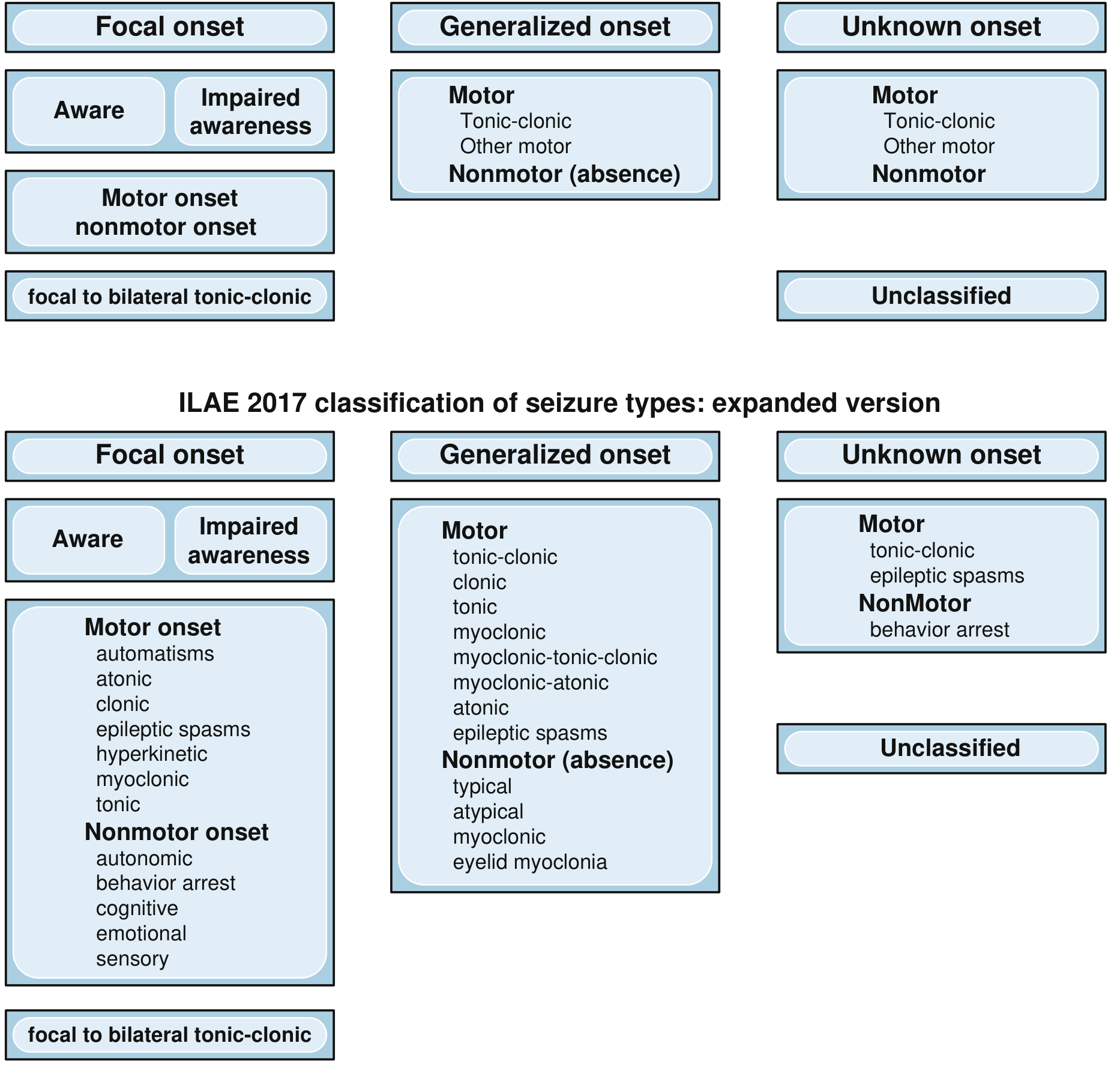
ILAE 2017 Operational Classification (Current Standard)
This is the currently used classification, replacing the older 1981 classification. It has three main classes based on site of onset:
CLASS 1: FOCAL ONSET SEIZURES
Originate within networks limited to one hemisphere (may be discretely localized or widely distributed, and may involve subcortical structures).
Step 1: Awareness classifier (optional but preferred):
- Focal Aware Seizure (FAS): Awareness fully preserved throughout [replaced "simple partial"]
- Focal Impaired Awareness Seizure (FIAS): Any alteration in awareness at any point [replaced "complex partial"]
- Unknown awareness: When awareness cannot be assessed
Step 2: First clinical manifestation:
Motor Onset:
- Automatisms (lip smacking, hand fumbling)
- Atonic
- Clonic
- Epileptic spasms
- Hyperkinetic
- Myoclonic
- Tonic
Nonmotor Onset:
- Autonomic (heart rate change, flushing, piloerection)
- Behavior arrest
- Cognitive (dysphasia, deja vu)
- Emotional (fear, laughing - gelastic)
- Sensory (visual, auditory, somatosensory)
Focal to Bilateral Tonic-Clonic: Replaces old "secondarily generalized" - focal seizure spreading to engage both hemispheres.
CLASS 2: GENERALIZED ONSET SEIZURES
Originate at some point within, and rapidly engage, bilaterally distributed networks. Do NOT necessarily include the entire cortex; may be asymmetrical.
Motor:
- Tonic-clonic (GTC / "grand mal")
- Clonic
- Tonic
- Myoclonic
- Myoclonic-tonic-clonic
- Myoclonic-atonic (formerly "myoclonic-astatic")
- Atonic (drop attacks)
- Epileptic spasms
Nonmotor (Absence):
- Typical absence: Abrupt onset, EEG 3 Hz spike-wave; stare, automatisms possible; <20 sec
- Atypical absence: Slower onset/offset, EEG <2.5 Hz; seen in Lennox-Gastaut
- Myoclonic absence
- Eyelid myoclonia (Jeavons syndrome)
CLASS 3: UNKNOWN ONSET SEIZURES
When onset cannot be determined:
- Motor: tonic-clonic, epileptic spasms
- Nonmotor: behavior arrest
CLASS 4: UNCLASSIFIED
Inadequate information or unusual presentation.
Comparison: 1981 vs 2017 Terminology
| 1981 ILAE Term | 2017 ILAE Term |
|---|
| Partial seizure | Focal seizure |
| Simple partial seizure | Focal aware seizure (FAS) |
| Complex partial seizure | Focal impaired awareness seizure (FIAS) |
| Secondarily generalized seizure | Focal to bilateral tonic-clonic seizure (FBTCS) |
| Benign | Self-limited or pharmacoresponsive |
- Bradley and Daroff's Neurology in Clinical Practice, Table 100.1
4. CLASSIFICATION OF EPILEPSIES (ILAE 2017 - Three-Level Framework)
The ILAE 2017 classification of epilepsies (Scheffer et al., 2017) operates at THREE levels:
Level 1: Seizure Types
(As classified above - focal, generalized, unknown, unclassified)
Level 2: Epilepsy Types
| Epilepsy Type | Description |
|---|
| Focal epilepsy | Consistently focal-onset seizures; epileptogenic zone in one hemisphere |
| Generalized epilepsy | Generalized onset seizures; bilateral epileptiform discharges on EEG |
| Combined generalized & focal | Patient has BOTH focal AND generalized onset seizures (e.g., Dravet syndrome) |
| Unknown | Seizure type cannot be determined |
Level 3: Epilepsy Syndrome
A syndrome is a cluster of features (seizure type, age of onset, EEG, imaging, genetics, prognosis) that together define a recognizable entity.
Level 4: Etiology (runs ACROSS all three levels)
The ILAE 2017 framework mandates considering etiology at EVERY level:
| Etiology | Examples |
|---|
| Structural | Hippocampal sclerosis, cortical dysplasia, tumor, stroke, tuberous sclerosis |
| Genetic | SCN1A (Dravet), KCNQ2 (BFNE), SCN2A, CDKL5, DEPDC5; chromosomal (TSC, Angelman) |
| Infectious | Neurocysticercosis (MOST COMMON in India!), TB, viral encephalitis, SSPE |
| Metabolic | Pyridoxine deficiency, GLUT1 deficiency, phenylketonuria, organic acidurias |
| Immune | Anti-NMDAR encephalitis, LGI1, CASPR2, Rasmussen encephalitis |
| Unknown | No identifiable cause despite thorough workup |
More than one etiology category may apply. - Bradley and Daroff's Neurology
5. PAEDIATRIC EPILEPSY SYNDROMES - CLASSIFIED BY AGE OF ONSET
This is particularly important for paediatric practice. About 50% of children can be characterized as having a specific epilepsy syndrome. - Kaplan & Sadock
NEONATAL PERIOD (0-28 days)
| Syndrome | Key Features |
|---|
| Self-limited familial neonatal epilepsy (BFNE) | KCNQ2/KCNQ3 mutation; day 2-3 of life; clonic seizures; remits by 6 months |
| Early Myoclonic Encephalopathy | Burst-suppression EEG; fragmentary myoclonus; very poor prognosis |
| Ohtahara Syndrome (EIEE) | Tonic spasms + burst-suppression EEG; onset first weeks; now called "EIEE" |
INFANCY (1 month - 2 years)
| Syndrome | Key Features |
|---|
| Febrile Seizures | 6 months-5 years; with fever; most common seizure disorder in paediatrics (4% incidence) |
| West Syndrome (Infantile Spasms) | 3-12 months; epileptic spasms + hypsarrhythmia EEG; developmental regression; triad |
| Dravet Syndrome (SMEI) | SCN1A mutation; onset <1 year; febrile-triggered initially; sodium channel blocker-worsened |
| Myoclonic Epilepsy of Infancy | Benign; myoclonic jerks; normal development; good prognosis |
| EIMSF (Migrating focal seizures) | Severe; multifocal; KCNT1 mutations |
CHILDHOOD (2-12 years)
| Syndrome | Key Features |
|---|
| Childhood Absence Epilepsy (CAE) | 4-10 years; 3 Hz spike-wave; staring spells; girl > boys; usually remits |
| BECTS (Self-limited epilepsy with centrotemporal spikes) | 3-13 years; nocturnal; unilateral facial/limb clonic; centrotemporal spikes; self-limited |
| Lennox-Gastaut Syndrome (LGS) | 1-7 years; multiple seizure types (atonic, tonic, atypical absence); slow spike-wave <2.5 Hz; intellectual disability |
| Panayiotopoulos Syndrome | 3-6 years; occipital; vomiting + ictal; prolonged; self-limited |
| Epilepsy with Myoclonic-Atonic Seizures (Doose) | 1-5 years; myoclonic-atonic drop attacks; variable prognosis |
| CSWS/ESES | Epileptic encephalopathy with continuous spike-wave during sleep; acquired aphasia |
| Landau-Kleffner Syndrome | Acquired epileptic aphasia + CSWS |
ADOLESCENCE
| Syndrome | Key Features |
|---|
| Juvenile Absence Epilepsy (JAE) | Absences + GTCs; persists into adulthood |
| Juvenile Myoclonic Epilepsy (JME) | Most common genetic generalized epilepsy; morning myoclonus + GTC; lifelong (valproate-responsive) |
| Epilepsy with GTCs alone | GTCs on awakening; genetic generalized |
6. ILAE 2025 UPDATED SEIZURE CLASSIFICATION (Newest Update)
The ILAE published an
updated seizure classification in 2025 (Beniczky, Trinka, Wirrell et al.,
Epileptic Disorders, 2025 -
PMID: 41081650). Key changes from 2017:
- "Awareness" replaced by "Consciousness" - now defined by BOTH awareness (recall) and responsiveness
- Seizure described by chronological sequence of signs/symptoms, not just first sign
- "Awareness" as optional but preferred classifier remains; focal seizures may be classified without it in resource-limited settings
- Observable vs. non-observable features explicitly distinguished
- Epileptic negative myoclonus added as a new recognized seizure type
- Epileptic spasms given special attention given urgency of early intervention in infants
- Basic + Expanded versions retained for low-resource vs. advanced settings
A Basic version allows classification in resource-limited or primary care environments, while the Expanded version supports surgical decision-making.
7. ILAE 2022 EPILEPSY SYNDROME CLASSIFICATION (India-Relevant Update)
The ILAE 2022 diagnostic criteria for epilepsy syndromes were validated in a major Indian study from Lady Hardinge Medical College and AIIMS New Delhi (Seizure Journal, 2024):
- 1,550 children with epilepsy analyzed
- 55.4% classified into epilepsy syndromes using new ILAE 2022 criteria
- 11.5% newly classified under a syndrome who were previously unclassified
- Application led to change in diagnosis name alone in 77.8% - showing evolution of nomenclature
8. INDIA-SPECIFIC CONSIDERATIONS
Epidemiology in India
- Epilepsy prevalence in India: 5-10 per 1,000 (higher in rural areas)
- India has approximately 10-12 million people with epilepsy (one of the largest burdens globally)
- Treatment gap in rural India: >80% (most with epilepsy never receive adequate treatment)
Common Etiologies in Indian Children
| Priority Etiology | Significance |
|---|
| Neurocysticercosis (NCC) | MOST COMMON cause of new-onset epilepsy in older children in India; calcified granuloma |
| Febrile seizures | Very common; ~4% incidence; FIRES (Febrile Infection-Related Epilepsy Syndrome) important in young children |
| Hypoxic-Ischemic Encephalopathy | Leading cause of neonatal seizures and subsequent epilepsy |
| Tuberculous meningitis | Significant cause of structural epilepsy |
| SSPE | Subacute sclerosing panencephalitis (measles-related); still seen in India |
| Genetic/Metabolic | Increasing recognition; SCN1A, pyridoxine dependency |
Indian Paediatrics Journal - Key Publications
- ILAE Task Force report on neonatal seizures (2022) was reprinted in Indian Pediatrics (2023) highlighting Indian applicability
- IAP (Indian Academy of Pediatrics) guidelines on febrile seizures recommend NO prophylactic ASMs for simple febrile seizures
9. QUICK REFERENCE - KEY DIFFERENTIATING POINTS (Exam-Oriented)
| Point | Remember |
|---|
| Seizure = single event | Epilepsy = brain disease with enduring predisposition |
| Provoked seizure ≠ epilepsy | NCC is #1 cause of new-onset epilepsy in Indian children |
| ILAE 2017: "partial" → "focal"; "complex partial" → "FIAS" | ILAE 2025 update: awareness→consciousness; adds negative myoclonus |
| West syndrome triad | Spasms + Hypsarrhythmia + Developmental regression |
| BECTS = most common focal epilepsy in children | Self-limited; no treatment needed if mild |
| JME = most common IGE | Lifelong; morning myoclonus; valproate-sensitive |
| LGS triad | Multiple seizure types + slow spike-wave (<2.5Hz) + cognitive regression |
| Dravet: avoid | Lamotrigine and phenytoin (Na+ channel blockers worsen it) |
Sources:
- Adams and Victor's Principles of Neurology, 12th Ed. - Seizure epidemiology and definitions
- Bradley and Daroff's Neurology in Clinical Practice - ILAE 2017 classification, terminology table, epilepsy syndrome framework
- Kaplan & Sadock's Comprehensive Textbook of Psychiatry - Paediatric seizure syndromes, ILAE 2014 definition
- Harrison's Principles of Internal Medicine, 22nd Ed. (2025) - Seizure types, focal/generalized
- Beniczky S et al. "A practical guide to the updated seizure classification 2025." Epileptic Disorders 2025 [PMID: 41081650] - ILAE 2025 updates
- Sharma S et al. "Applicability of ILAE 2022 diagnostic criteria for epilepsy syndromes in children: A retrospective review of 1550 children." Seizure Journal 2024 - India validation study (LHMC & AIIMS New Delhi)