
Interpeduncular fossa
The interpeduncular fossa is a diamond-shaped depression on the ventral surface of the midbrain, located between the two cerebral peduncles. Key contents include the posterior perforated substance (through which central branches of the posterior cerebral artery enter), and it's where the oculomotor nerves (CN III) emerge from the medial aspect of the cerebral peduncles. The basilar artery bifurcates just superior to this region.
Searching sources...
Reading File
Searching Images
interpeduncular fossa midbrain anatomy
Finding Sources
Finding Sources
Reading File
Reading File
Reading File
Reading File
Searching Images
interpeduncular fossa cerebral peduncles oculomotor nerve CN III midbrain ventral
Loading Image
Finding Sources
Finding Sources
Searching Images
midbrain ventral surface anatomy diagram cerebral peduncles oculomotor nerve
Reading File
Here is a detailed account of the interpeduncular fossa drawn from multiple authoritative textbooks:
Interpeduncular Fossa
Definition and Location
The interpeduncular fossa is a deep, diamond-shaped (rhomboid) depression on the ventral (anterior) surface of the midbrain, bounded on either side by the crura cerebri (cerebral peduncles). It is also known as the interpeduncular cistern because it is a widened region of the subarachnoid space filled with CSF. - Neuroanatomy through Clinical Cases, 3rd Ed.; Gray's Anatomy for Students
Boundaries
| Wall | Structure |
|---|---|
| Anterolateral (sides) | Crura cerebri (crus cerebri) |
| Posterior (floor) | Posterior perforated substance |
| Anterior (roof, in life) | Mamillary bodies and posterior perforated substance |
Floor: The Posterior Perforated Substance
The floor of the fossa is formed by the posterior perforated substance, a region of grey matter pierced by numerous small perforating branches of the posterior cerebral artery (P1 segment) and the posterior communicating artery. These vessels - the thalamoperforating arteries and thalamogeniculate arteries - enter the brain here to supply the thalamus, subthalamus, and midbrain tegmentum. - Grainger & Allison's Diagnostic Radiology
Key Contents and Relations
CN III - Oculomotor Nerve (most important)
The oculomotor nerve fascicles originate from the oculomotor nucleus (ventral to the periaqueductal grey, at the level of the superior colliculus). The fascicle traverses the midbrain, passing through/near the red nucleus and close to the cerebral peduncles, before emerging as rootlets in the lateral interpeduncular fossa. Within the fossa, the rootlets converge into the third nerve trunk. - Bradley and Daroff's Neurology in Clinical Practice
The CN III trunk then passes:
- Through the subarachnoid space toward the cavernous sinus
- Between the superior cerebellar artery (SCA) and the posterior cerebral artery (PCA) - this relationship is key for aneurysm compression
- Parallel to the posterior communicating artery (PCOM) - the junction of PCOM with the internal carotid artery is a classic aneurysm site
Vascular Relations
- The basilar artery bifurcates just above the dorsum sellae, forming the two posterior cerebral arteries (PCAs) at the top of the fossa
- CN III passes between the PCA and SCA on each side - Grainger & Allison's
Clinical Significance
1. PCOM Aneurysm and CN III Palsy
The most common cause of oculomotor dysfunction in the interpeduncular fossa / subarachnoid segment is compression by a posterior communicating artery (PCOM) aneurysm. The pupillomotor fibers run superomedially on the nerve surface and are particularly vulnerable to extrinsic compression, producing a "surgical" (pupil-involving) CN III palsy. - Bradley and Daroff's Neurology
Key rule: Any CN III palsy - whether pupil-involving or pupil-sparing incomplete - requires immediate imaging to exclude a PCOM aneurysm, given the risk of subarachnoid hemorrhage. Pupil-sparing incomplete palsies may still progress to pupillary involvement within ~1 week.
2. Subarachnoid Hemorrhage (SAH)
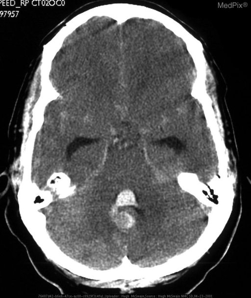
Blood from ruptured aneurysms or perimesencephalic bleeding pools in the interpeduncular cistern. On CT, hyperdensity in the interpeduncular fossa is a classic finding of perimesencephalic (non-aneurysmal) SAH, a generally benign form - Grainger & Allison's Diagnostic Radiology
3. Herniation Syndromes
Transtentorial (uncal) herniation compresses CN III in the interpeduncular fossa, causing ipsilateral fixed dilated pupil with progressive ophthalmoplegia - a neurosurgical emergency.
4. Named Brainstem Syndromes involving CN III emerging from this region
| Syndrome | Structures Involved | Clinical Features |
|---|---|---|
| Weber | CN III fascicle + cerebral peduncle | Ipsilateral CN III palsy + contralateral hemiparesis |
| Benedikt | CN III fascicle + red nucleus | Ipsilateral CN III palsy + contralateral chorea/tremor |
| Nothnagel | CN III fascicle + superior cerebellar peduncle | Ipsilateral CN III palsy + ipsilateral hemiataxia |
| Claude | CN III + combined red nucleus + cerebellar peduncle | CN III + contralateral ataxia + tremor |
- Bradley and Daroff's Neurology in Clinical Practice
5. Inflammatory / Neoplastic Meningitis
CN III involvement in the interpeduncular fossa may occur with meningitis (infectious or carcinomatous), in which case it may be isolated or accompanied by other cranial nerve palsies and meningeal signs. - Bradley and Daroff's Neurology
Summary of Key Points
- Location: ventral midbrain, between the two crura cerebri
- Floor: posterior perforated substance (perforated by thalamoperforating branches of PCA/PCOM)
- CN III emerges in its lateral walls and converges into a trunk here
- Arterial relations: CN III passes between the SCA and PCA; PCOM is closely parallel
- Clinically most important for: PCOM aneurysm causing CN III palsy, SAH pooling, transtentorial herniation, and named midbrain syndromes
This is a shared conversation. Sign in to Orris to start your own chat.