
Kreb cycle
Reading File
The Krebs cycle (also called the citric acid cycle or TCA cycle) is a series of 8 enzymatic reactions in the mitochondrial matrix that oxidize acetyl-CoA to CO2 while generating NADH, FADH2, and GTP for ATP production.
Searching sources...
Finding Sources
Searching the Web
Krebs cycle steps enzymes products summary
Reading File
Reading File
Reading File
Loading Image
Reading File
Loading Image
Here is a thorough breakdown of the Krebs cycle, sourced primarily from Harper's Illustrated Biochemistry (32nd Ed).
The Krebs Cycle (Citric Acid Cycle / TCA Cycle)
Overview
The Krebs cycle is a cyclic sequence of 8 enzyme-mediated reactions occurring in the mitochondrial matrix. It is the central hub of aerobic metabolism, oxidizing acetyl-CoA (derived from carbohydrates, fats, and amino acids) to CO2 while generating reducing equivalents (NADH, FADH2) that drive ATP synthesis via the respiratory chain.
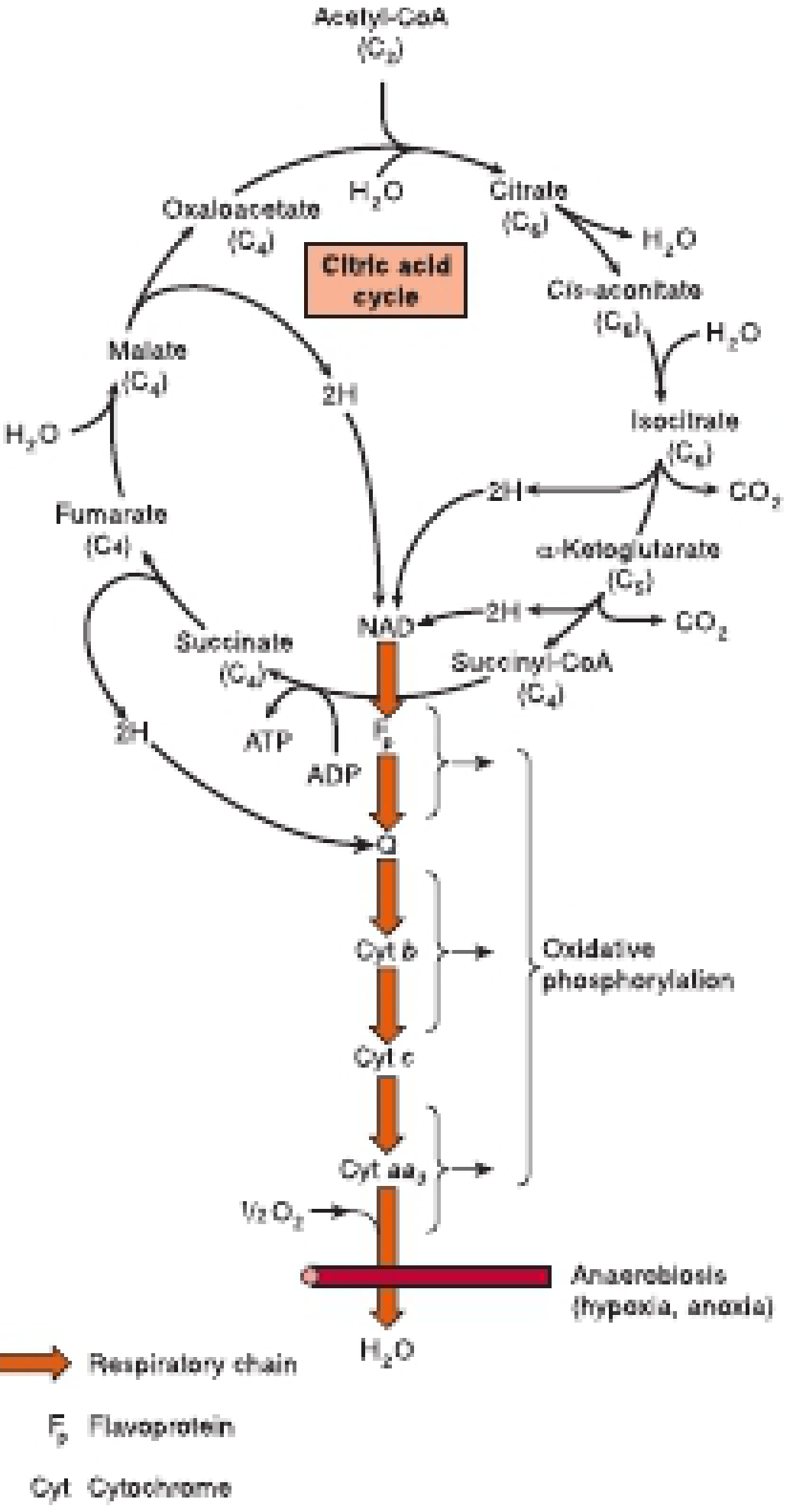
Entry into the Cycle: Acetyl-CoA
Before entering the cycle, pyruvate (from glycolysis) is converted to acetyl-CoA by the pyruvate dehydrogenase complex (PDC) in the mitochondrial matrix. This reaction requires:
- Thiamin diphosphate (B1), lipoate, NAD+, FAD, CoA
The 8 Steps
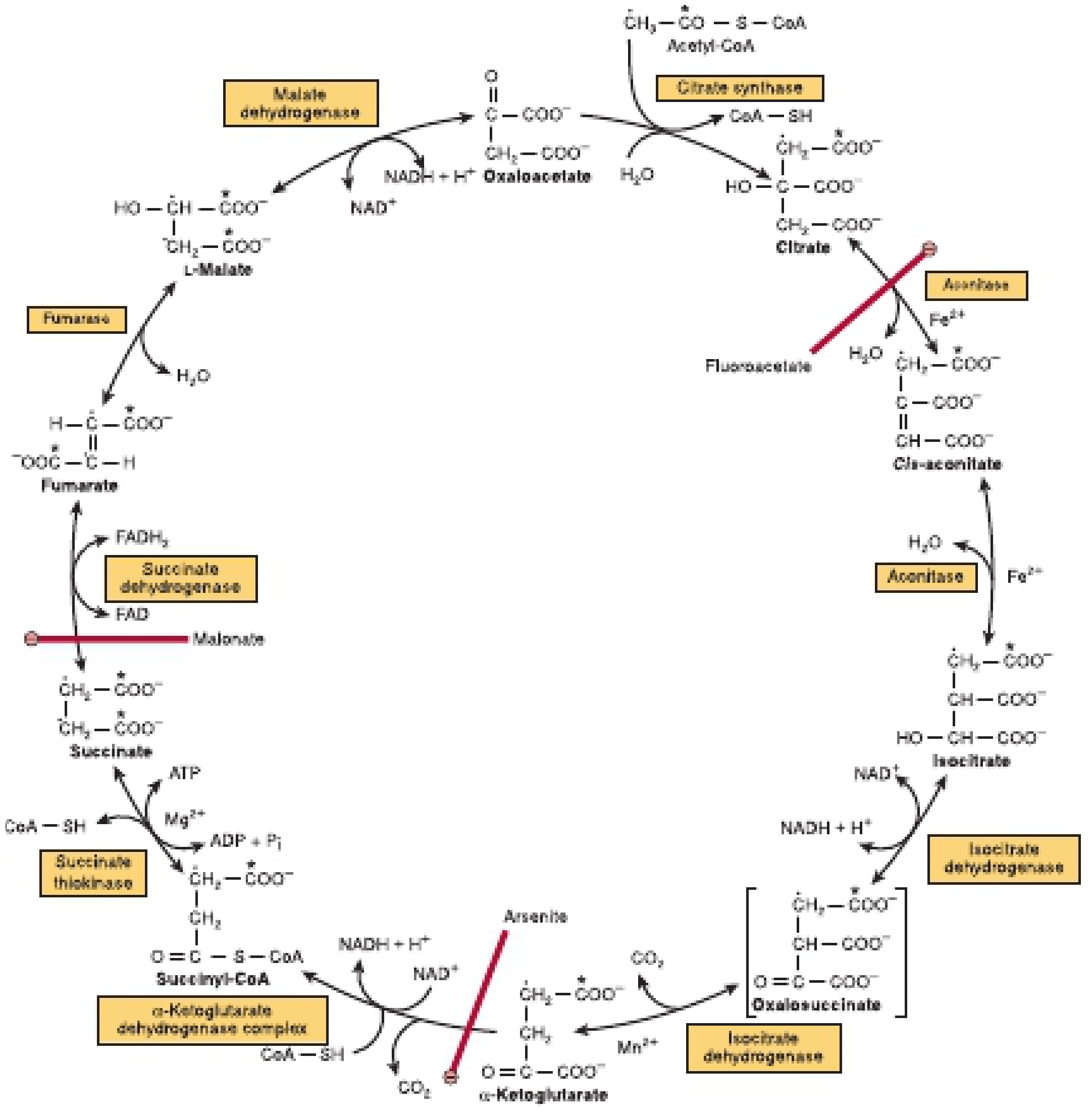
Here is the detailed diagram from Harper's showing all enzymes, intermediates, and inhibitors:
| Step | Substrate → Product | Enzyme | Key Output |
|---|---|---|---|
| 1 | Oxaloacetate (C4) + Acetyl-CoA (C2) → Citrate (C6) | Citrate synthase | Exothermic; CoA released |
| 2 | Citrate → Cis-aconitate → Isocitrate | Aconitase (requires Fe2+) | Dehydration then rehydration |
| 3 | Isocitrate → Oxalosuccinate → α-Ketoglutarate (C5) + CO2 | Isocitrate dehydrogenase (NAD+, Mn2+) | 1st NADH, 1st CO2 |
| 4 | α-Ketoglutarate → Succinyl-CoA (C4) + CO2 | α-Ketoglutarate dehydrogenase complex | 2nd NADH, 2nd CO2 |
| 5 | Succinyl-CoA → Succinate | Succinate thiokinase (succinyl-CoA synthetase, Mg2+) | 1 ATP or GTP (substrate-level phosphorylation) |
| 6 | Succinate → Fumarate | Succinate dehydrogenase (FAD, Fe-S protein) | 1 FADH2 |
| 7 | Fumarate + H2O → L-Malate | Fumarase (fumarate hydratase) | - |
| 8 | L-Malate → Oxaloacetate | Malate dehydrogenase (NAD+) | 3rd NADH |
Products per Turn
| Product | Number | ATP equivalents |
|---|---|---|
| NADH | 3 | ~2.5 each = 7.5 ATP |
| FADH2 | 1 | ~1.5 ATP |
| GTP/ATP (substrate level) | 1 | 1 ATP |
| CO2 | 2 | - |
| Total | ~10 ATP |
Regulation (3 Key Points)
- Citrate synthase - inhibited by ATP, NADH, succinyl-CoA, citrate; activated by ADP
- Isocitrate dehydrogenase - inhibited by ATP and NADH; activated by ADP and Ca2+
- α-Ketoglutarate dehydrogenase - inhibited by succinyl-CoA, NADH; activated by Ca2+
High [NADH] and [ATP] signal energy sufficiency and slow the cycle. High [ADP] and [Ca2+] (during muscle contraction) accelerate it.
Cofactors and B Vitamins
The cycle depends on 4 B vitamins:
- Thiamin (B1) - thiamin diphosphate, cofactor for α-ketoglutarate dehydrogenase
- Riboflavin (B2) - as FAD, cofactor for succinate dehydrogenase
- Niacin (B3) - as NAD+, electron acceptor for isocitrate DH, α-ketoglutarate DH, and malate DH
- Pantothenic acid (B5) - as CoA, esterified to form acetyl-CoA and succinyl-CoA
Key Inhibitors
| Inhibitor | Target | Effect |
|---|---|---|
| Fluoroacetate | Aconitase (via fluorocitrate) | Citrate accumulates |
| Malonate | Succinate dehydrogenase | Competitive inhibitor (structural analogue of succinate) |
| Arsenite | α-Ketoglutarate dehydrogenase | α-Ketoglutarate accumulates |
Amphibolic Role
The cycle is described as amphibolic - it serves both catabolic and anabolic roles:
- Gluconeogenesis: oxaloacetate exits as phosphoenolpyruvate
- Amino acid synthesis: intermediates undergo transamination (e.g., α-ketoglutarate → glutamate)
- Fatty acid synthesis: citrate is exported to the cytosol as an acetyl-CoA source
- Heme synthesis: succinyl-CoA is a precursor for porphyrin rings
Anaplerosis (carbon entering the cycle, e.g., pyruvate → oxaloacetate via pyruvate carboxylase) must equal cataplerosis (carbon leaving, e.g., oxaloacetate → PEP) to sustain the cycle.
Location
All enzymes are in the mitochondrial matrix except:
- Succinate dehydrogenase and aconitase - bound to the inner mitochondrial membrane
Sources: Harper's Illustrated Biochemistry, 32nd Ed., p. 168-171; Guyton and Hall Textbook of Medical Physiology
Formation and utilization of ketone bodies.
Ketone bodies (acetoacetate, β-hydroxybutyrate, acetone) are produced in the liver from acetyl-CoA during fasting, starvation, or uncontrolled diabetes, and are used as fuel by extrahepatic tissues including the brain and heart.
Searching sources...
Finding Sources
Searching the Web
ketone bodies formation utilization biochemistry ketogenesis
Finding Sources
Reading File
Finding Sources
Reading File
Loading Image
Reading File
Finding Sources
Reading File
Finding Sources
Finding Sources
Loading Image
Finding Sources
Reading File
I now have comprehensive information from the textbooks. Here is the complete answer.
Formation and Utilization of Ketone Bodies
The Three Ketone Bodies
| Compound | Nature | Notes |
|---|---|---|
| Acetoacetate | Primary ketone body | Main product released into blood |
| D-β-Hydroxybutyrate | Reduced form | Most abundant in blood (ratio ~3:1 to acetoacetate); not technically a ketone (has -OH group, not C=O) |
| Acetone | Volatile byproduct | Formed by spontaneous decarboxylation of acetoacetate; expired via lungs |
PART 1: Formation (Ketogenesis) - Occurs ONLY in Liver
Location
Mitochondrial matrix of hepatocytes only. All enzymes are present at sufficient levels only in liver.
Precursor
Acetyl-CoA, generated from:
- β-oxidation of fatty acids (major source)
- Catabolism of ketogenic amino acids (leucine, isoleucine, lysine, tryptophan, phenylalanine, tyrosine)
- Minor contribution from pyruvate oxidation
Why does ketogenesis occur?
When acetyl-CoA production overwhelms the TCA cycle capacity (due to low oxaloacetate - which is being diverted to gluconeogenesis during fasting), excess acetyl-CoA is channeled into ketone body synthesis.
Steps of Ketogenesis (HMG-CoA Pathway)
| Step | Reaction | Enzyme |
|---|---|---|
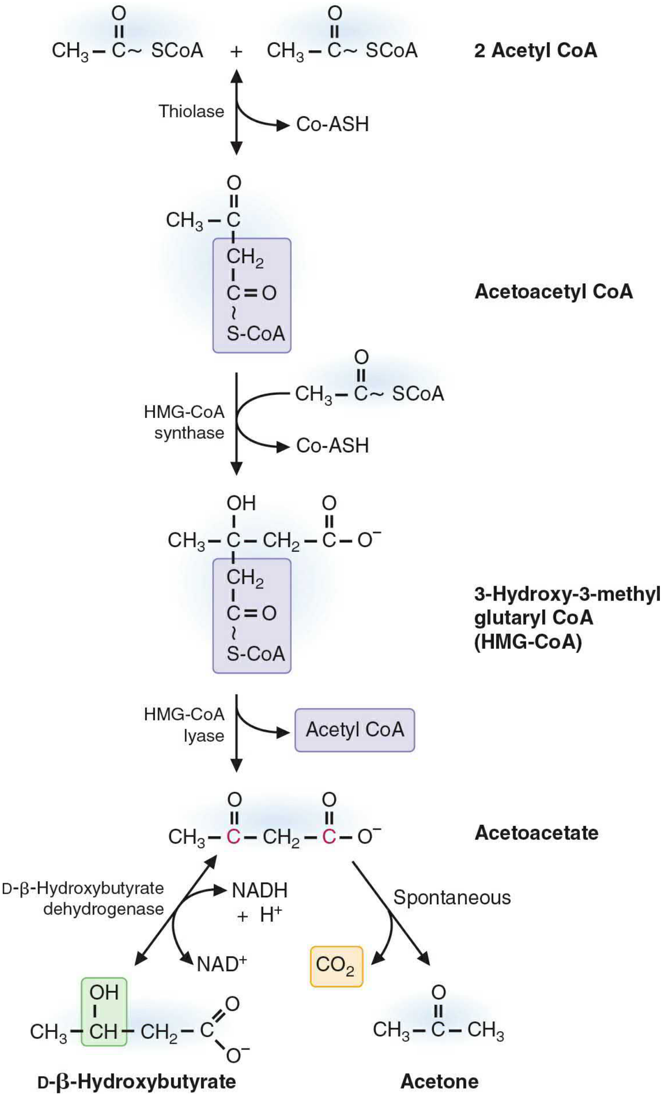
| 1 | 2 Acetyl-CoA → Acetoacetyl-CoA + CoA | Thiolase (reversible; favors acetyl-CoA normally, reverses when acetyl-CoA is high) |
| 2 | Acetoacetyl-CoA + Acetyl-CoA → HMG-CoA + CoA | HMG-CoA synthase (rate-limiting step; mitochondrial isoform - distinct from cytosolic HMG-CoA synthase used in cholesterol synthesis) |
| 3 | HMG-CoA → Acetoacetate + Acetyl-CoA | HMG-CoA lyase |
| 4a | Acetoacetate + NADH → D-β-Hydroxybutyrate + NAD+ | D-β-Hydroxybutyrate dehydrogenase (reversible; ratio determined by mitochondrial NADH/NAD+) |
| 4b | Acetoacetate → Acetone + CO2 | Spontaneous (non-enzymatic decarboxylation) |
The ratio of β-hydroxybutyrate to acetoacetate in blood (~3:1) is governed by the mitochondrial NADH/NAD+ ratio. During severe fasting/DKA, NADH is high, so β-hydroxybutyrate predominates.
PART 2: Utilization (Ketolysis) - Occurs in Extrahepatic Tissues
Tissues that USE ketone bodies
- Skeletal muscle, cardiac muscle, brain (after 2-3 days of starvation), kidney cortex, intestinal mucosa
Why the LIVER cannot use its own ketone bodies
The liver lacks succinyl-CoA:acetoacetate CoA transferase (thiotransferase) in sufficient quantities - the key enzyme needed to reactivate acetoacetate. This ensures ketone bodies are exported to peripheral tissues.
Steps of Ketolysis
| Step | Reaction | Enzyme |
|---|---|---|
| 1 | D-β-Hydroxybutyrate + NAD+ → Acetoacetate + NADH | D-β-Hydroxybutyrate dehydrogenase |
| 2 | Acetoacetate + Succinyl-CoA → Acetoacetyl-CoA + Succinate | Succinyl-CoA:acetoacetate CoA transferase (thiotransferase) - KEY STEP; costs 1 GTP equivalent |
| 3 | Acetoacetyl-CoA + CoA → 2 Acetyl-CoA | Thiolase |
| 4 | Acetyl-CoA → TCA cycle → ATP |
Energy Yield
| Ketone Body | Net ATP |
|---|---|
| Acetoacetate | ~20 ATP (2 × acetyl-CoA via TCA) minus 1 ATP equivalent for activation = ~19 ATP |
| β-Hydroxybutyrate | ~21.5 ATP (1 extra NADH from step 1) |
Regulation of Ketogenesis
The rate-limiting enzyme is HMG-CoA synthase (mitochondrial). Key regulators:
| Factor | Effect on Ketogenesis | Mechanism |
|---|---|---|
| Insulin (fed state) | Inhibits ↓ | Suppresses adipose lipolysis → less fatty acid to liver; promotes malonyl-CoA production → inhibits carnitine palmitoyltransferase-I (CPT-I) → blocks fatty acid entry into mitochondria |
| Glucagon/Fasting | Stimulates ↑ | Activates lipolysis; decreases malonyl-CoA → CPT-I active |
| High fatty acid supply | Stimulates ↑ | More acetyl-CoA delivered |
| Low oxaloacetate | Stimulates ↑ | OAA diverted to gluconeogenesis → TCA cycle slows → acetyl-CoA diverted to ketogenesis |
| High NADH/NAD+ | Shifts toward β-hydroxybutyrate | Favors reduction of acetoacetate |
Kinetics During Fasting
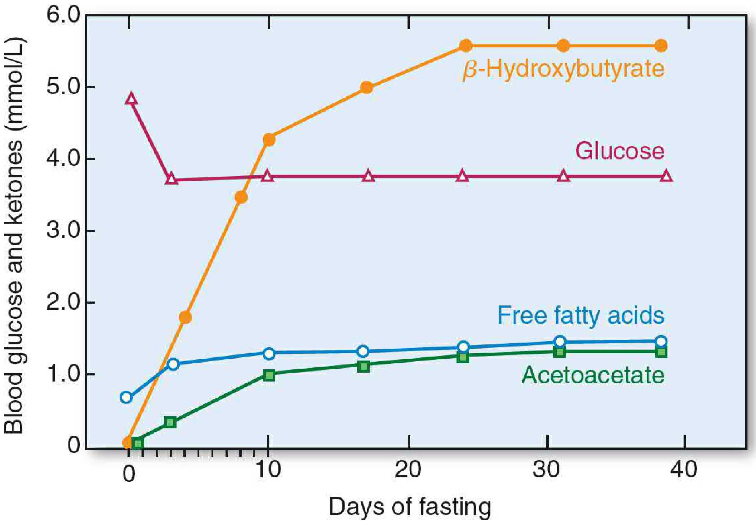
- 3-4 hours post-meal: fatty acids begin rising
- Day 2-3 of fasting: ketone bodies reach levels sufficient to cross blood-brain barrier
- Prolonged starvation: ketone bodies can supply up to 2/3 of brain's energy - sparing muscle protein from gluconeogenesis
Ketosis vs. Diabetic Ketoacidosis (DKA)
| State | Cause | Blood pH | Ketone Level |
|---|---|---|---|
| Physiologic ketosis (fasting) | Starvation; insulin present but low | Normal | Mildly elevated |
| Diabetic ketoacidosis | Absent insulin (Type 1 DM); unrestrained lipolysis + ketogenesis | Acidotic (<7.3) | Severely elevated |
In DKA: absence of insulin → maximal lipolysis + maximal ketogenesis → ketonemia → ketonuria → metabolic acidosis. β-Hydroxybutyrate predominates (may be missed by dipstick tests which detect only acetoacetate/acetone via nitroprusside reaction).
Alternative Fates of Ketone Bodies
- Cytosolic acetyl-CoA source: Acetoacetate can be activated in the cytosol → acetoacetyl-CoA → acetyl-CoA for acetylcholine synthesis (neuronal cells) and cholesterol synthesis
- Ketogenic diet: High fat:carbohydrate (3:1 ratio) diet induces therapeutic ketosis - used in refractory epilepsy in children and pyruvate dehydrogenase deficiency
- Ketogenic amino acids: Leucine, isoleucine, lysine, tryptophan, phenylalanine, tyrosine are catabolized to acetyl-CoA/acetoacetyl-CoA, providing an additional route to ketone bodies
Sources: Basic Medical Biochemistry: A Clinical Approach, 6th Ed., p. 1097-1101; Schwartz's Principles of Surgery, 11th Ed., p. 92
This is a shared conversation. Sign in to Orris to start your own chat.