
Give comprehensive notes on tuberculosis, especially in the context of the Pakistani population and it's guidelines followed in Pakistan and also compare with International guidelines: - its diagnosis - treatment and management - what its different types are - how to diagnose each - how to give information on each type - what different protocols and guidelines there are for Pakistani populations - and also give recent updates regarding diagnsosis and management
Give comprehensive notes on tuberculosis, especially in the context of the Pakistani population and it's guidelines followed in Pakistan and also compare with International guidelines: - its diagnosis - treatment and management - what its different types are - how to diagnose each - how to give information on each type - what different protocols and guidelines there are for Pakistani populations - and also give recent updates regarding diagnsosis and management
Give comprehensive notes on tuberculosis, especially in the context of the Pakistani population and it's guidelines followed in Pakistan and also compare with International guidelines: - its diagnosis - treatment and management - what its different types are - how to diagnose each - how to give information on each type - what different protocols and guidelines there are for Pakistani populations - and also give recent updates regarding diagnsosis and management.. What are the different types of emergencies in a tuberculosis case? How to diagnose and treat each? What are the protocols for a latent case of tuberculosis? What PACT questions about history of tuberculosis are to be taken? What examination findings are to be expected in a case of tuberculosis? Based on each case scenario, also give algorithms, illustrations, diagrams, and images from the latest guidelines. Textbooks from Davidson, Harrison, Kumar, and Clark textbook of medicine, and the latest Pakistani local tuberculosis guidelines. Also give memorable mnemonics in an easy way to remember different information wherever necessary. Don't make unnecessary mnemonics.
Pakistan National TB Program guidelines 2023 2024 NTP diagnostic treatment protocol
tuberculosis Pakistan diagnosis treatment guidelines
tuberculosis chest X-ray pulmonary TB cavitation upper lobe
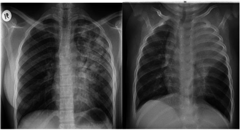
This side-by-side comparison displays two posteroanterior (PA) chest X-rays illustrating the diverse radiographic presentations of pulmonary tuberculosis (TB) in adult versus pediatric patients. The adult image (left) demonstrates extensive, heterogeneous, patchy infiltrates and areas of consolidation predominantly involving the left lung field, typical of post-primary or reactivation TB. Visible radiolucencies suggest cavitation within the upper lobe. In contrast, the pediatric image (right) shows a different pattern characterized by diffuse, relatively homogeneous opacification throughout both lung fields. Notable features in the pediatric X-ray include significant widening of the superior mediastinum, which is highly suggestive of bilateral mediastinal lymphadenopathy, a hallmark of primary TB in children. Both cases exhibit reduced lung clarity, but the pediatric case shows more pronounced bronchial involvement and hilar prominence without the distinct cavitary lesions seen in the adult. These images serve as a comparative educational resource for identifying age-specific diagnostic markers of Mycobacterium tuberculosis infection.
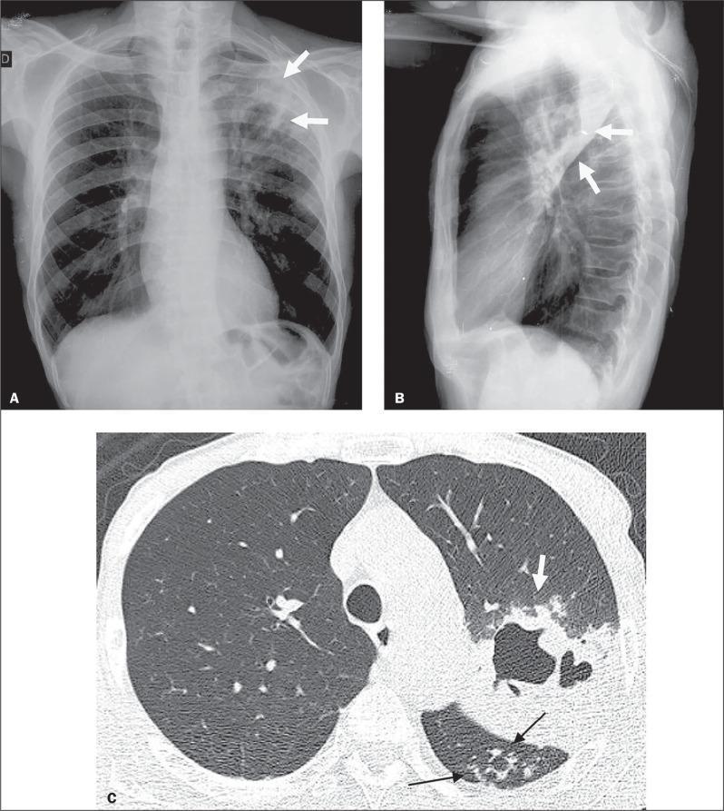
Diagnostic imaging composite of a 66-year-old male with active pulmonary tuberculosis (TB), comprising a posteroanterior chest X-ray (A), a lateral chest X-ray (B), and an axial High-Resolution Computed Tomography (HRCT) scan (C). Panels A and B demonstrate increased opacity in the left upper lung zone indicative of consolidation, with radiolucent areas representing cavitation (white arrows). Panel C, the HRCT at the level of the lower lobes, provides a detailed view of the pulmonary pathology. It reveals a focal area of dense consolidation containing a central air-filled cavity (white arrow) in the left lung. Additionally, characteristic 'tree-in-bud' patterns are visible (black arrows), consisting of small centrilobular nodules and branching linear opacities that indicate endobronchial spread of infection within the upper segment of the left lower lobe. These imaging findings are hallmark radiological features of secondary or active tuberculosis, illustrating parenchymal destruction and small airway involvement.
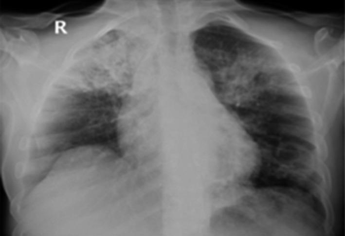
This diagnostic image is a frontal (posteroanterior) chest X-ray demonstrating significant bilateral pulmonary pathology. In the right lung, there is a dense, heterogeneous lobar consolidation primarily occupying the right upper lobe. Within this opacification, a radiolucent area is visible, highly suggestive of cavitation. The left lung displays sublobar, patchy infiltrates and consolidation, also predominantly in the upper zone, with subtle features indicating potential central cavitation. The cardiac silhouette appears within normal limits, though the mediastinal borders are partially obscured by the adjacent pulmonary opacities. These findings are classically associated with infectious processes such as pulmonary tuberculosis (TB) or necrotizing pneumonia. In the provided clinical context of a co-infection involving COVID-19 and tuberculosis, the imaging illustrates the characteristic upper-lobe predilection and cavitary nature of reactivated TB, while the more diffuse patchy elements may represent overlapping viral pneumonia patterns.
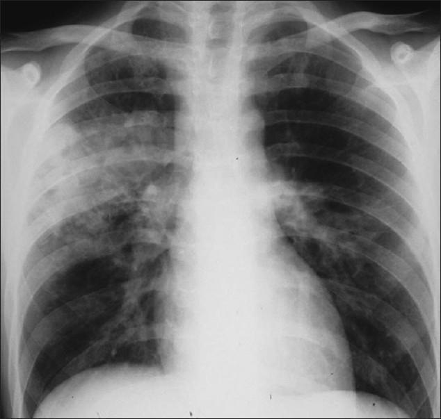
This diagnostic image is a posteroanterior (PA) chest X-ray demonstrating features consistent with primary Mycobacterium tuberculosis in an immunocompromised patient. The radiograph reveals focal areas of dense opacification, specifically consolidation in the right upper lobe and the left midzone. These opacities appear as increased white density that obscures the underlying pulmonary vasculature. In addition to parenchymal disease, there is evidence of mediastinal adenopathy, characterized by widening and contour abnormality of the hilar and paratracheal regions. Notably, despite the significant consolidation, there is a total lack of cavitation (hollowing out of the lung tissue), which is a common radiographic feature of primary TB in patients with low CD4 counts. The pleural spaces appear clear with no visible pleural effusions or pneumothorax. The image illustrates the atypical presentation of tuberculosis often seen in advanced HIV/AIDS, where lymphatic involvement and absence of cavitation are prominent features.
tuberculosis histology granuloma Langhans giant cells caseous necrosis
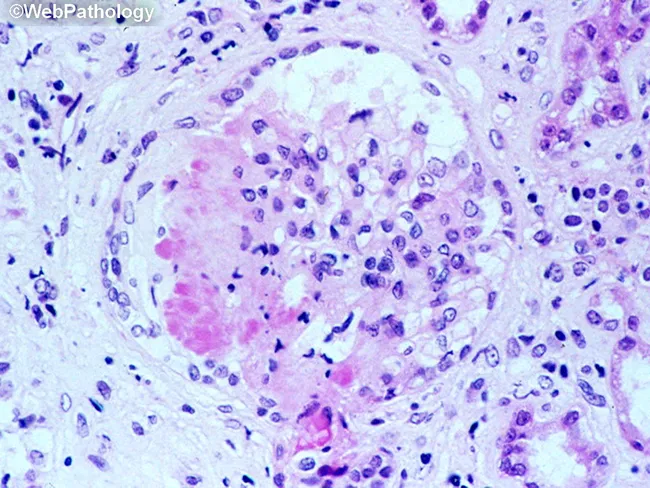
This is a light microscopy image of a formalin-fixed, paraffin-embedded tissue section stained with Hematoxylin and Eosin (H&E). The sample reveals a well-formed granulomatous lesion characterized by a surrounding cuff of lymphocytes and numerous epithelioid histiocytes with elongated, crescent-shaped nuclei. Within the granuloma, multinucleated giant cells (Langhans-type) are visible, and a central amorphous pink necrotic area (caseous necrosis) is evident. The tissue architecture demonstrates organized granulomatous inflammation with a necrotic core, consistent with infectious granulomas. The differential includes mycobacterial disease (tuberculosis) and certain fungal infections (histoplasmosis, coccioidomycosis), though non-necrotizing granulomas can occur in sarcoidosis. The staining highlights chromatin-rich nuclei and cytoplasmic eosinophilia; occasional fibroblasts and mild edema extend into the perigranulomatous region. Clinically, granulomatous tissue with caseation is highly suggestive of TB in the appropriate clinical context and warrants further diagnostic workup (acid-fast bacilli staining, fungal stains, culture, PCR). This image serves as teaching material for histopathology education, differential diagnosis of granulomatous inflammation, and correlation with radiologic and microbiologic findings in suspected infectious etiologies. Additional keywords for indexing: epithelioid macrophages, granuloma, necrosis, caseation, tuberculosis, mycobacteria, fungal infection, lymphohistiocytic rim, tissue biopsy, pathology slide, H&E, histology teaching. Clinical correlation with imaging and sputum analysis improves diagnostic yield.
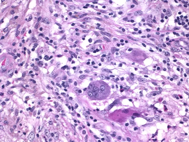
Histology of granulomatous mastitis on bright-field light microscopy. A dense inflammatory infiltrate forms granulomas centered on Langhans-type giant cells surrounded by epithelioid histiocytes, scattered lymphocytes, and occasional eosinophils. The image depicts a high-power view of the center of a granuloma within breast tissue biopsy; multinucleated giant cells with horseshoe-shaped nuclei are evident. The granulomatous architecture includes a peripheral rim of lymphocytes and histiocytes with minimal necrosis described. Staining by Hematoxylin and Eosin highlights basophilic nuclei and eosinophilic cytoplasm; pink collagenous stroma and adipose breast tissue may be visible in the background. Clinically, granulomatous mastitis can mimic infectious etiologies or neoplasia; the presence of Langhans giant cells strongly suggests a granulomatous process and warrants infectious workup for tuberculosis and fungal pathogens, as well as consideration of sarcoidosis or granulomatous reaction to foreign material. This image is diagnostic for granulomatous inflammation in breast tissue when correlated with clinical data and microbiologic studies; useful for education, differential diagnosis, and research on breast granulomatous disease. Additional educational value lies in distinguishing nonnecrotizing granulomas from caseating TB patterns, correlating histology with mammography and ultrasound, and guiding microbiologic testing and patient management in suspected granulomatous breast disease. The image supports teaching of granuloma biology and pathology.
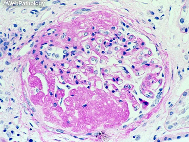
This is a histopathology image obtained with light microscopy from a formalin-fixed paraffin-embedded tissue section stained with Hematoxylin and Eosin. The field demonstrates a granulomatous inflammatory lesion, typically well circumscribed. The central area shows eosinophilic, acellular to granular necrotic material (caseous necrosis), surrounded by a dense mantle of epithelioid histiocytes with elongated, pale-staining nuclei and abundant pale cytoplasm. Multinucleated giant cells of the Langhans type may be present at the periphery. Scattered lymphocytes and occasional plasma cells form a peripheral lymphohistiocytic cuff around the granuloma. The surrounding stroma contains collagen fibers and occasional fibroblasts, with minimal additional inflammatory cells in this field. The morphology is classic for granulomatous inflammation, most commonly associated with infectious etiologies such as Mycobacterium tuberculosis or fungal infections, but can be seen in sarcoidosis or foreign body reactions depending on clinical context. Diagnostic significance: identification of a caseating granuloma raises suspicion for mycobacterial or fungal infection and warrants targeted microbiologic stains (Ziehl-Neelsen for acid-fast bacilli; GMS or PAS for fungi), cultures, and molecular testing. Clinical correlation is essential, including TB exposure history, immune status, and presentation—cough, weight loss, fever, pulmonary nodules or granulomatous lymphadenitis. This image is educational for teaching, differential diagnosis, and research into granulomatous disease pathology.
tuberculosis WHO guidelines 2023 2024 treatment drug resistant
https://www.cmu.gov.pk/national-tb-control-programme
tuberculosis algorithm diagnosis treatment flowchart GeneXpert sputum
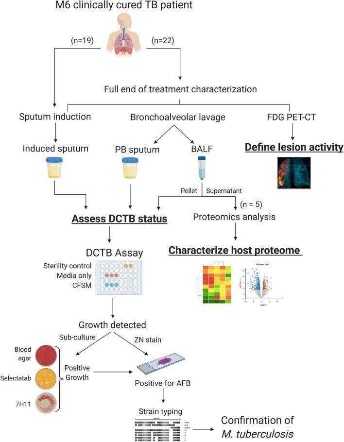
This medical flowchart details the research workflow for investigating a sterilizing cure in clinically cured Tuberculosis (TB) patients at the 6-month (M6) post-treatment milestone. The diagram outlines two patient cohorts (n=19 and n=22), with the latter undergoing 'Full end of treatment characterization.' Key diagnostic and investigative modalities include FDG PET-CT for defining lung lesion activity and sample collection via sputum induction (induced sputum) and bronchoscopy (post-bronchoscopy (PB) sputum and Bronchoalveolar Lavage Fluid (BALF)). The methodology pathways show the assessment of Differentially Culturable Mycobacterium tuberculosis (DCTB) status and host proteome characterization via TMT-based proteomics (Heatmap and Volcano Plot). The DCTB assay branch describes the laboratory progression from growth detection in Culture Filtrate Supplemented Media (CFSM) to sub-culturing on blood agar, Selectatab, and 7H11 agar. Confirmatory steps include Ziehl-Neelsen (ZN) staining to identify Acid-Fast Bacilli (AFB) and strain typing via spoligotyping for the final confirmation of M. tuberculosis. This schematic serves as an educational tool for pulmonary medicine and clinical microbiology research.
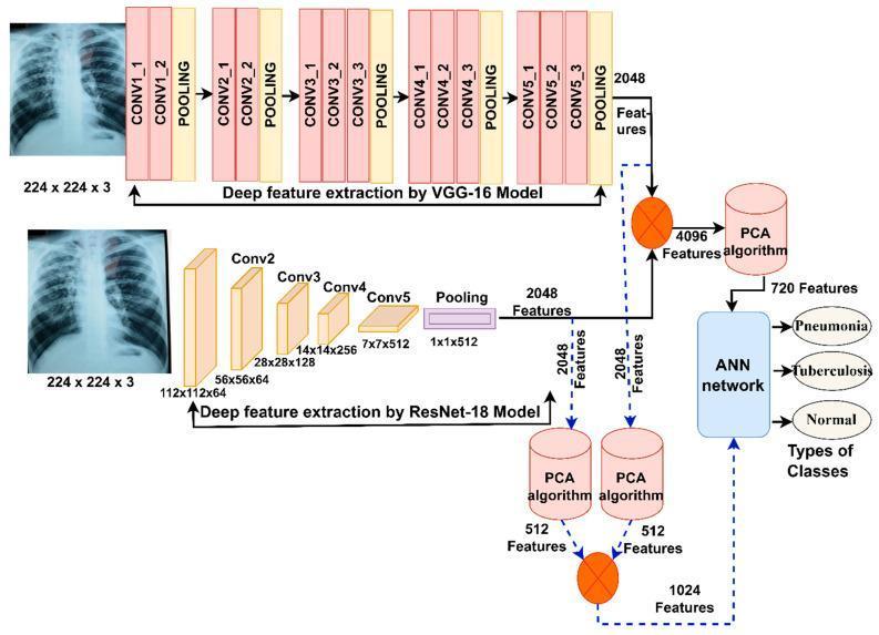
This medical flow diagram illustrates a deep learning architecture for the differential diagnosis of pneumonia and tuberculosis using chest X-rays. The technical algorithm utilizes two parallel feature extraction pathways: a VGG-16 model and a ResNet-18 model. Each model processes an input X-ray image (224x224x3) into 2048 deep features. The VGG-16 pathway details sequential convolutional layers (CONV1_1 through CONV5_3) interspersed with pooling layers, while the ResNet-18 pathway shows its specific block architecture (Conv2 to Conv5). Following feature extraction, two data processing routes are depicted. The first route merges features into a 4096-feature vector, which is then reduced to 720 features via Principal Component Analysis (PCA). The second route applies independent PCA to each model's features (reducing to 512 each) before combining them into a 1024-feature vector. Both routes converge at an Artificial Neural Network (ANN) for final classification. The clinical output categorizes findings into three distinct classes: Pneumonia, Tuberculosis, or Normal. This flowchart highlights advanced diagnostic informatics for respiratory infectious diseases.
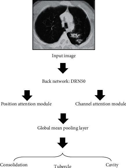
A medical flowchart detailing a deep learning diagnostic algorithm for pulmonary tuberculosis detection from computerized tomography (CT). The process begins with an 'Input image' showing an axial cross-section of the thorax, where the right lung displays a relatively healthy appearance with clear branching airways, while the left lung exhibits abnormal pathology characterized by increased opacities, mottling, and potential cavitary lesions. The image data is processed through a 'Back network: DRN50' for feature extraction. This is followed by parallel processing via a 'Position attention module' and a 'Channel attention module' to enhance spatial and feature-specific focus. These outputs are integrated at a 'Global mean pooling layer' before the final classification stage. The clinical outcomes classified by the DADRN algorithm are categorized into three primary diagnostic findings related to pulmonary tuberculosis: 'Consolidation', 'Tubercle' (nodules), and 'Cavity'. This flowchart serves as a technical overview for the application of artificial intelligence in radiological lesion localization and disease classification.
tuberculosis treatment BPaL bedaquiline pretomanid linezolid 2024