
Explain source of rda , functions, disorders of iron for 10 mark
Reading File
Now let me search the medical library for iron - RDA, sources, functions, and disorders simultaneously.
Finding Sources
Finding Sources
Found a great biochemistry source. Let me read the iron section from Basic Medical Biochemistry and also search for iron functions and disorders in parallel.
Reading File
Finding Sources
Finding Sources
Excellent content found. Let me now read more from the biochemistry textbook (iron section) and also from Robbins for disorders.
Reading File
Reading File
Now let me read more from the biochemistry textbook for iron functions and hemochromatosis (iron overload), and get the iron metabolism diagram.
Reading File
Finding Sources
Loading Image
I now have comprehensive information. Let me also quickly get Harper's iron overload table and read a bit more about hemochromatosis clinical features.
Reading File
I now have all the information needed for a thorough 10-mark answer. Let me compose a complete, well-structured response.
IRON: RDA, Sources, Functions, and Disorders
1. Recommended Dietary Allowance (RDA) of Iron
Iron requirements vary based on physiological state:
| Population Group | RDA |
|---|---|
| Adult men | 10 mg/day |
| Postmenopausal women | 10 mg/day |
| Premenopausal women | 15 mg/day |
| Pregnant women | 27 mg/day (increased demand for fetal supply) |
| Infants / growing children | Increased needs (relative to body weight) |
The average daily US diet contains 10-50 mg of iron, but only 10-15% is normally absorbed. Daily iron loss in adult men is approximately 1 mg/day (via desquamation, bile, feces, urine, and sweat).
- Basic Medical Biochemistry, 6e, p.1526
2. Sources of Iron
Dietary Sources
Iron exists in two forms in the diet:
a) Heme Iron (animal sources - readily absorbed)
- Red meat (beef, pork, lamb)
- Poultry and fish
- Organ meats (liver, kidney)
- Heme iron is directly absorbed in the gut and is not affected by dietary inhibitors.
b) Non-Heme Iron (plant sources - less bioavailable)
- Leafy green vegetables (spinach, fenugreek)
- Legumes (lentils, beans, chickpeas)
- Cereals and fortified grains
- Dried fruits (raisins, apricots)
- Seeds and nuts
Factors Affecting Iron Absorption
Enhancers of absorption:
- Vitamin C (ascorbic acid) - converts Fe³⁺ to Fe²⁺ and increases uptake of non-heme iron
- Heme iron itself
- Acidic gastric environment (HCl)
Inhibitors of absorption:
- Oxalates, phytates, tannins (tea, coffee), and other phenolic compounds in plants - these chelate iron or form insoluble precipitates
- Antacids, achlorhydria
- Phosphates, carbonates
Absorption Mechanism
Iron is absorbed in the ferrous (Fe²⁺) state by intestinal epithelial cells via the divalent metal transporter-1 (DMT-1). Inside the cell, it is oxidized to Fe³⁺ by the ferroxidase ceruloplasmin (a copper-containing enzyme) for transport through the blood.
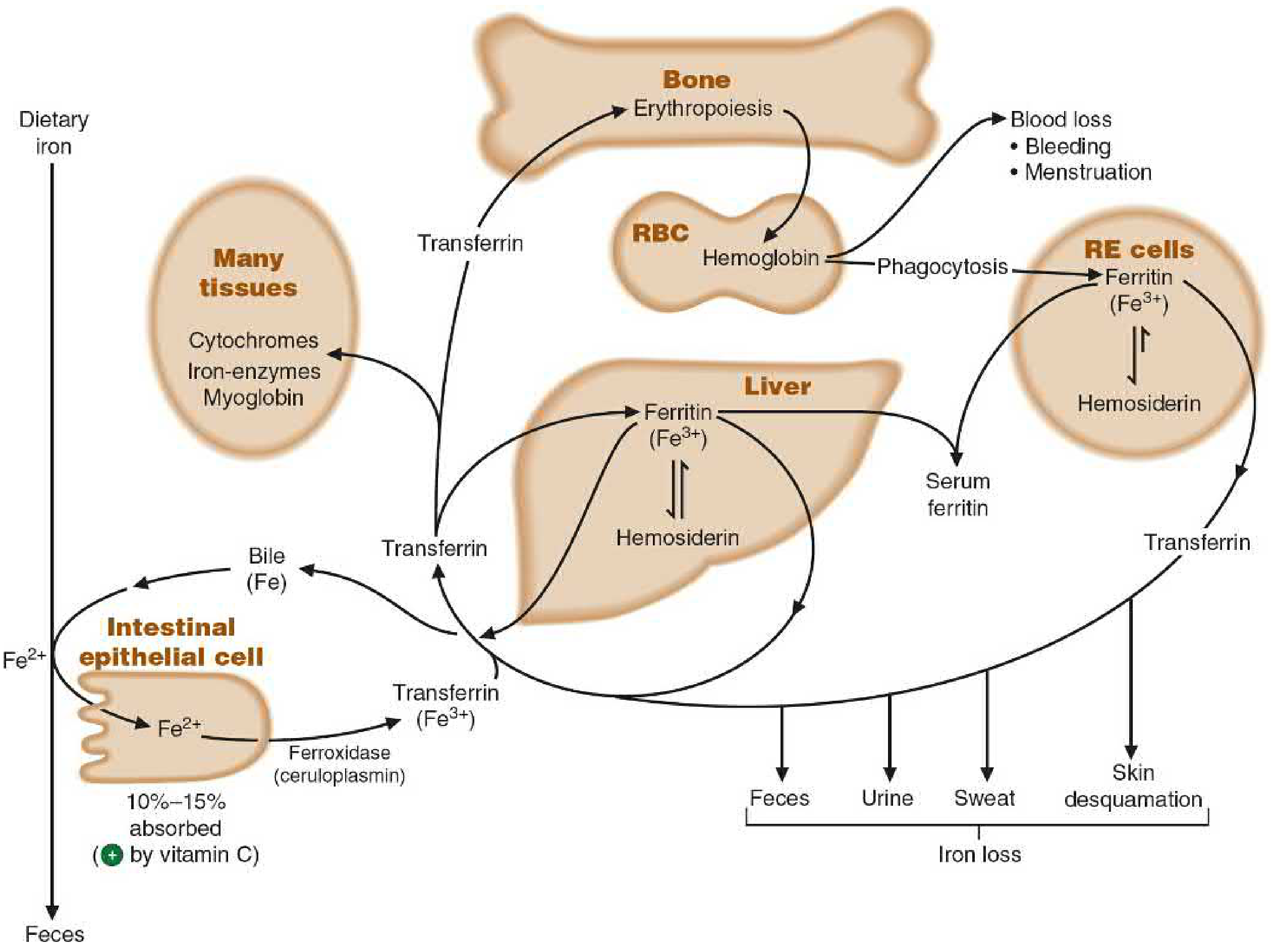
Iron metabolism: absorbed from the diet, transported by transferrin, stored in ferritin/hemosiderin, used for cytochromes, iron-containing enzymes, hemoglobin, and myoglobin - Basic Medical Biochemistry, 6e
3. Functions of Iron
Iron is an essential trace mineral with wide-ranging biochemical roles:
3.1 Oxygen Transport - Hemoglobin
- Iron (Fe²⁺) is the core of the heme moiety in hemoglobin in red blood cells.
- It binds and transports oxygen from the lungs to the tissues.
- Each hemoglobin molecule contains 4 heme groups, each with one iron atom.
3.2 Oxygen Storage - Myoglobin
- Iron in myoglobin stores oxygen in muscle tissue.
- Myoglobin releases oxygen during intense muscular activity.
3.3 Electron Transport Chain - Cytochromes
- Iron-containing cytochromes (cytochrome a, b, c, cytochrome P450) are essential components of the mitochondrial electron transport chain.
- They enable ATP synthesis via oxidative phosphorylation.
3.4 Enzymatic Functions
Iron is a cofactor or component of many enzymes:
- Catalase and peroxidase - antioxidant enzymes that decompose hydrogen peroxide
- Ribonucleotide reductase - essential for DNA synthesis
- Aconitase - TCA cycle enzyme
- Succinate dehydrogenase - links TCA cycle to ETC
- Various oxidase and hydroxylase enzymes
3.5 Transport in Blood
- Iron is transported in blood as transferrin (Fe³⁺ bound to apotransferrin).
- Transferrin is normally only one-third saturated; total iron-binding capacity (TIBC) is ~300 µg/dL.
- Transferrin-iron complex binds to transferrin receptors on cells and is internalized by receptor-mediated endocytosis.
3.6 Storage
- Ferritin: primary storage form - apoferritin binds Fe²⁺ in the liver, spleen, and bone marrow. Serum ferritin is the most sensitive indicator of body iron stores.
- Hemosiderin: when iron is in excess, it forms this less-mobilizable storage complex (ferritin + additional iron).
3.7 Immune Function and Cognitive Performance
- Adequate iron is required for normal immunocompetence and lymphocyte proliferation.
- Iron deficiency impairs cognitive performance and work capacity.
4. Disorders of Iron
A. Iron Deficiency and Iron Deficiency Anemia (IDA)
Causes:
- Chronic blood loss (most common in higher-resource countries): GI bleeding (peptic ulcers, colon cancer, hemorrhoids), menorrhagia, metrorrhagia
- Inadequate dietary intake: vegetarian diets, food insecurity, exclusively milk-fed infants, elderly
- Increased demand: pregnancy, infancy, adolescence
- Malabsorption: celiac disease, gastritis, post-gastrectomy
Pathogenesis (sequential stages):
- Stage 1 - Iron depletion: Iron stores depleted; serum ferritin falls; no stainable iron in bone marrow macrophages
- Stage 2 - Iron-deficient erythropoiesis: Serum iron falls; TIBC rises; transferrin saturation falls below 20%
- Stage 3 - Iron deficiency anemia: Hemoglobin synthesis fails; microcytic, hypochromic anemia develops
Lab findings in IDA:
| Test | Normal | IDA |
|---|---|---|
| Serum ferritin | 50-200 µg/dL | Decreased (<15) |
| Serum iron | 50-150 µg/dL | Decreased (<30) |
| TIBC | 300-360 µg/dL | Increased (>400) |
| Transferrin saturation | 30-50% | Decreased (<10%) |
| RBC morphology | Normal | Microcytic, hypochromic |
Clinical Features:
-
Mild cases: asymptomatic
-
Severe cases: weakness, fatigue, pallor, dyspnea on exertion
-
Koilonychia (spoon-shaped nails): thinning, flattening of fingernails
-
Pica: compulsion to eat non-food items (dirt, clay, ice) - a curious neurobehavioral complication
-
Angular stomatitis, glossitis
-
Reduced immunocompetence and cognitive performance
-
Robbins & Kumar Basic Pathology, p.1971-1977
B. Anemia of Chronic Disease (ACD)
- Most common anemia in hospitalized patients
- Occurs in chronic infections (osteomyelitis, endocarditis), chronic immune disorders (rheumatoid arthritis, Crohn's disease), and cancers
- Pathogenesis: Pro-inflammatory cytokines (IL-6) elevate hepcidin, which blocks ferroportin and traps iron in marrow macrophages, preventing its transfer to erythroid precursors
- Laboratory: low serum iron but normal/elevated ferritin (iron stores are present but inaccessible)
C. Iron Overload - Hemochromatosis
Hereditary Hemochromatosis is an autosomal recessive condition caused most often by mutations in the HFE gene (C282Y homozygosity accounts for 80-90% of cases in northern European descent). Other genetic causes include mutations in TFR2, ferroportin, hemojuvelin, and hepcidin itself.
Pathophysiology: Defective hepcidin regulation leads to uncontrolled iron absorption, with progressive iron deposition in:
- Liver (cirrhosis)
- Pancreas ("bronze diabetes")
- Heart (cardiomyopathy, arrhythmias)
- Pituitary/gonads (hypogonadism)
- Joints (arthropathy)
- Skin (bronze discoloration)
Classic triad: Cirrhosis + Diabetes mellitus + Skin bronzing ("bronze diabetes")
Secondary Hemochromatosis causes include:
- Repeated blood transfusions (transfusional iron overload)
- Ineffective erythropoiesis (e.g., thalassemia major)
- Excessive parenteral iron therapy
D. DMT-1 Mutation (Rare)
An inherited mutation in SLC11A2 (encoding DMT-1) leads to iron being trapped in endosomal vesicles, unable to reach cytoplasm. Result: refractory hypochromic microcytic anemia despite normal or elevated tissue iron. - Basic Medical Biochemistry, 6e
Summary Table
| Aspect | Key Points |
|---|---|
| RDA | 10 mg (men/postmenopausal women); 15 mg (premenopausal women) |
| Best dietary source | Heme iron from red meat; enhanced by vitamin C |
| Major function | O₂ transport (Hb), O₂ storage (Mb), ETC cytochromes, enzymes |
| Transport protein | Transferrin (Fe³⁺); stored as ferritin/hemosiderin |
| Deficiency disorder | IDA - microcytic, hypochromic anemia; koilonychia; pica |
| Excess disorder | Hemochromatosis - cirrhosis, diabetes, bronzing |
| Key regulatory hormone | Hepcidin (blocks ferroportin, reduces iron release) |
Sources: Basic Medical Biochemistry - A Clinical Approach, 6e; Robbins & Kumar Basic Pathology; Harper's Illustrated Biochemistry, 32nd ed.
This is a shared conversation. Sign in to Orris to start your own chat.