
Lathyrism
I'll look this up in the medical library first.
Reading File
Now let me search the library and PubMed in parallel.
Finding Sources
Finding Sources
Searching PubMed
lathyrism neurotoxicity
Good results. Now let me read the key sections in parallel.
Reading File
Reading File
Reading File
Reading File
Now let me read the Park's section more fully (including prevention/treatment) and also the forensic medicine section.
Reading File
Reading File
I now have comprehensive information from multiple authoritative textbooks. Here is a thorough overview of lathyrism:
Lathyrism
Definition
Lathyrism is a chronic toxic nutritional neurological disease caused by prolonged ingestion of flour or dhal made from the drought-resistant grass pea (Lathyrus sativus, also called chickling vetch, khesari dhal, or vetch pea). In humans it is called neurolathyrism; in animals (horses, cattle) it is called osteolathyrism, where skeletal deformities predominate.
- Adams & Victor's Principles of Neurology, 12th Ed.
- Bradley & Daroff's Neurology in Clinical Practice
Historical Background
The disease was known to Hippocrates, Pliny, Galen, Avicenna, and described in ancient Hindu texts. The term "lathyrism" was coined by Cantani (Italy) when the relationship to L. sativus consumption was recognized. It occurred in epidemic form in concentration camps during World War II.
Epidemiology
- Endemic regions: India (Madhya Pradesh - Rewa and Satna districts had 25,000 and 32,000 cases respectively; also UP, Bihar, Odisha, Maharashtra, West Bengal, Rajasthan, Assam, Gujarat), Bangladesh, Ethiopia, China, Romania, Spain, Algeria, North Africa.
- Trigger: Outbreaks occur during famine and drought, when L. sativus (cheap, hardy, drought-resistant) becomes the dietary staple.
- At-risk group: Mainly young men aged 15-45 years; a higher incidence has been noted in boys aged 10-14 in Ethiopian epidemics.
- Dose threshold: Diets containing over 30% of this dhal, taken over 2-6 months, result in neurolathyrism.
- Park's Textbook of Preventive and Social Medicine
The Pulse
Lathyrus sativus (khesari dhal) is known by local names: Teora dhal, Lak dhal, Batra, Gharas, Matra. The seeds are triangularly shaped and grey; when dehusked they resemble red gram or bengal gram dhal. It is consumed predominantly by poor agricultural laborers because of its low cost and resilience as a crop.
Toxin: BOAA
The neurotoxin is beta-N-oxalylamino-L-alanine (BOAA), also written as beta-oxalyl amino alanine.
- A free, water-soluble amino acid found in seed cotyledons (approximately 1%)
- Acts as an AMPA (alpha-amino-3-hydroxy-5-methyl-4-isoxazolepropionate) glutamate receptor agonist - this is the key excitotoxic mechanism
- Stimulation of AMPA receptors increases intracellular reactive oxygen species (ROS) and impairs mitochondrial oxidative phosphorylation
- There is a blood-brain barrier to the toxin; large amounts must be ingested over months to overcome it
- Water-soluble property is exploited for detoxification (soaking in hot water)
The selective degeneration is most prominent in:
-
Betz cells of the motor cortex (especially those subserving lower-extremity function)
-
The longest corresponding pyramidal (corticospinal) tracts
-
Loss of ascending and descending tracts - corticospinal and direct spinocerebellar tracts
-
Loss of myelinated fibers in lateral and posterior columns
-
Anterior horn cells are relatively spared (hence an upper motor neuron picture)
-
Bradley & Daroff's Neurology in Clinical Practice
-
Adams & Victor's Principles of Neurology, 12th Ed.
Clinical Features (Stages)
The disease is a pure upper motor neuron (UMN) syndrome affecting predominantly the lower limbs. Park's classifies the clinical course into 5 stages:
| Stage | Description |
|---|---|
| (a) Latent stage | Apparently healthy; ungainly gait on physical stress. Neurological signs present. Complete remission possible if pulse withdrawn at this stage. |
| (b) No-stick stage | Walks with short, jerky steps without a stick. Most patients found here. |
| (c) One-stick stage | Crossed scissor gait, tendency to walk on tiptoes; needs one stick. |
| (d) Two-stick stage | Excessive knee bending, crossed legs; needs two crutches. Slow, clumsy gait. |
| (e) Crawler stage | Cannot maintain erect posture; crawls by throwing weight on hands. Atrophy of thigh and leg muscles. |
Key signs and symptoms:
- Severe, agonizing pain in calf muscles
- Weakness in legs; difficulty sitting and standing
- Spastic paraplegia of lower limbs (bilateral)
- Scissor (crossed) gait, tiptoe walking
- Increased knee jerks, ankle clonus, extensor plantar response (Babinski positive)
- Paresthesias, numbness, formication in legs
- Frequency and urgency of micturition, erectile dysfunction, sphincteric spasms
- Coarse tremor and involuntary movements of upper extremities (occasionally)
- No sensory loss, no loss of consciousness (classically)
- No atrophy initially, no reaction of degeneration, muscle tone maintained
Once established, the condition is irreversible but non-progressive (unless ingestion continues), and lifespan is not significantly affected.
Prevention and Control
(a) Vitamin C prophylaxis
Daily administration of 500-1000 mg ascorbic acid may repair damage in certain instances and prevent it in lathyrogenic diets - demonstrated in guinea pigs and monkeys.
(b) Banning the crop
- India's Prevention of Food Adulteration Act has banned Lathyrus in all forms (whole, split, flour).
- The ban is poorly enforced in endemic states (MP, Bihar, Odisha, Gujarat).
- If unavoidable, khesari dhal should not exceed 25% of total cereals and pulses per day.
(c) Removal of toxin (Detoxification)
Since BOAA is water-soluble, it can be removed:
- Steeping method: Soak pulse in hot (boiling) water for 2 hours, drain, wash with clean water, dry in sun. Drawback: some vitamin/mineral loss.
- Parboiling: Soak in lime water overnight then boil - destroys BOAA and trypsin inhibitors. Suitable for large-scale operations.
(d) Genetic approach
Some strains of Lathyrus contain very low toxin levels (~0.1%). Selective propagation of low-toxin strains is considered the most effective long-term solution.
(e) Public education
Educating populations about dangers of excess consumption and detoxification methods.
Treatment
There is no specific treatment once the condition is established.
- Exclude L. sativus from the diet (early withdrawal may cause remission in the latent stage)
- Diet rich in Vitamin A, carotene, and other vitamins
- Physiotherapy for muscular and neurological involvement
- Vitamin C supplementation
Comparison with Konzo
Lathyrism is closely compared to Konzo (African cyanide poisoning from cassava), as both are:
- Toxic nutritional UMN diseases
- Affect longest corticospinal tracts and Betz cells
- Occur in famine conditions in developing countries
- Irreversible but non-progressive
| Feature | Lathyrism | Konzo |
|---|---|---|
| Toxin | BOAA (AMPA agonist) | Cyanide/thiocyanate (AMPA agonist) |
| Source | L. sativus (grass pea) | Insufficiently processed cassava |
| Geography | India, Africa, China | Sub-Saharan Africa |
| Onset | Gradual (chronic/subacute) | Rapid |
Key Exam Points
- Toxin: BOAA (Beta-N-Oxalylamino-L-Alanine) - AMPA receptor agonist
- Mechanism: Excitotoxic; selective UMN / corticospinal tract degeneration
- Clinical: Spastic paraplegia, scissor gait, tiptoe walking - pure UMN, no sensory loss
- Population: Young men, famine conditions, diet >30% L. sativus for >2 months
- Reversible only at latent stage (withdraw the pulse)
- Detoxification: Hot water soaking (BOAA is water-soluble)
- India: Banned under Prevention of Food Adulteration Act
- Local name: Khesari dhal (triangular grey seeds)
Causalgia
Causalgia is a severe burning pain following partial peripheral nerve injury, now classified as Complex Regional Pain Syndrome Type II (CRPS-II). It involves sympathetically maintained pain, allodynia, and autonomic changes.
Searching sources...
Finding Sources
Finding Sources
Good - multiple relevant sections found. Reading all key sources in parallel.
Reading File
Reading File
Reading File
Reading File
Now let me read the Bradley & Daroff section further for diagnosis/treatment, and also the Rockwood & Green section for CRPS stages.
Reading File
Reading File
I now have comprehensive, multi-source coverage. Let me also fetch the CRPS clinical image.
Loading Image
Excellent. I now have everything needed for a comprehensive answer.
Causalgia (CRPS Type II)
Definition and Terminology
Causalgia - from the Greek kausos (heat/burning) and algos (pain) - is the term coined by Silas Weir Mitchell in the 1870s to describe severe, progressive, burning distal limb pain occurring after partial injury to a major peripheral nerve. It is now classified as Complex Regional Pain Syndrome Type II (CRPS-II).
The condition has accumulated a large number of synonyms over its history, reflecting evolving theories of pathogenesis:
| Historical name | Association |
|---|---|
| Causalgia | Weir Mitchell, 1870s - nerve injury |
| Sudeck's atrophy | Sudeck (1900s) - osteoporotic changes |
| Reflex Sympathetic Dystrophy (RSD) | Evans, 1946 - sympathetic hyperactivity |
| Minor causalgia | Homans, 1940 - without major nerve injury |
| Shoulder-hand syndrome | Steinbrocker - upper limb/shoulder variant |
| Algodystrophy / Algoneurodystrophy | French rheumatologists - "painful progressive degeneration" |
| Mimocausalgia | CNS-origin causalgic pain |
In 1994, the IASP (International Association for the Study of Pain) standardized the terminology at a consensus workshop in Orlando, introducing the term Complex Regional Pain Syndrome:
-
CRPS I = no definable nerve lesion (formerly RSD)
-
CRPS II = definable nerve lesion (formerly causalgia)
-
Adams & Victor's Principles of Neurology, 12th Ed.
-
Bradley & Daroff's Neurology in Clinical Practice
-
Rockwood & Green's Fractures in Adults, 10th Ed.
Nerves Commonly Involved
Causalgia most often follows partial (not complete) interruption of:
- Median nerve (most common)
- Ulnar nerve
- Sciatic nerve
- Peroneal nerve
Complete nerve section rarely produces causalgia - it is the partial, incomplete injury that sets up the pain circuit.
Clinical Features
Pain (Cardinal Feature)
- Severe, persistent burning pain - the hallmark
- Located in the hand or foot, most pronounced in the digits, palm, or sole
- Radiates beyond the territory of a single peripheral nerve
- Severity is disproportionate to the initiating injury
- Not limited to the area of injury or a specific nerve distribution
Sensory Abnormalities
- Allodynia - innocuous stimuli (light touch, clothing, drafts of air) cause severe, long-lasting pain
- Hyperalgesia - exaggerated pain to minor stimuli (e.g., safety pin prick)
- Thermal allodynia - even a drop of alcohol or ambient heat/cold intensifies pain
- Even emotional stimuli, noise can worsen the pain
- The patient keeps the extremity protected and immobile, often wrapped in cool, moist cloth
Autonomic/Vasomotor Changes
- Sudomotor changes: excessive sweating (~60% of patients)
- Vasomotor instability: skin alternates between red, blue, purple, or pale within minutes to hours
- Livedo reticularis - reticulated red network pattern on skin
- Temperature asymmetry between limbs (>1°C)
- Early: warm, moist skin; Later: cold, cyanotic limb
Trophic Changes (later)
- Shiny, smooth, sometimes scaly skin devoid of hair
- Abnormal nail growth (increased or decreased)
- Hyperkeratosis
- Osteoporosis of underlying bone (Sudeck's atrophy)
- Muscle atrophy
Motor Dysfunction
- Mild weakness
- Decreased range of motion
- Tremor, dystonia, myoclonus
Edema
- Mild early (may mimic cellulitis)
- Severe late (may require Doppler to exclude DVT)
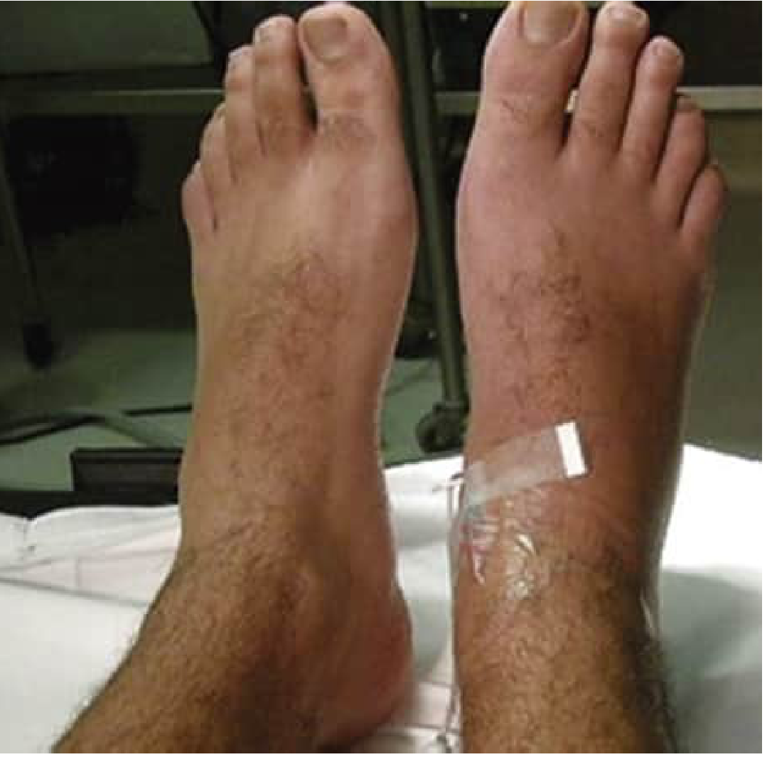
Clinical photo - CRPS I of the right foot showing erythema and swelling:
Three Stages (RSD Classification - Firestein/Kelley Rheumatology)
| Stage | Features | Duration |
|---|---|---|
| Stage 1 (Acute) | Sympathetic overflow; diffuse swelling, pain, increased vascularity, radiographic demineralization | 0-3 months |
| Stage 2 (Dystrophic) | Atrophy; cold, shiny skin; muscle and skin atrophy | 3-6 months |
| Stage 3 (Atrophic) | Irreversible trophic changes; flexion contracture; pale, cold, painful extremity; controlled by higher thalamic centers | >6 months |
Recovery is possible while vasomotor activity (swelling and hyperemia) persists (Stage 1). After Stage 2-3, prognosis is poor.
Pathophysiology
Multiple mechanisms are implicated:
-
Ephaptic transmission (short-circuiting): Abnormal direct electrical connection between efferent sympathetic and somatic afferent pain fibers at the nerve injury site.
-
Adrenergic chemical coupling: Depletion of neurotransmitters at sympathetic adrenergic endings abolishes causalgic pain - suggesting chemical (not electrical) cross-excitation at nerve terminals. Injured nociceptors develop abnormal adrenergic sensitivity and respond to circulating sympathetic neurotransmitters.
-
Central sensitization: Sustained sensory pain bombardment sensitizes central structures. Molecular changes include:
- NMDA receptor alterations
- Induction of cyclooxygenase and prostaglandin synthesis
- Changes in GABAergic inhibition in dorsal horns
-
Neuroinflammation: Elevated CGRP (calcitonin gene-related peptide) drives neurogenic inflammation, edema, vasodilation, and sweating.
-
Immunological mechanisms: Altered HLA expression, substance P, cytokines, and interleukins contribute to edema and pain.
-
Cortical reorganization: Disinhibition of motor cortex; reorganization of primary somatosensory cortex.
- Bradley & Daroff's Neurology in Clinical Practice
- Adams & Victor's Principles of Neurology, 12th Ed.
IASP Diagnostic Criteria (Original)
- Presence of an initiating noxious event or cause of immobilization (not required - 5-10% have none)
- Continuing pain, allodynia, or hyperalgesia disproportionate to any known inciting event
- Evidence of edema, skin blood flow abnormality, or abnormal sudomotor activity in the region of pain at some time
- Diagnosis excluded by other conditions that would otherwise account for the degree of pain and dysfunction
Diagnosis is clinical. EMG/NCV studies are not sensitive (and cause severe pain). Triple-phase bone scan may show abnormal absorption but is not a primary diagnostic tool.
Special Presentations
- Causalgia-dystonia syndrome (Bhatia et al.): A fixed dystonic posture engrafted on a site of causalgic pain. Injury is often trivial or absent; both the causalgia and dystonia can spread to distant sites. Does not respond consistently to treatment; some spontaneous recovery occurs.
- Algodystrophy post-DVT: Causalgia and RSD following deep venous thrombosis in a leg.
- Mimocausalgia: Causalgic pain arising from CNS lesions rather than peripheral nerve injury.
Treatment
Treatment remains challenging with limited high-quality evidence.
Physical/Occupational Therapy
- First-line in early CRPS - movement, desensitization
Pharmacological
| Drug class | Notes |
|---|---|
| Bisphosphonates | Best evidence in RCTs for clear benefit |
| Tricyclic antidepressants | Used empirically (amitriptyline) |
| Antiepileptics | Gabapentin, pregabalin - used empirically |
| Corticosteroids | Some benefit in RCTs |
| DMSO (dimethyl sulfoxide) | Topical; RCT evidence of improvement |
| Methadone/narcotics | Used empirically |
| Ketamine infusions | Subanesthetic infusions - promising for selected refractory cases |
| Epidural clonidine | RCT evidence |
| Intrathecal baclofen | RCT evidence (especially for dystonia) |
Sympathetic Interventions
- Sympathetic nerve block - can reverse the syndrome, especially in Stage 1
- Intravenous regional sympathetic block (Bier block)
- Surgical sympathectomy - Leriche (1916) was the first to demonstrate that sympathectomy could alter the clinical features
Psychological
- Psychological evaluation for patients with CRPS >2 months
- Cognitive behavioral therapy, biofeedback, relaxation therapy, group therapy, self-hypnosis
Neuromodulation
- Spinal cord stimulation for refractory cases
Key Exam Points
- Term coined by Weir Mitchell (1870s); renamed CRPS by IASP in 1994
- CRPS I = no nerve lesion (RSD); CRPS II = nerve lesion present (causalgia)
- Most common nerves: median > ulnar > sciatic > peroneal
- Requires partial nerve injury (not complete)
- Pain is burning, disproportionate, beyond nerve territory, with allodynia and autonomic changes
- Patient keeps limb wrapped in cool moist cloth
- Three stages: acute (warm) → dystrophic (atrophy) → atrophic (irreversible contracture)
- Best pharmacological evidence: bisphosphonates
- Sympathetic block can produce dramatic relief
Jendrassik manoeuvre
The Jendrassik manoeuvre is a technique to reinforce deep tendon reflexes - the patient hooks their flexed fingers together and pulls outward while the knee jerk is elicited. It works by reducing gamma motor neuron inhibition, amplifying the reflex arc.
Searching sources...
Finding Sources
Finding Sources
Good - found relevant sections in Harrison's, Ganong's, and Firestein & Kelley's. Reading all three in parallel.
Reading File
Reading File
Reading File
Let me also search for more detailed physiological context - specifically the gamma motor neuron and spindle arc, and also check Adams & Victor for any deeper neurological context.
Finding Sources
Finding Sources
Let me look at the broader context of Ganong's stretch reflex section for the full physiological explanation of gamma motor neurons and the reflex arc.
Reading File
I now have all the information needed from authoritative sources. Let me compile the complete answer.
Jendrassik Manoeuvre
Definition
The Jendrassik manoeuvre is a reflex reinforcement technique used during neurological examination to enhance (augment) deep tendon reflexes (DTRs) that appear absent or diminished on initial testing.
It was described by Erno Jendrassik, a Hungarian physician, in 1885.
How It Is Performed
The classic (lower limb) technique:
- The patient flexes the fingers of both hands into a hook-like form
- The two hands are interlocked at the fingers
- The patient is asked to pull the hands apart isometrically (as hard as possible, without actually unlocking the fingers)
- While this sustained effort is maintained, the patellar (knee jerk) or Achilles reflex is elicited by striking the tendon
Upper limb reinforcement variants:
-
Upper limb reflexes (biceps, triceps, brachioradialis) can be reinforced by asking the patient to voluntarily clench their teeth
-
The Achilles reflex can also be reinforced by the same finger-hooking technique
-
Harrison's Principles of Internal Medicine, 22nd Ed.
-
Firestein & Kelley's Textbook of Rheumatology
Mechanism of Action
The mechanism is explained through gamma (γ) motor neuron physiology:
Normal Stretch Reflex Arc (brief recap)
- Tapping the tendon stretches the muscle spindle (intrafusal fibers)
- This activates Ia afferent fibers (large, fast-conducting) from the spindle
- Ia fibers synapse directly (monosynaptically) on alpha (α) motor neurons in the anterior horn
- Alpha motor neurons activate extrafusal (skeletal) muscle fibers → visible reflex jerk
Role of Gamma Motor Neurons
- Gamma (γ) motor neurons innervate the intrafusal fibers of the muscle spindle
- When γ-motor neuron discharge increases → intrafusal fibers contract → spindle is "loaded" (put under tension)
- A loaded spindle is more sensitive to stretch - it fires more readily and more vigorously when the tendon is tapped
- This amplifies Ia afferent traffic → stronger alpha motor neuron activation → brisker reflex
How the Manoeuvre Works
- The isometric pulling effort of the hands activates afferent impulses from the hand muscles and tendons
- These afferent signals travel to the spinal cord and increase γ-motor neuron discharge to the lower limb muscles
- The spindles become more sensitive (loaded)
- When the tendon is then struck, the reflex arc is amplified - a previously undetectable reflex becomes visible
"Trying to pull the hands apart when the flexed fingers are hooked together facilitates the knee jerk reflex (Jendrassik maneuver), and this may also be due to increase γ-motor neuron discharge initiated by afferent impulses from the hands."
- Ganong's Review of Medical Physiology, 26th Ed.
A secondary explanation is that the manoeuvre acts as a distraction, reducing voluntary (cortical) suppression of the reflex - patients often unconsciously suppress reflexes by tensing the tested muscle. The mental and physical effort of the manoeuvre prevents this.
- Ganong's Review of Medical Physiology, 26th Ed.
- Firestein & Kelley's Textbook of Rheumatology
Clinical Significance
When to Use It
- Any time a reflex appears absent or diminished on initial testing
- Should be routinely attempted before recording a reflex as absent (0)
- Especially important for the patellar and Achilles reflexes, which are most commonly suppressed by voluntary muscle tension or patient anxiety
Interpretation
| Finding | Interpretation |
|---|---|
| Reflex appears with reinforcement | Hypoactive but present (grade 1); note the need for reinforcement |
| Reflex still absent with reinforcement | Truly absent (grade 0) - suggests lower motor neuron lesion, peripheral neuropathy, or muscle disease |
| Reflex dramatically exaggerated | Upper motor neuron lesion (hyperreflexia) |
Grading Scale (Harrison's)
| Grade | Meaning |
|---|---|
| 0 | Absent |
| 1 | Present but diminished |
| 2 | Normoactive |
| 3 | Increased (without clonus) |
| 4 | Clonus |
Additional Physiological Notes
- Anxiety independently increases γ-motor neuron discharge, explaining why anxious patients may have hyperactive reflexes
- Unexpected movement also increases γ-motor neuron efferent discharge
- Noxious skin stimulation increases γ-motor neuron activity to ipsilateral flexor spindles while decreasing it to extensors (explains flexor withdrawal patterning)
- The manoeuvre works because of α-γ co-activation: descending excitatory input simultaneously activates both α and γ motor neurons, keeping the spindle sensitive throughout muscle contraction
Key Exam Points
- Named after Erno Jendrassik (Hungarian, 1885)
- Technique: interlock flexed fingers of both hands and pull apart while the knee/ankle jerk is tested
- Mechanism: increases γ-motor neuron discharge → loads muscle spindle → amplifies Ia afferent input → stronger reflex
- Also works via distraction (reduces voluntary cortical suppression)
- Upper limb equivalent: voluntary teeth-clenching
- A reflex absent even with reinforcement = truly absent (lower motor neuron pathology)
- Anxiety → increased γ-motor discharge → hyperreflexia (same underlying mechanism)
This is a shared conversation. Sign in to Orris to start your own chat.