
Histopathology of chronic cholecystitis (include h & e slide)
Reading File
Now let me search the medical library for chronic cholecystitis histopathology, including images.
Finding Sources
Finding Sources
Good. Now let me read the Robbins Pathology and Robbins/Cotran sections (most likely to have H&E images), plus search for images.
Reading File
Reading File
Finding Sources
I can see image references in both Robbins texts. Let me get the full image URLs and read the Robbins/Cotran text further.
Reading File
Reading File
I have both H&E slide images. Now let me fetch them to display properly.
Loading Image
Loading Image
Excellent - I have both H&E slides. Now I have all the material needed for a comprehensive answer.
Histopathology of Chronic Cholecystitis
Overview
Chronic cholecystitis is the most common disorder of the gallbladder and is almost always associated with gallstones (>90% of cases). The morphologic changes are extremely variable and sometimes subtle - the diagnosis is ultimately a pathologic one made on the resected specimen.
Gross Morphology
- The gallbladder may be contracted, normal-sized, or enlarged
- The serosa is usually smooth and glistening but may be dulled by subserosal fibrosis
- Dense fibrous adhesions may be present (sequelae of prior acute inflammation)
- On section, the wall is variably thickened with an opaque gray-white appearance
- The lumen typically contains green-yellow mucoid bile and gallstones
- The mucosa itself is generally preserved
H&E Histology - Key Features
Figure A - Chronic inflammatory infiltrate:
Chronic cholecystitis (A): gallbladder mucosa infiltrated by chronic inflammatory cells with preserved mucosal architecture.
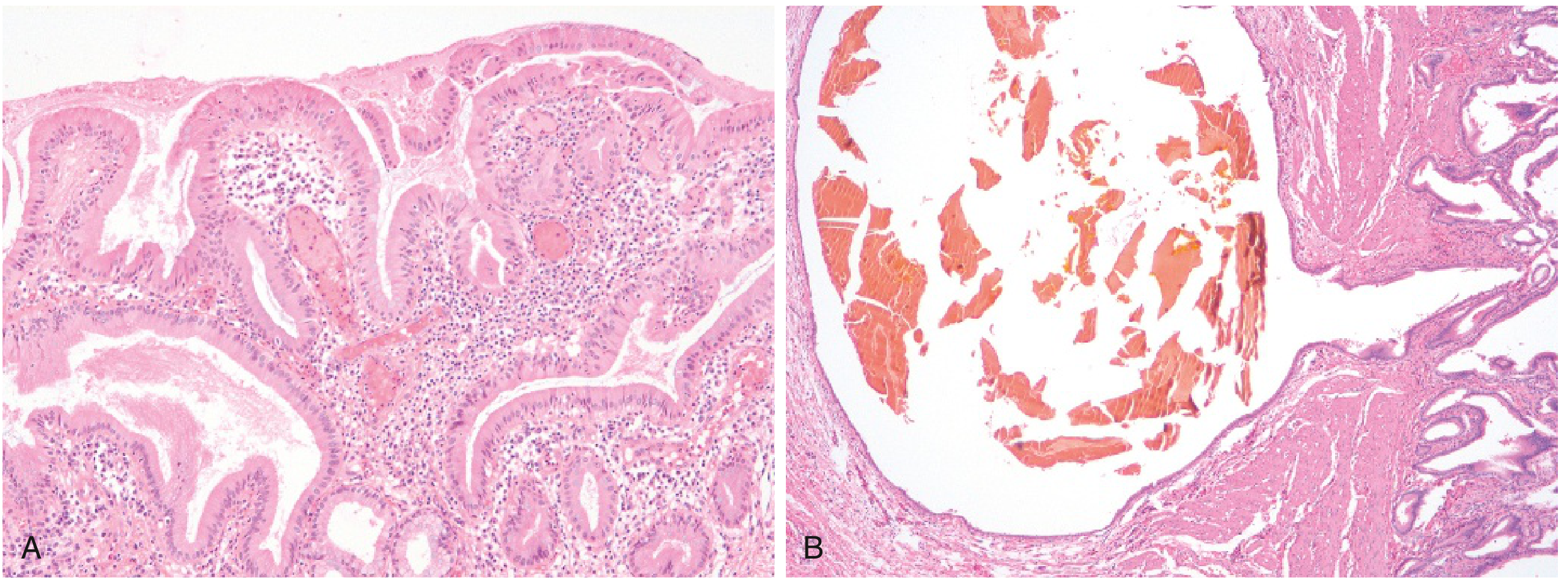
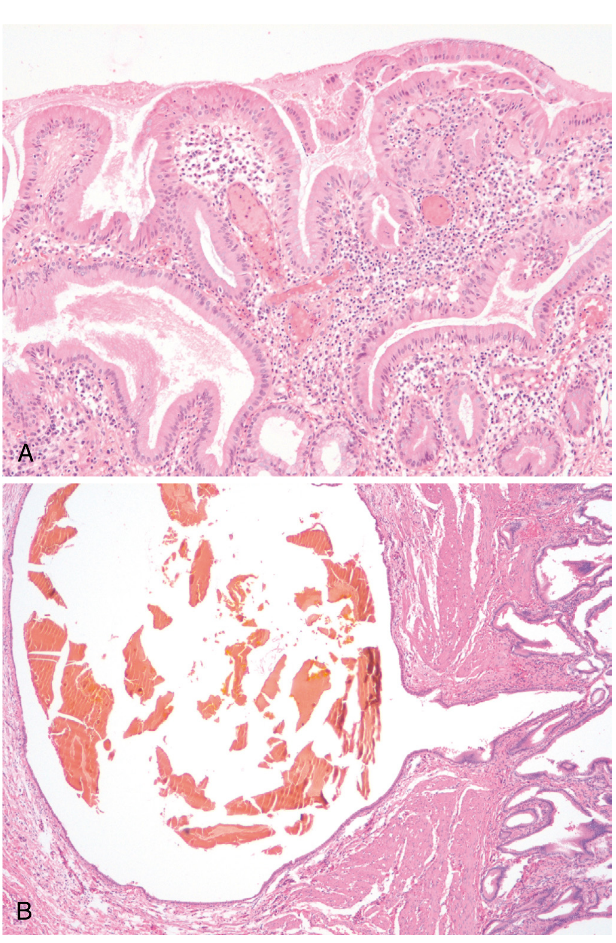
Microscopic findings in detail:
| Feature | Detail |
|---|---|
| Inflammatory cells | Scattered lymphocytes, plasma cells, and macrophages in mucosa and subserosal fibrous tissue (mildest cases) |
| Fibrosis | Marked subepithelial and subserosal fibrosis in more advanced cases |
| Mononuclear infiltration | Chronic inflammatory cells throughout the wall; in the absence of superimposed acute cholecystitis, lymphocytes alone may be the only sign |
| Mucosal changes | Reactive proliferation of mucosa; fusion of mucosal folds; buried crypts of epithelium within the gallbladder wall |
| Rokitansky-Aschoff sinuses | Outpouchings of mucosal epithelium through the muscular wall - a hallmark feature (Fig. B above) |
Special Variants (also seen on H&E)
- Porcelain gallbladder - Extensive dystrophic calcification of the wall (rare); associated with increased risk of carcinoma
- Hyalinizing cholecystitis - Complete replacement of the wall and mucosa by dense fibrosis, with or without calcification; also linked to gallbladder carcinoma
- Xanthogranulomatous cholecystitis - Rupture of Rokitansky-Aschoff sinuses leads to accumulation of lipid-laden foamy macrophages (xanthoma cells) in the wall; massively thickened, shrunken, nodular gallbladder
- IgG4-related sclerosing cholecystitis - A manifestation of IgG4-related fibrosing disease; storiform fibrosis with IgG4+ plasma cell infiltrates
Key Diagnostic Points
- The hallmark of uncomplicated chronic cholecystitis on H&E is lymphocytic infiltration of the mucosa and submucosa with subepithelial and subserosal fibrosis
- Rokitansky-Aschoff sinuses (mucosal herniations through the muscularis) are highly characteristic
- No neutrophils in pure chronic cholecystitis (their presence suggests superimposed acute-on-chronic disease)
- Microorganisms (E. coli, enterococci) can be cultured from bile in ~1/3 of cases
Clinical Significance
- Chronic cholecystitis does not have the dramatic presentation of acute forms
- Characterized by recurrent right upper quadrant or epigastric pain, nausea, vomiting, and fatty food intolerance
- The main importance lies in the association with gallstones + chronic inflammation → gallbladder carcinoma
Sources: Robbins & Kumar Basic Pathology, p. 633 | Robbins, Cotran & Kumar Pathologic Basis of Disease, p. 813
This is a shared conversation. Sign in to Orris to start your own chat.