
Give easy explanation of digastric triangle and diagram drawn in the exam according to vishram singh and a gold medal scoring answer in easy way
Digastric Triangle (Submandibular Triangle) - Easy Explanation + Gold Medal Answer
What is the Digastric Triangle?
"Two legs of a V (the digastric muscle) under your jaw, holding everything important for your mouth and face."
LOCATION
BOUNDARIES (How to Remember: "M-AP")
| Boundary | Structure |
|---|---|
| Superior (roof/base) | Inferior border of the Mandible |
| Anteroinferior | Anterior belly of digastric muscle |
| Posteroinferior | Posterior belly of digastric + stylohyoid muscle |
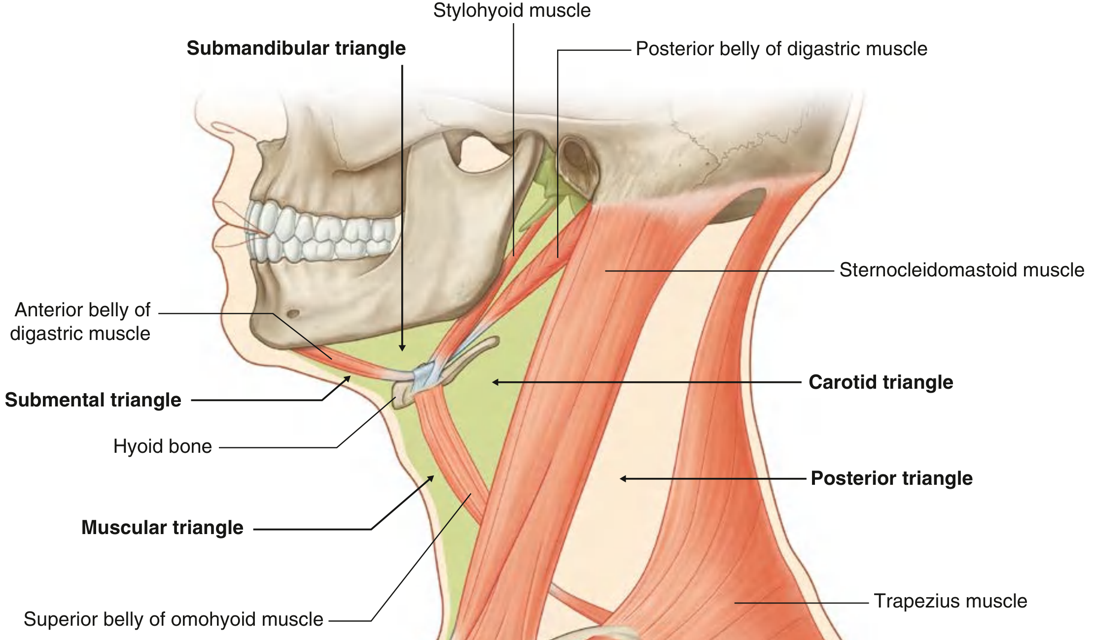
ROOF (what covers the triangle from outside)
- Skin
- Superficial fascia - contains platysma muscle + cervical branch of facial nerve (VII)
- Deep cervical fascia (investing layer)
FLOOR (what forms the base of the triangle inside)
- Anteriorly: Mylohyoid muscle
- Posteriorly: Hyoglossus muscle (and a small part of middle constrictor)
CONTENTS - The Most Important Part!
1. Gland
- Submandibular salivary gland - the main occupant; superficial part lies in the triangle, deep part wraps around the posterior border of mylohyoid into the floor of the mouth
2. Vessels (Arteries & Veins)
- Facial artery - enters the triangle deep to the posterior belly of digastric + stylohyoid, grooves the submandibular gland, then hooks over the lower border of mandible to reach the face
- Facial vein - passes superficial to the submandibular gland
- Submental artery - a branch of facial artery
3. Nerves
- Marginal mandibular branch of facial nerve (VII) - runs in the roof, just deep to investing fascia, superficial to the facial vein. Most important surgically - at risk during submandibular gland surgery
- Mylohyoid nerve - runs in the floor, supplies mylohyoid + anterior belly of digastric
- Hypoglossal nerve (XII) - enters the triangle deep to the posterior belly of digastric; runs on the hyoglossus; supplies all muscles of the tongue
4. Lymph Nodes
- Submandibular lymph nodes (Level IB) - 3-6 nodes; drain tongue, teeth, gums, cheek, lips, submandibular gland itself
DIAGRAM TO DRAW IN EXAM (Vishram Singh style)
MANDIBLE (inferior border)
/ \
/ ant. belly digastric \ post. belly digastric
/ \ + stylohyoid
/________________________________\
FLOOR:
Mylohyoid (ant) + Hyoglossus (post)
- Roof layers: Skin → Platysma (+ cervical branch VII) → Deep cervical fascia
- Floor: Mylohyoid (ant.) + Hyoglossus (post.)
- Contents: Submandibular gland, Facial artery (curves around gland), Facial vein, Hypoglossal nerve (XII) on hyoglossus, Marginal mandibular branch of VII (in roof), Submandibular lymph nodes
GOLD MEDAL EXAM ANSWER FORMAT
Q. Describe the digastric triangle. (Vishram Singh pattern)
- Base (superiorly): Inferior border of body of mandible
- Anteroinferior side: Anterior belly of digastric
- Posteroinferior side: Posterior belly of digastric + stylohyoid muscle
- Skin
- Superficial fascia with platysma and cervical branch of facial nerve
- Investing layer of deep cervical fascia
- Mylohyoid muscle (anteriorly)
- Hyoglossus muscle (posteriorly)
- Part of middle constrictor (posteriorly, small portion)
| Structure | Notes |
|---|---|
| Submandibular salivary gland | Largest content; wraps around free border of mylohyoid |
| Facial artery | Enters deep, grooves the gland, crosses mandible onto face |
| Facial vein | Runs superficial to gland |
| Submental artery | Branch of facial artery |
| Marginal mandibular nerve (VII) | In roof; at risk in surgery |
| Mylohyoid nerve | Supplies floor muscles |
| Hypoglossal nerve (XII) | On hyoglossus; supplies tongue muscles |
| Submandibular lymph nodes | Level IB; drain oral cavity and face |
- Submandibular sialadenitis and calculi: gland swelling in this triangle
- Ludwig's angina: dangerous cellulitis that starts in this space and spreads to the floor of the mouth
- Marginal mandibular nerve (VII) injury during submandibular gland excision causes drooping of corner of the mouth
- Submandibular lymph nodes are Level IB nodes - important in head and neck cancer staging
KEY MNEMONICS FOR REVISION
| "MAP" | Mandible (base), Anterior belly, Posterior belly |
|---|---|
| "My Hyoid floor" | Mylohyoid (ant) + Hyoglossus (post) |
| "SAF-MHL" | Submandibular gland, Artery (facial), Facial vein, Marginal mandibular VII, Hypoglossal XII, Lymph nodes |
Give answer about oesophagus relations and diagram drawn in exam easy way of relations and gold medal scoring answer For 8 marks
Oesophagus - Relations + Diagram + Gold Medal 8-Mark Answer
QUICK ORIENTATION FIRST
DIAGRAM TO DRAW IN EXAM
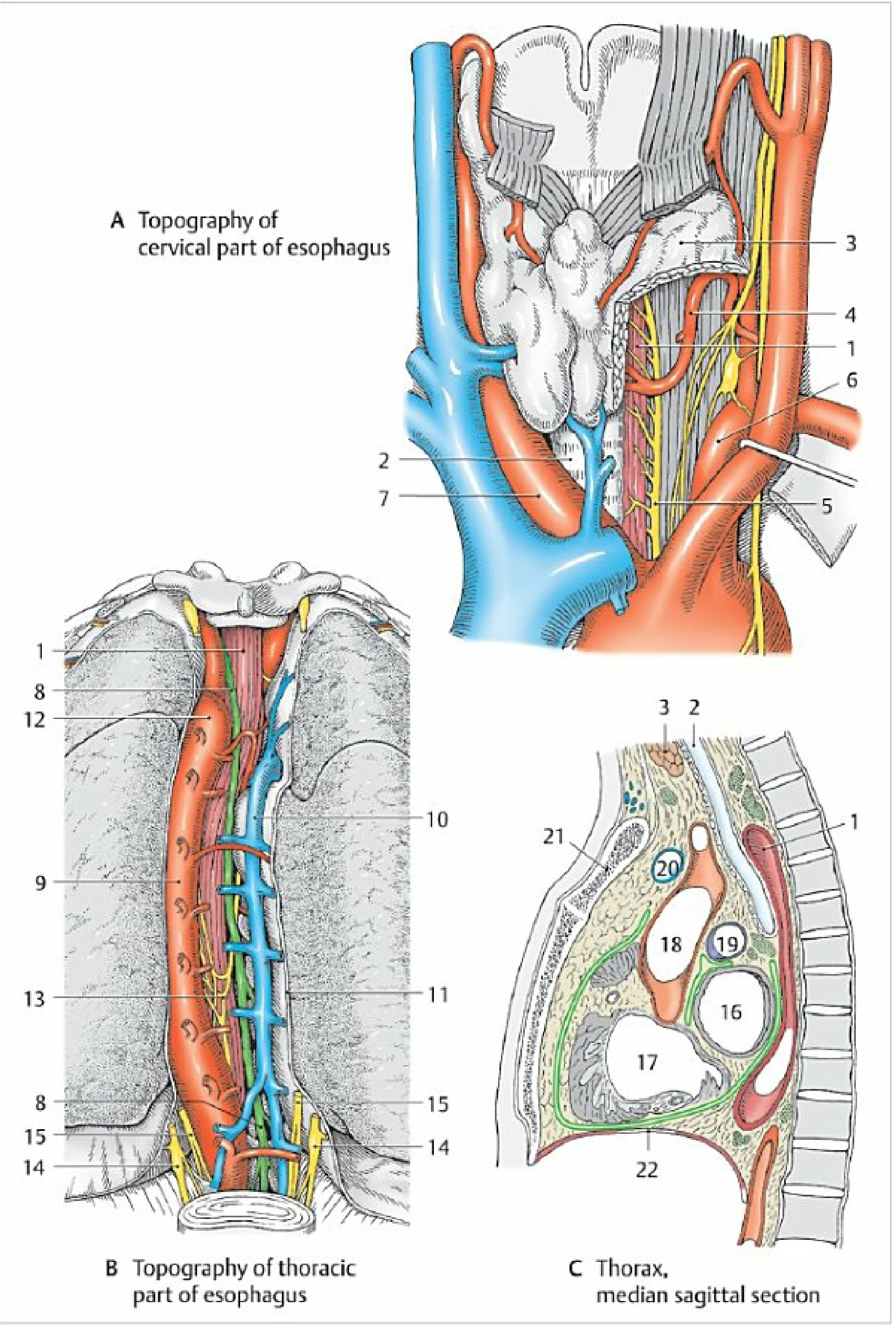
Your Exam Diagram - Draw a Simple Cross Section at 3 Levels:
CERVICAL (C6-T1):
Trachea
↑ (ANTERIOR)
LEFT ← OESOPHAGUS → RIGHT
(thyroid lobe, (thyroid lobe,
L recurrent R recurrent
laryngeal n.) laryngeal n.)
↓ (POSTERIOR)
Vertebral column
(Longus colli + prevertebral fascia)
THORACIC - SUPERIOR MEDIASTINUM (T1-T4):
Trachea + L recurrent laryngeal n.
↑
LEFT ← OESOPHAGUS → RIGHT
(Aortic arch, (Azygos vein,
L subclavian a., R pleura)
thoracic duct,
L pleura)
↓
Thoracic vertebrae (T1-T4)
Thoracic duct
THORACIC - POSTERIOR MEDIASTINUM (T4-T10):
L main bronchus → Pericardium (left atrium)
↑
LEFT ← OESOPHAGUS → RIGHT
(Descending aorta) (Azygos vein,
R pleura)
↓
Vertebral bodies + Thoracic duct
+ R posterior intercostal arteries
GOLD MEDAL 8-MARK ANSWER
Q. Describe the relations of the oesophagus.
INTRODUCTION
PART 1 - CERVICAL OESOPHAGUS (C6 to T1)
| Direction | Relation |
|---|---|
| Anterior | Trachea (adherent via loose connective tissue) |
| Posterior | Vertebral column - bodies of C6, C7 covered by prevertebral fascia and longus colli muscle |
| Left | Left lobe of thyroid gland; left recurrent laryngeal nerve (in tracheo-oesophageal groove) |
| Right | Right lobe of thyroid gland; right recurrent laryngeal nerve |
| Lateral (both) | Common carotid arteries within carotid sheaths |
Note: The oesophagus lies slightly to the left of the midline at this level.
Clinical point: The recurrent laryngeal nerves ascend in the tracheo-oesophageal grooves on both sides - injury during thyroid surgery causes hoarseness.
PART 2 - THORACIC OESOPHAGUS (T1 to T10)
- (a) Superior mediastinum (T1-T4, above tracheal bifurcation)
- (b) Posterior mediastinum (T4-T10, below tracheal bifurcation)
(a) Superior Mediastinum Relations:
| Direction | Relation |
|---|---|
| Anterior | Trachea; left recurrent laryngeal nerve |
| Left | Aortic arch; left subclavian artery; thoracic duct; left pleura |
| Right | Azygos vein; right pleura and lung |
| Posterior | Vertebral bodies T1-T4; thoracic duct (runs along right side up to T5, then crosses to the left) |
(b) Posterior Mediastinum Relations:
| Direction | Relation |
|---|---|
| Anterior | Left main bronchus; tracheobronchial lymph nodes; pericardium (left atrium - important!) |
| Left | Descending thoracic aorta (oesophagus is to the right of aorta above T7, then lies anterior to aorta below T7) |
| Right | Azygos vein; right pleura and lung |
| Posterior | Thoracic vertebrae; thoracic duct; right posterior intercostal arteries; hemiazygos + accessory azygos veins |
Clinical point 1: The left atrium is directly anterior to the thoracic oesophagus - used for transesophageal echocardiography (TEE).
Clinical point 2: The thoracic duct ascends on the right side of the oesophagus below T5, then crosses behind the oesophagus to the left above T5 - at risk in thoracic oesophageal surgery.
Clinical point 3: The right and left vagus nerves form the oesophageal plexus around the thoracic oesophagus below the tracheal bifurcation. Below this, they condense into anterior vagal trunk (mainly left vagus) and posterior vagal trunk (mainly right vagus) which travel with the oesophagus into the abdomen.
PART 3 - ABDOMINAL OESOPHAGUS (T10 to T11)
| Direction | Relation |
|---|---|
| Anterior and Right | Left lobe of liver; anterior and right vagal trunks |
| Posterior | Left crus of diaphragm; posterior vagal trunk; descending aorta (now posterior) |
| Left | Fundus of stomach |
CONSTRICTIONS OF THE OESOPHAGUS (Bonus - gets extra marks!)
| Constriction | Level | Distance from incisor teeth |
|---|---|---|
| Cricopharyngeal (Upper sphincter) | C6 | 15 cm |
| Aortic arch | T4 | 22.5 cm |
| Left main bronchus | T5 | 27.5 cm |
| Oesophageal hiatus | T10 | 40 cm |
These are sites where foreign bodies lodge and where carcinoma is common.
BLOOD SUPPLY
| Part | Artery | Vein |
|---|---|---|
| Cervical | Inferior thyroid artery | Inferior thyroid vein → brachiocephalic vein |
| Thoracic | Oesophageal branches of aorta | Azygos + hemiazygos veins |
| Abdominal | Left gastric + inferior phrenic arteries | Left gastric vein → portal vein |
Clinical point: Portal-systemic anastomosis at lower oesophagus (left gastric vein + azygos vein) - dilates in portal hypertension to form oesophageal varices.
LYMPHATIC DRAINAGE
| Part | Drains to |
|---|---|
| Upper 1/3 (above tracheal bifurcation) | Deep cervical + paratracheal nodes (drains upward) |
| Lower 2/3 (below tracheal bifurcation) | Posterior mediastinal nodes → coeliac nodes (drains downward) |
This explains why oesophageal cancer can have unusual lymph node spread patterns.
CLINICAL IMPORTANCE SUMMARY (for 8-mark answer clincher)
- Recurrent laryngeal nerve injury - in thyroid/oesophageal surgery → hoarseness
- Left atrium relation - used for TEE; left atrial enlargement can compress oesophagus and cause dysphagia
- Oesophageal varices - portal hypertension at the porto-systemic anastomosis
- Foreign body impaction - at the 4 constriction sites
- Thoracic duct injury - during oesophagectomy → chylothorax
- Oesophageal carcinoma - spreads to surrounding structures (aorta, trachea, left atrium) making it inoperable
MEMORY TRICK for Thoracic Relations
- Trachea (anterior, superior)
- Aortic arch (left, superior)
- Left main bronchus (anterior, inferior)
- Thoracic duct (posterior, crosses T5)
- Azygos vein (right)
- Descending aorta (left, inferior)
- Pericardium/left atrium (anterior, inferior)
- Azygos (right throughout)
- Vagus plexus (surrounds it)
- Abdominal relations (liver, fundus, crura)

Give a detailed explanation for understanding relations of every structure of thorax give the concepts and I am feeling difficulty in understanding and remembering the relations and course of the following structured give a detailed linked answers for 8 marks questions that I will not mug up and understand the concepts and give in such a way that I can score gold medal in exams and easy way that I can underdtsnd
THE COMPLETE THORAX ANATOMY MASTER GUIDE
Concept-Based Understanding for Gold Medal Answers
THE BIG PICTURE FIRST - How to think about the Thorax: The thorax is like a packed suitcase - everything is sandwiched tightly together. The key to understanding ALL relations is to first memorize the STERNAL ANGLE (T4/T5 level) - because almost EVERYTHING important happens at this one level. Once you understand the "neighbourhood" of each structure, relations come automatically.
THE ONE LEVEL YOU MUST KNOW: STERNAL ANGLE (T4/T5)
- Superior mediastinum ends / Inferior mediastinum begins
- Ascending aorta ends / Arch of aorta begins AND ends
- Trachea bifurcates into left and right main bronchi
- Azygos vein arches forward to join SVC
- Thoracic duct crosses from right to left
- 2nd rib articulates with sternum (surface marking)
Think of it as "The Grand Central Station of the Thorax" - everything meets here.
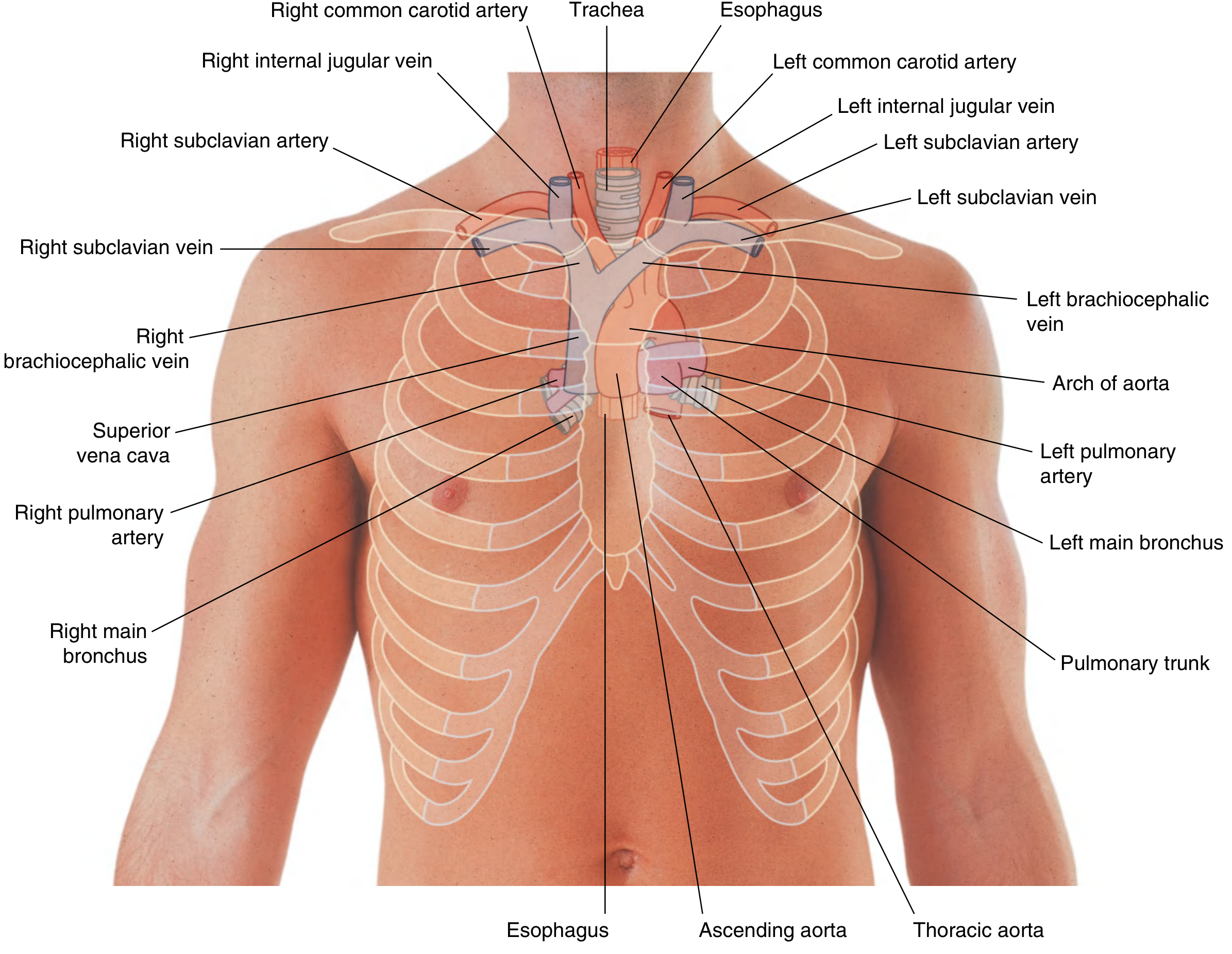
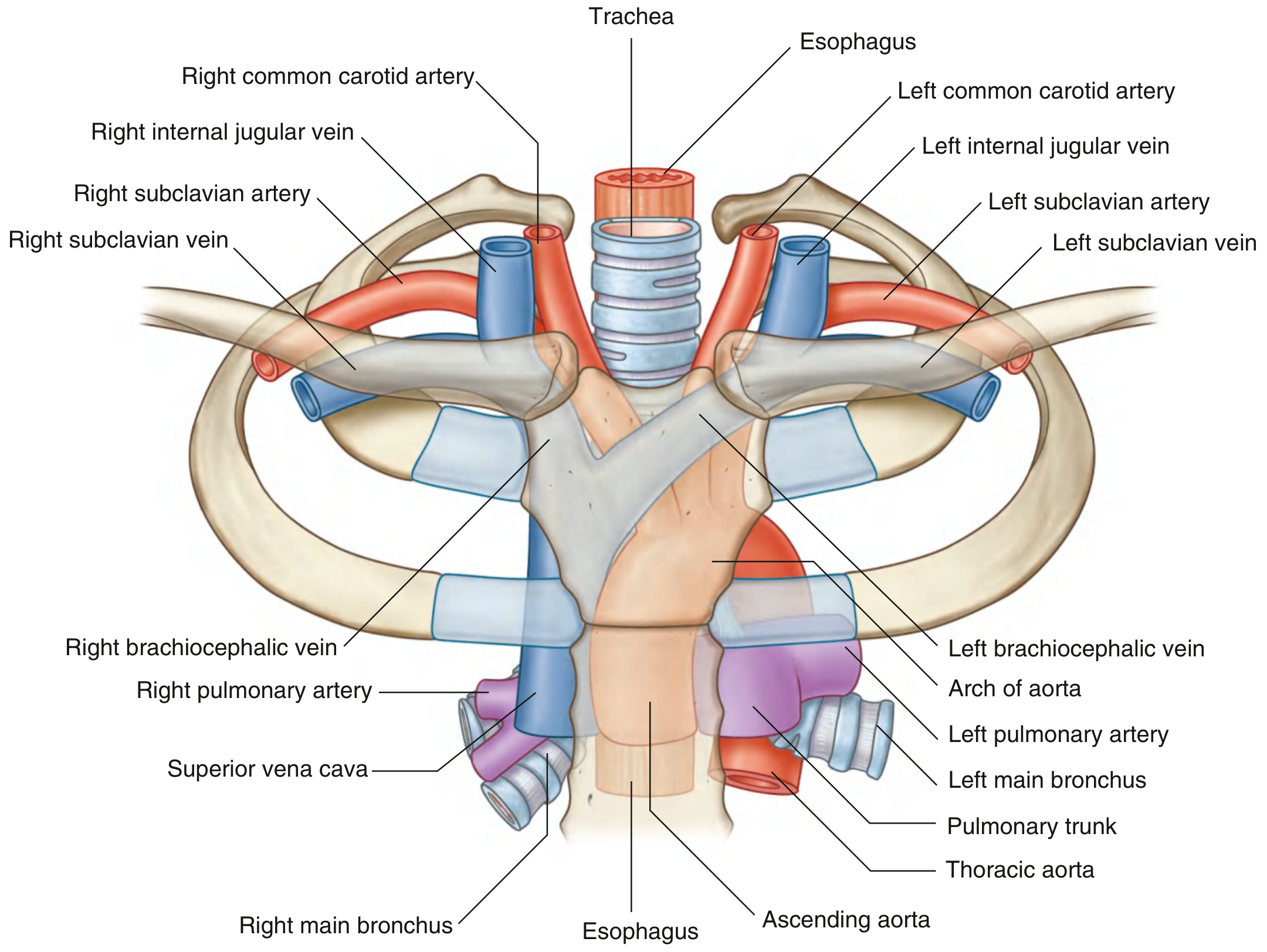
TOPIC 1 - THORACO-ABDOMINAL DIAPHRAGM ⭐⭐⭐⭐⭐
Concept to Understand First:
The 3 Openings - THE MOST ASKED PART:
| Opening | Level | Structures passing through |
|---|---|---|
| Caval opening (in central tendon) | T8 | Inferior vena cava + right phrenic nerve + lymphatics |
| Oesophageal hiatus (in right crus) | T10 | Oesophagus + anterior & posterior vagal trunks + oesophageal branches of left gastric artery + lymphatics |
| Aortic hiatus (between crura, behind diaphragm - actually NOT in diaphragm, so aortic pulse is NOT transmitted to abdomen - clinically important!) | T12 | Aorta + thoracic duct + azygos vein |
Why is the aortic hiatus at T12 and not in the diaphragm? Because it is behind the median arcuate ligament - the aorta doesn't pierce the diaphragm. So aortic pulsation is NOT affected by diaphragm contractions - important in aortic aneurysm palpation.
Surfaces (Relations):
- Superior surface (thoracic): Heart (on central tendon), Lungs and pleura (on either side), Pericardium
- Inferior surface (abdominal):
- Right dome: Liver (right lobe)
- Left dome: Stomach, Spleen, Left lobe of liver
Nerve Supply:
| Nerve | Supply | Memory |
|---|---|---|
| Phrenic nerve (C3,4,5) | Motor to whole diaphragm + sensory to central part | "C3,4,5 keeps the diaphragm alive" |
| Lower 6 intercostal nerves | Sensory to peripheral part only | "Periphery = peripheral nerves" |
Clinical: Referred pain from diaphragm goes to the SHOULDER TIP (C4 dermatome). So a subphrenic abscess, ruptured spleen, or ectopic pregnancy gives shoulder tip pain!
Blood Supply:
- Superior phrenic arteries (from aorta), Inferior phrenic arteries (from abdominal aorta), Pericardiacophrenic arteries (from internal thoracic arteries), Musculophrenic arteries
Exam Diagram to Draw:
Right Phrenic Left Phrenic
↓ ↓
___________T8 (IVC)___________
| T10 (Oesophagus + Vagus) |
| T12 (Aorta + Thoracic duct) |
|_____________________________________________|
Right crus (C-lumbar) Left crus (C-lumbar)
(Stronger - forms
oesophageal hiatus)
TOPIC 2 - INTERNAL THORACIC ARTERY ⭐⭐⭐
Concept First:
Origin, Course, Termination:
- Origin: From the first part of subclavian artery (inferior surface)
- Course: Descends 1 cm lateral to the sternum, posterior to the costal cartilages, anterior to the pleura
- Terminates at the 6th intercostal space by dividing into:
- Superior epigastric artery (enters rectus sheath)
- Musculophrenic artery (runs along costal margin)
Branches:
- Anterior intercostal arteries (to intercostal spaces 1-6)
- Perforating branches (to breast - medial mammary branches 2nd-4th spaces - important in breast surgery)
- Pericardiacophrenic artery (accompanies phrenic nerve, supplies pericardium and diaphragm)
- Mediastinal branches
Relations:
- Medially: Sternum
- Laterally: Pleura (internal thoracic vein runs beside it)
- Anteriorly: Costal cartilages + internal intercostal muscles
- Posteriorly: Transversus thoracis muscle (lower half), pleura (upper half)
Clinical Points:
- Used as LIMA (Left Internal Mammary Artery) graft in CABG - best long-term patency
- Injury in sternal fracture causes haemothorax
- Anastomoses with inferior epigastric artery (branch of external iliac) - important collateral route in aortic coarctation
TOPIC 3 - AZYGOS SYSTEM OF VEINS ⭐⭐⭐⭐⭐
Concept First - The "Ladder Analogy":
The System:
Azygos Vein (RIGHT SIDE):
- Origin: Ascending lumbar vein (L4/L5) on right side
- Enters thorax: Through aortic hiatus (T12) or behind right crus
- Ascends: Along right side of vertebral column (T12 to T4)
- Arches forward at T4 over the right main bronchus and root of lung
- Drains into: SVC (from behind, at T4 level) - the azygos arch
- Right superior intercostal vein (intercostal spaces 2 & 3)
- Right posterior intercostal veins (4th-11th)
- Hemiazygos vein (crosses from left at T8-T9)
- Accessory hemiazygos vein (crosses from left at T6-T7)
- Bronchial, oesophageal, mediastinal, pericardial veins
Hemiazygos Vein (LEFT SIDE, LOWER):
- Origin: Left ascending lumbar vein
- Ascends left side of vertebral column
- Crosses to the right at T8-T9 to drain into azygos
- Drains: Left posterior intercostal veins 9, 10, 11
Accessory Hemiazygos Vein (LEFT SIDE, UPPER):
- Drains left posterior intercostal veins 4-8
- Crosses to the right at T6-T7 to join azygos
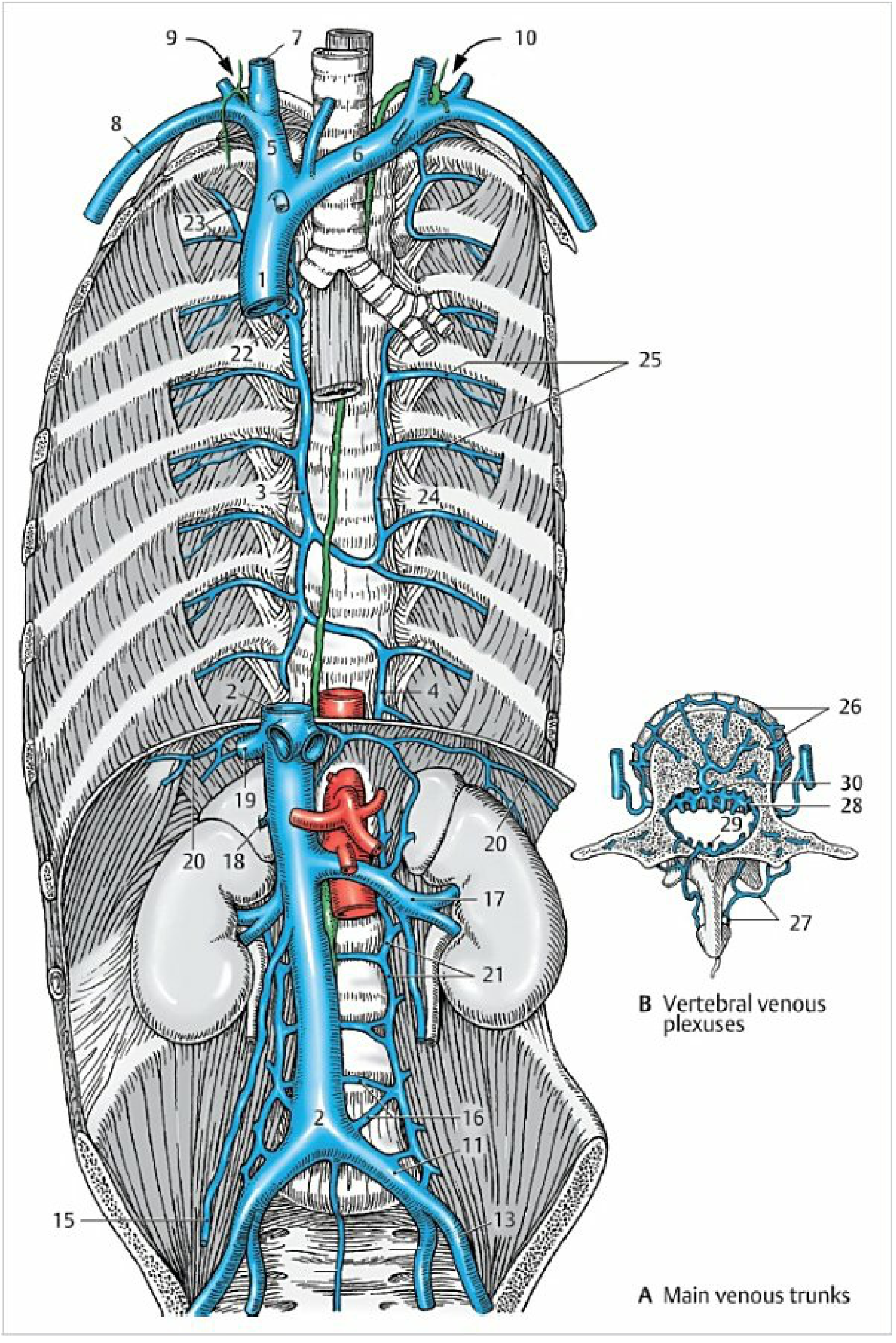
Memory Trick - "The Crossing Levels":
- Accessory hemiazygos crosses at T6-T7 (upper left)
- Hemiazygos crosses at T8-T9 (lower left)
- Both cross behind the aorta and thoracic duct
Clinical Importance:
- Collateral pathway: If SVC is blocked, blood can drain via azygos backwards into IVC (collateral circulation)
- Portal hypertension: Oesophageal veins (tributary) → dilate → oesophageal varices
- Azygos continuation of IVC: Rare anomaly where IVC drains via azygos into SVC - important to know before surgery
TOPIC 4 - THORACIC DUCT ⭐⭐⭐⭐⭐
Concept First - "The Snake that Crosses":
Course - Step by Step:
| Segment | Location | Level |
|---|---|---|
| Origin | Cisterna chyli (dilated sac at L1-L2) | L1-L2, right of aorta |
| Enters thorax | Through aortic hiatus with aorta and azygos vein | T12 |
| Lower thorax | Ascends on RIGHT side - between aorta (left) and azygos vein (right), in front of vertebral bodies | T12-T5 |
| CROSSES to LEFT | Crosses behind the oesophagus, in front of vertebral column | At T5 |
| Upper thorax | Ascends on LEFT side - behind left common carotid artery and subclavian artery | T5 upwards |
| Enters neck | Arches over left subclavian artery | |
| Terminates | At junction of left subclavian vein + left internal jugular vein (left venous angle) |
Memory trick for crossing: "The duct starts Right, crosses at T5 (5 fingers), ends Left"
Relations in Thorax:
- Anteriorly: Oesophagus (whole length - remember this for oesophageal surgery risk)
- Posteriorly: Vertebral bodies, right posterior intercostal arteries
- Right: Azygos vein (below T5)
- Left: Descending aorta (below T5)
- Above T5 (left side): Left common carotid + left subclavian artery on left; oesophagus + trachea medially
Clinical Importance:
- Chylothorax: Injury to thoracic duct (oesophagectomy, left neck dissection) → milky lymph in pleural cavity
- Why is chylothorax on the LEFT? Because the duct runs on the left in the upper chest
- Thoracic duct terminates on the LEFT - so left supraclavicular lymph node enlargement (Virchow's node / Troisier's sign) indicates visceral cancer spread via the thoracic duct
TOPIC 5 - PLEURA AND ITS DIVISIONS ⭐⭐⭐⭐⭐
Concept First - "A Glove Around the Lung":
Divisions of Parietal Pleura (4 Parts):
| Part | Lines | Important relation |
|---|---|---|
| Costal pleura | Inner surface of ribs, intercostal muscles, sternum | Separated from chest wall by endothoracic fascia |
| Diaphragmatic pleura | Upper surface of diaphragm | Closely adherent |
| Mediastinal pleura | Lateral surface of mediastinum | Contains hilum of lung; Phrenic nerve runs between mediastinal pleura and pericardium |
| Cervical pleura (Dome of pleura) | Extends 2.5 cm above medial 1/3 of clavicle into the neck | AT RISK in neck surgery and subclavian line insertion |
Pleural Recesses (Most Important for Exams!):
| Recess | Where | Clinical importance |
|---|---|---|
| Costodiaphragmatic recess | Between costal and diaphragmatic pleura | Deepest recess - first to fill with fluid (pleural effusion). Located at rib 8 (midclavicular), rib 10 (midaxillary), rib 12 (paravertebral) |
| Costomediastinal recess | Between costal and mediastinal pleura (anterior, near sternum on left side near cardiac notch) | Fills during deep inspiration |
Memory for Costodiaphragmatic Recess Levels:
Nerve Supply:
- Parietal pleura: Sensitive (intercostal nerves for costal; phrenic nerve for mediastinal and diaphragmatic) - PAINFUL when inflamed → pleurisy
- Visceral pleura: Insensitive to pain (autonomic) - lung cancer can grow without pain until it involves parietal pleura
Clinical Points:
- Pleural effusion settles in costodiaphragmatic recess first (gravity)
- Safe triangle for chest drain: Bordered by anterior border of latissimus dorsi, lateral border of pectoralis major, apex below axilla, base at level of nipple - avoids neurovascular bundle
- Pneumothorax: Air in pleural cavity collapses lung; treat with chest drain in 2nd ICS midclavicular line
TOPIC 6 - ARCH OF AORTA ⭐⭐⭐⭐⭐
Concept First - "The Rollercoaster":
Branches (3 branches from right to left):
- Brachiocephalic trunk (also called innominate artery) - right side
- Left common carotid artery - middle
- Left subclavian artery - left
Relations of the Arch of Aorta:
Anterior and Left side (structures looping over/under the arch):
- Left vagus nerve - crosses the arch from lateral to medial, then gives off the left recurrent laryngeal nerve which hooks under the arch (around the ligamentum arteriosum) and ascends in the tracheo-oesophageal groove
- Left phrenic nerve - crosses anterior to the arch (lateral to the vagus)
- Left superior intercostal vein - drains left 2nd & 3rd intercostal spaces, crosses over the arch
- Left brachiocephalic vein - crosses above the arch from left to right
Posterior to the arch:
- Trachea (bifurcation at T4/T5)
- Oesophagus (behind trachea)
- Left recurrent laryngeal nerve (after it hooks under)
Right side of the arch:
- SVC (the arch is to the left of the SVC)
- Trachea
- Deep cardiac plexus (between tracheal bifurcation and arch)
Above the arch:
- The 3 branches (brachiocephalic, left common carotid, left subclavian)
Below the arch:
- Left main bronchus
- Left pulmonary artery
- Ligamentum arteriosum (connects arch to left pulmonary artery - remnant of ductus arteriosus)
Ligamentum Arteriosum - Why It's Important:
- Connects the arch of aorta (inferior surface, between left common carotid and subclavian origins) to the left pulmonary artery
- Left recurrent laryngeal nerve hooks around it
- In patent ductus arteriosus (PDA) - this remains open after birth → left-to-right shunt
Memory trick for the nerve relationships:
- Left phrenic nerve: Anterior to aortic arch
- Left vagus nerve: Posterior to aortic arch
TOPIC 7 - SUPERIOR MEDIASTINUM ⭐⭐⭐⭐⭐
Boundaries:
- Superior: Thoracic inlet (line from jugular notch to T1)
- Inferior: Sternal angle (T4/T5) - the KEY LEVEL
- Anterior: Manubrium sternum
- Posterior: T1-T4 vertebral bodies
- Lateral: Mediastinal pleura
Contents - Arranged FRONT to BACK (How to Remember):
| Layer | Structure |
|---|---|
| Most anterior | Thymus (or thymic remnant in adults) |
| Veins | Left brachiocephalic vein (horizontal, in front of arch), SVC (right side) |
| Arteries | Arch of aorta + 3 branches |
| Central | Trachea (midline, tilts slightly right) |
| Nerves | Phrenic nerves (on each side), Vagus nerves, Left recurrent laryngeal |
| Behind trachea | Oesophagus |
| Most posterior | Thoracic duct |
The KEY relationship: Left brachiocephalic vein crosses from left to right ANTERIOR to the 3 branches of the arch. This is why you can see it pulsate during CPR and it's at risk during median sternotomy.
TOPIC 8 - MIDDLE MEDIASTINUM ⭐⭐⭐
What's in the Middle Mediastinum?
- Pericardium (fibrous + serous)
- Heart (all 4 chambers)
- Ascending aorta (lower half - upper half is in superior mediastinum)
- Pulmonary trunk + right and left pulmonary arteries
- Lower SVC and upper IVC
- Pulmonary veins (4 total - 2 right, 2 left)
- Phrenic nerves + pericardiacophrenic vessels (run in middle mediastinum, between pericardium and mediastinal pleura)
- Tracheal bifurcation and main bronchi
Why the Phrenic Nerve Is in the Middle Mediastinum:
- Pericarditis can give referred shoulder tip pain (C4 via phrenic)
- Mediastinal tumors can trap and paralyze the phrenic nerve
TOPIC 9 - POSTERIOR MEDIASTINUM ⭐⭐⭐⭐⭐
Boundaries:
- Anterior: Pericardium (middle mediastinum) + bifurcation of trachea
- Posterior: T5-T12 vertebral bodies
- Superior: T4/T5 (sternal angle plane)
- Inferior: Diaphragm
- Lateral: Mediastinal pleura
Contents - FRONT to BACK again:
| Structure | Position |
|---|---|
| Oesophagus | Central/anterior part; vagal plexus around it |
| Thoracic aorta | Left of oesophagus initially, then posterior at lower levels |
| Azygos vein | Right of oesophagus and aorta |
| Hemiazygos + accessory hemiazygos | Left of aorta |
| Thoracic duct | Between aorta and azygos vein below T5; crosses to left at T5 |
| Thoracic sympathetic chain | On either side of vertebral column (on heads of ribs) |
| Greater, lesser, least splanchnic nerves | From sympathetic chain through diaphragm |
| Posterior intercostal arteries & veins | In intercostal spaces |
| Lymph nodes | Posterior mediastinal (paraesophageal) nodes |
Concept link: The posterior mediastinum is like a highway corridor - aorta takes traffic down, azygos brings venous blood up, thoracic duct drains lymph up, oesophagus carries food down, sympathetic chain controls everything.
TOPIC 10 - CORONARY SINUS ⭐⭐⭐⭐⭐
Concept First:
Location:
- Lies in the posterior part of the atrioventricular (coronary) sulcus between the left atrium (above) and left ventricle (below)
- Opens into the right atrium between the IVC opening and the tricuspid valve - guarded by a semilunar valve (valve of Thebesius)
Length: ~3 cm
Tributaries (draining into coronary sinus):
| Tributary | Drains | Location |
|---|---|---|
| Great cardiac vein | Anterior heart | Runs with anterior interventricular artery (LAD), then in anterior AV groove |
| Middle cardiac vein | Posterior heart | Runs with posterior interventricular artery, in posterior interventricular groove |
| Small cardiac vein | Right heart margin | Runs with right marginal artery |
| Posterior vein of left ventricle | Posterior LV | |
| Oblique vein of left atrium (vein of Marshall) | Left atrium | Remnant of embryonic left SVC |
Clinical Importance:
- Cardiac resynchronization therapy (CRT): Pacemaker leads placed in coronary sinus branches to pace the left ventricle
- Retrograde cardioplegia: Cardioplegia solution delivered via coronary sinus during bypass surgery
- Coronary venous system imaging on angiography
- The oblique vein (Marshall) is important in atrial fibrillation ablation procedures
TOPIC 11 - SUPERIOR VENA CAVA (SVC) ⭐⭐⭐⭐⭐
Formation:
Course and Relations:
- Length: ~7 cm
- Course: Descends vertically on the RIGHT side of the mediastinum
- Enters right atrium at T3 level (right 3rd costal cartilage)
- Lower half is intrapericardial (covered by pericardium)
| Direction | Relation |
|---|---|
| Right | Right pleura and lung, right phrenic nerve (between SVC and pleura) |
| Left | Ascending aorta |
| Anterior | Thymus/thymic fat, anterior mediastinum |
| Posterior | Right pulmonary artery, right main bronchus (at hilum level) |
Tributaries:
- Right and left brachiocephalic veins (form it)
- Azygos vein (enters from behind at T4 - the azygos arch)
- Small mediastinal and pericardial veins
Clinical - SVC Syndrome:
- Facial and arm swelling ("cape distribution")
- Dilated neck veins and chest wall veins
- Headache, dyspnoea
- Non-pulsatile venous engorgement (distinguish from JVP pulsation)
TOPIC 12 - STERNAL ANGLE (ANGLE OF LOUIS) ⭐⭐⭐⭐⭐
What is it?
At the Level of Sternal Angle (T4/T5), ALL of these occur:
- 2nd rib articulates with sternum (count ribs clinically starting here)
- Superior mediastinum separates from inferior mediastinum
- Ascending aorta → Arch of aorta begins (and arch also ends here)
- Trachea bifurcates (carina) into right and left main bronchi
- Azygos vein arches forward to join SVC
- Thoracic duct crosses from right to left
- Lower border of thymus reaches here
- Left recurrent laryngeal nerve hooks under ligamentum arteriosum (at arch)
Clinical Importance:
- Count ribs for intercostal space identification (start from 2nd rib at sternal angle)
- Upper limit of cardiac dullness
- Level for central venous catheter tip position monitoring on X-ray
TOPIC 13 - OESOPHAGUS ⭐⭐⭐
Quick Summary:
- C6 to T11, 25 cm total
- Cervical: Behind trachea, recurrent laryngeal nerves in tracheo-oesophageal grooves
- Thoracic superior: Behind trachea, left to aortic arch, right to azygos
- Thoracic inferior: Behind pericardium (LEFT ATRIUM directly anterior), left to descending aorta, right to azygos, behind - thoracic duct
- Constrictions: C6 (15cm), T4/aortic arch (22.5cm), T5/left bronchus (27.5cm), T10/hiatus (40cm)
- Key clinical: Left atrium relation → transesophageal echo; Portal-systemic anastomosis → varices
TOPIC 14 - PERICARDIAL SINUSES ⭐⭐⭐
Concept First - "The Two Hidden Spaces":
Transverse Pericardial Sinus:
- Think of it as: A tunnel you can pass your finger through from right to left, behind the great arteries
- How it forms: During development, when the arterial end (aorta + pulmonary trunk) and venous end (venae cavae) fuse, they leave a gap = transverse sinus
- Boundaries:
- Anteriorly: Ascending aorta + pulmonary trunk
- Posteriorly: Left atrium + SVC
- Superiorly: Reflection of serous pericardium
Oblique Pericardial Sinus:
- Shape: Like an inverted J or a horseshoe opening downward
- Boundaries: Surrounded by pulmonary veins (laterally), left atrium (anteriorly), pericardial wall (posteriorly)
- Formed by: The pericardial reflection around the pulmonary veins
Memory Trick:
- Transverse = Trans = Goes ACROSS (you can pass finger through)
- Oblique = Blind = Dead end (you cannot pass through)
TOPIC 15 - TRACHEA ⭐
Concept: "The flexible pipe leading to the lungs"
Course:
- Starts at C6 (lower border of cricoid cartilage)
- Ends at T4/T5 (sternal angle) → bifurcates into right and left main bronchi
- Length: ~10-11 cm, 16-20 C-shaped cartilage rings (deficient posteriorly - membranous wall)
- Slightly to the right of midline (pushed by aortic arch on left)
Relations:
In the Neck (Cervical Trachea):
- Anterior: Skin, fascia, thyroid isthmus (crosses rings 2-4), thyroid veins, inferior thyroid arteries
- Posterior: Oesophagus
- Lateral: Thyroid lobes, common carotid arteries, recurrent laryngeal nerves (in tracheo-oesophageal groove)
In the Thorax (Intrathoracic Trachea - Superior Mediastinum):
- Anterior: Manubrium, brachiocephalic trunk (right), left brachiocephalic vein (crossing), aortic arch, thymus
- Posterior: Oesophagus (throughout)
- Right: Azygos vein, right vagus nerve, right pleura
- Left: Aortic arch (pushes trachea to right), left common carotid artery, left recurrent laryngeal nerve
Carina: The bifurcation point at T4/T5
- Angle of bifurcation normally ~60-70°
- Widened carina (>70°) on CXR or bronchoscopy indicates left atrial enlargement or subcarinal lymph node enlargement
Right vs Left Main Bronchus - Why Right Lung Gets More Foreign Bodies:
| Feature | Right main bronchus | Left main bronchus |
|---|---|---|
| Length | Short (~2.5 cm) | Long (~5 cm) |
| Angle with trachea | More vertical (~25°) | More horizontal (~45°) |
| Foreign body | More common | Less common |
| Relation above | Azygos vein arches over | Aortic arch passes above |
INTEGRATING EVERYTHING - THE BIG PICTURE MAP
SUPERIOR MEDIASTINUM (Manubrium to Sternal Angle T4/T5):
FRONT → BACK order:
Thymus → L brachiocephalic vein → Aortic arch (+ 3 branches) → Trachea → Oesophagus → Thoracic duct
STERNAL ANGLE = T4/T5 = GRAND CENTRAL STATION
POSTERIOR MEDIASTINUM (T4/T5 to Diaphragm):
FRONT → BACK order:
Oesophagus (with vagal plexus) → Descending aorta (left) + Azygos (right) → Thoracic duct → Sympathetic chain (on sides) → Vertebral bodies
DIAPHRAGM OPENINGS (Bottom of Thorax):
T8 = IVC + R phrenic T10 = Oesophagus + Vagus T12 = Aorta + Thoracic duct + Azygos
MASTER MEMORY TABLE - For Quick Revision
| Structure | Key Number | Most Important Relation | Clinical Point |
|---|---|---|---|
| Diaphragm | T8-T10-T12 | Phrenic nerve (motor) | Referred shoulder tip pain |
| Internal thoracic A | 1 cm from sternum | Subclavian origin, bifurcates at 6th ICS | CABG graft (LIMA) |
| Azygos vein | Crosses T4 → SVC | Between aorta and vertebrae | SVC collateral; varices |
| Thoracic duct | Crosses right→left at T5 | Between aorta and azygos | Chylothorax; Virchow's node |
| Pleura | Dome 2.5 cm above clavicle | Costodiaphragmatic recess 8-10-12 | Pneumothorax; effusion |
| Arch of aorta | T4/T5 | Left vagus hooks under (RLN) | Hoarseness in arch aneurysm |
| Superior mediastinum | T1-T4/T5 | Thymus-Veins-Aorta-Trachea-Oesophagus-Duct | Thymic tumors |
| Posterior mediastinum | T5-T12 | Oesophagus + Aorta + Azygos + Thoracic duct | Mediastinitis, descending necrotising |
| Coronary sinus | AV groove, posterior | Right atrium (opens between IVC and tricuspid) | CRT pacemaker lead placement |
| SVC | Right mediastinum, T3 | Ascending aorta on left, azygos from behind | SVC syndrome |
| Sternal angle | T4/T5 | EVERYTHING changes here | Count ribs clinically |
| Oesophagus | C6-T11 | Left atrium (posterior thoracic) | TEE; varices |
| Pericardial sinuses | Behind great vessels | Transverse: behind aorta/PT; Oblique: behind LA | Cardiac surgery |
| Trachea | C6-T4/T5 | Oesophagus posterior; Aortic arch on left | Right bronchus gets foreign bodies |
Give a explanation answer on the intrinsic muscles of larynx and lateral ventricles of brain easy to understand and a gold medal answer anf easy to understand and easy to temember and diagrams
PART 1: INTRINSIC MUSCLES OF THE LARYNX
Gold Medal Answer - Concept Based
THE BIG CONCEPT FIRST - Understand This and You'll Never Forget
| Job | What it means | Muscle(s) |
|---|---|---|
| 1. OPEN the glottis (abduct cords) | Pull cords apart → breathe | Only PCA |
| 2. CLOSE the glottis (adduct cords) | Bring cords together → speak/protect | LCA + IA + TA |
| 3. TIGHTEN cords (increase tension) | Stretch cords → high pitch | Cricothyroid |
| 4. RELAX cords (decrease tension) | Shorten cords → low pitch | Thyroarytenoid (Vocalis) |
THE MOST IMPORTANT RULE IN LARYNX ANATOMY: "PCA is the ONLY ABDUCTOR. Everything else is an ADDUCTOR or tension changer." PCA = Posterior Crico-Arytenoid
UNDERSTAND THE CARTILAGE SYSTEM FIRST
- The arytenoid cartilages sit on top of the cricoid ring like two chairs
- When arytenoids rotate OUTWARD → vocal cords OPEN (abduction)
- When arytenoids rotate INWARD → vocal cords CLOSE (adduction)
- The cricoid cartilage can tilt relative to thyroid → changes distance between thyroid angle and arytenoids → changes cord tension
EXAM DIAGRAM - Larynx from Above (Bird's Eye View)
ANTERIOR
(Thyroid angle)
↑
Anterior commissure
/ \
Left vocal cord Right vocal cord
| |
Left arytenoid Right arytenoid
| |
Interarytenoid muscle (bridges both)
↓
POSTERIOR
(Cricoid lamina)
OPEN (abducted): CLOSED (adducted):
/ \ ||
/ \ ||
Arytenoids Arytenoids
spread apart brought together
THE 9 INTRINSIC MUSCLES - GROUPED BY FUNCTION
GROUP 1: ABDUCTOR (Opens glottis) - Only 1 muscle
1. POSTERIOR CRICOARYTENOID (PCA) - THE MOST IMPORTANT MUSCLE IN THE BODY
- Origin: Lower and medial surface of the back of cricoid lamina
- Insertion: Muscular process of arytenoid (fans out)
- Action: Pulls muscular process POSTERIORLY and DOWNWARD → arytenoid rotates OUTWARD → vocal processes separate → ABDUCTION
- Nerve: Recurrent laryngeal nerve
- Memory: "PCA = Posterior = Pull Apart" - the only muscle keeping your airway open!
Clinical GOLD: Bilateral PCA paralysis (bilateral RLN palsy) = immediate airway emergency. Both cords fall to midline → stridor → may need emergency tracheostomy. This is why surgeons are terrified of bilateral recurrent laryngeal nerve injury in thyroid surgery!
GROUP 2: ADDUCTORS (Close glottis) - 3 muscles
2. LATERAL CRICOARYTENOID (LCA)
- Origin: Superior border of the lateral arch of cricoid
- Insertion: Muscular process of arytenoid
- Action: Pulls muscular process ANTERIORLY and DOWNWARD → arytenoid rotates INWARD → vocal processes come together → ADDUCTION
- Nerve: Recurrent laryngeal nerve
- Memory: "LCA is the OPPOSITE of PCA" - same bone, same insertion, but opposite fibre direction → opposite action
3. TRANSVERSE ARYTENOID (Interarytenoid) - ONLY UNPAIRED MUSCLE
- Origin: Posterior surface of one arytenoid
- Insertion: Posterior surface of the OTHER arytenoid (crosses the midline)
- Action: Pulls the two arytenoids TOGETHER → closes the posterior glottis (the posterior commissure)
- Nerve: Recurrent laryngeal nerve
- Memory: "Trans = Across = bridges the gap" - the only unpaired muscle
4. OBLIQUE ARYTENOID
- Origin: Muscular process of one arytenoid
- Insertion: APEX of the other arytenoid (crosses over the transverse arytenoid like an X)
- Action: Assists adduction; its fibres continue as...
5. ARYEPIGLOTTICUS
- Continuation of oblique arytenoid fibres that continue to the epiglottis
- Action: Closes the laryngeal inlet during swallowing - narrows the aditus
- Memory: "Oblique + Aryepiglotticus = X-shape crossover - they protect airway during swallowing"
GROUP 3: TENSION CHANGERS - 2 muscles
6. CRICOTHYROID - THE EXCEPTION MUSCLE
- Origin: Lateral surface of the anterior arch of cricoid
- Insertion: Lower border + inferior cornu of thyroid cartilage (in two groups: straight anterior fibres + oblique posterior fibres)
- Action: Rocks the cricoid backwards (or tilts thyroid forward) → INCREASES the distance between thyroid angle and arytenoids → STRETCHES/LENGTHENS vocal cords → INCREASES TENSION → higher pitch
- Nerve: EXTERNAL branch of SUPERIOR LARYNGEAL NERVE (NOT the recurrent!) ← the ONE exception
- Location: The ONLY intrinsic muscle that lies OUTSIDE the cartilaginous framework, on the OUTER surface
Why is cricothyroid supplied by SLN and not RLN? Because embryologically it develops from the 4th arch (SLN territory), while all other intrinsic muscles develop from the 6th arch (RLN territory). Understanding embryology = never forgetting the exception!
Clinical: Singer's nerve = external SLN. Injury during thyroid surgery (superior thyroid artery ligation) → loss of high notes, voice fatigue, monotone voice.
7. THYROARYTENOID (VOCALIS MUSCLE)
- Origin: Inner surface of thyroid angle + cricothyroid ligament
- Insertion: Vocal process and body of arytenoid
- Action: Pulls arytenoid TOWARD thyroid → SHORTENS cords → DECREASES tension → lower pitch
- The medial fibres = VOCALIS MUSCLE - the true vocal cord itself
- Nerve: Recurrent laryngeal nerve
- Memory: "Thyroepi-ACTIVE: the Vocalis IS the cord"
8. THYROEPIGLOTTICUS
- Extension of thyroarytenoid fibres to the epiglottis
- Action: Widens the laryngeal inlet (pulls aryepiglottic folds apart)
COMPLETE SUMMARY TABLE (Perfect for Exam)
| Muscle | Origin | Insertion | Action | Nerve |
|---|---|---|---|---|
| PCA | Back of cricoid lamina | Muscular process of arytenoid | ABDUCTION (opens) | RLN |
| LCA | Lateral arch of cricoid | Muscular process of arytenoid | ADDUCTION (closes) | RLN |
| Transverse arytenoid | Posterior arytenoid | Opposite arytenoid | ADDUCTION (closes posterior) | RLN |
| Oblique arytenoid | Muscular process | Apex of opposite arytenoid | ADDUCTION + inlet closure | RLN |
| Aryepiglotticus | Apex of arytenoid | Epiglottis | Closes inlet | RLN |
| Cricothyroid | Anterior cricoid arch | Thyroid cartilage | TENSES cords (high pitch) | EXTERNAL SLN |
| Thyroarytenoid (Vocalis) | Inner thyroid angle | Arytenoid vocal process | RELAXES cords (low pitch) | RLN |
| Thyroepiglotticus | Thyroid cartilage | Epiglottis | Widens inlet | RLN |
NERVE SUPPLY - GOLD MEDAL RULE
"ALL intrinsic muscles supplied by RLN... EXCEPT cricothyroid (external SLN)"
- Recurrent laryngeal nerve (branch of vagus): ALL intrinsic muscles (PCA, LCA, IA, OA, TA)
- External branch of Superior Laryngeal Nerve: ONLY cricothyroid
CLINICAL APPLICATIONS - What Examiners Always Ask
| Clinical Scenario | Muscle/Nerve affected | Result |
|---|---|---|
| Unilateral RLN palsy | All muscles on one side paralyzed | Cord on affected side lies in PARAMEDIAN position (partially adducted) - hoarse voice, not complete airway obstruction |
| Bilateral RLN palsy | Both PCA paralyzed (other muscles still have some tone) | Both cords drawn to midline (remaining cricothyroid tone) → STRIDOR, EMERGENCY |
| External SLN palsy | Cricothyroid paralyzed | Loss of high notes, voice fatigue, monotone - affects professional singers most |
| "Cadaveric position" | Complete nerve + muscle paralysis | Cord lies in intermediate abducted position (halfway between adduction and abduction) |
PART 2: LATERAL VENTRICLES OF THE BRAIN
Gold Medal Answer - Concept Based
THE BIG CONCEPT - Understand the Shape First
CORPUS CALLOSUM (roof of all parts)
↑
ANTERIOR HORN → BODY → ATRIUM → POSTERIOR HORN
↘
INFERIOR HORN (into temporal lobe)
EXAM DIAGRAM - Lateral Ventricle Parts
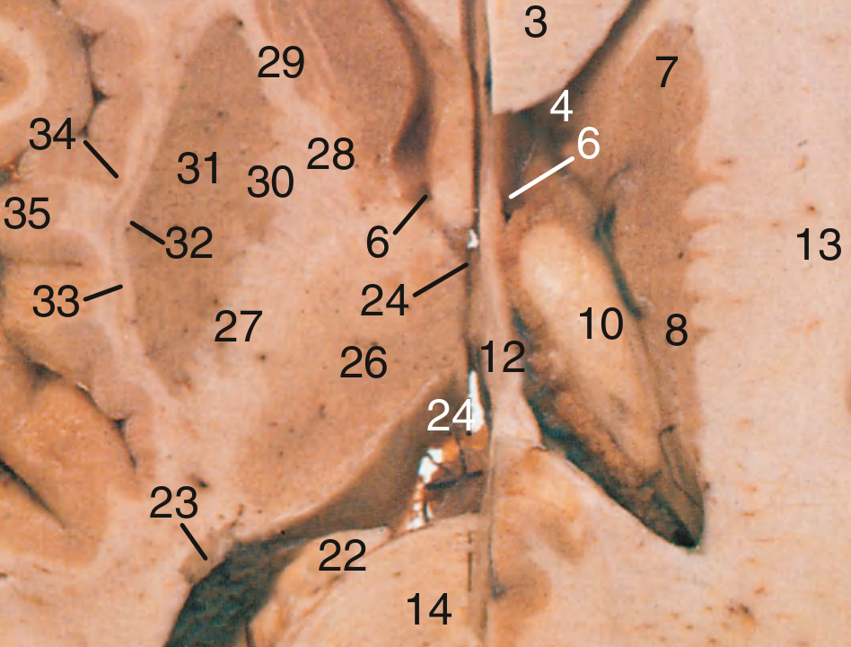
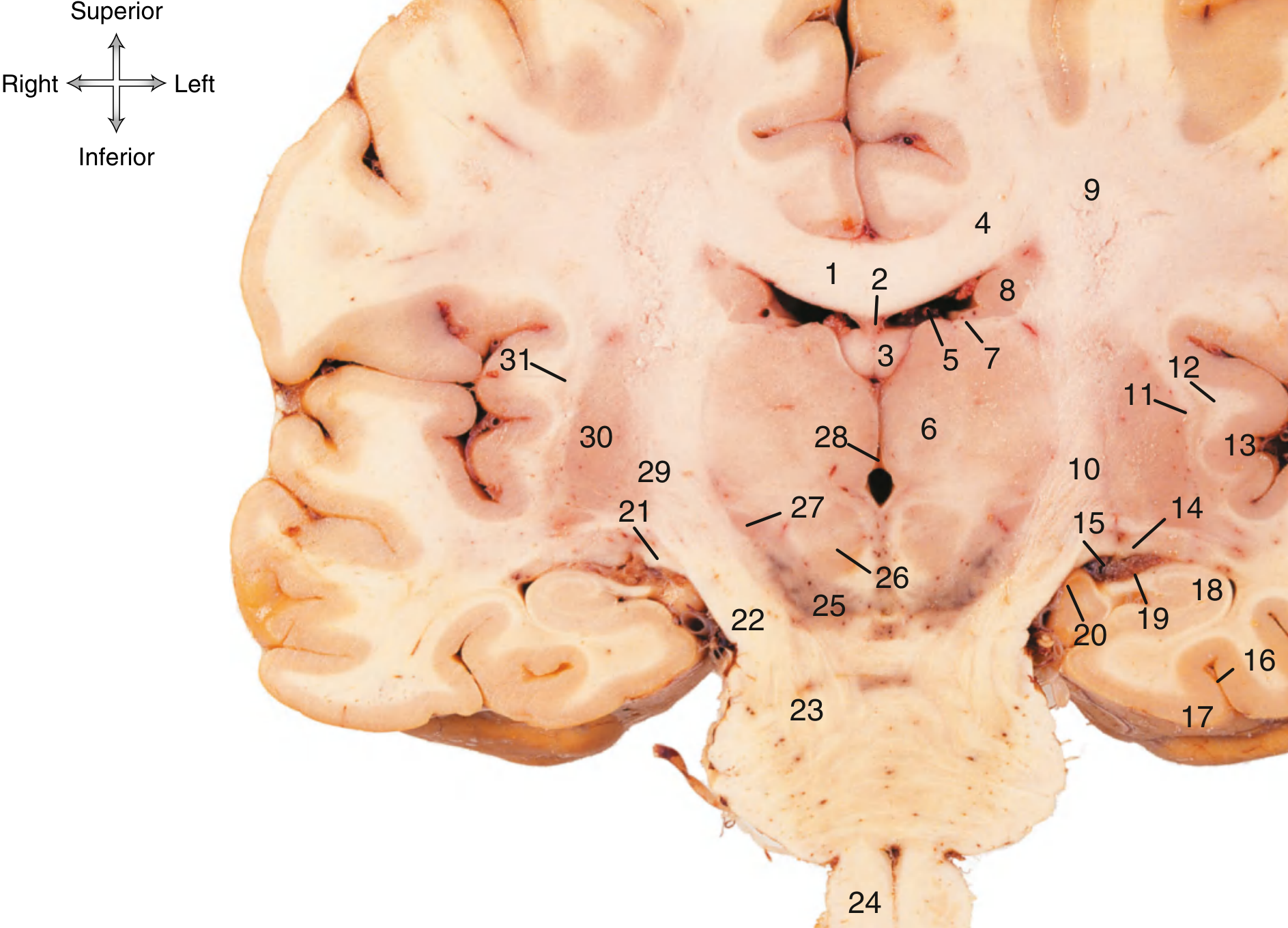
PART-BY-PART BREAKDOWN - Relations
KEY RULE to remember ALL relations:
The CAUDATE NUCLEUS follows the lateral ventricle like a loyal shadow at ALL levels.
- Head of caudate = lateral wall of anterior horn
- Body of caudate = floor of body
- Tail of caudate = roof of inferior horn
- Amygdala = where the tail ends
PART 1: ANTERIOR HORN (Frontal Horn)
| Wall | Structure |
|---|---|
| Roof | Corpus callosum (genu and body) |
| Floor | Head of caudate nucleus (bulging in) |
| Medial wall | Septum pellucidum (thin membrane separating the two lateral ventricles) |
| Anterior wall | Genu of corpus callosum |
| Lateral wall | Head of caudate nucleus |
PART 2: BODY (Central Part)
| Wall | Structure |
|---|---|
| Roof | Body of corpus callosum |
| Medial wall | Septum pellucidum (anteriorly) → Body of fornix (posteriorly) |
| Floor | Body of caudate nucleus (laterally) + Thalamus (medially) + Stria terminalis + Thalamostriate vein (in the groove between caudate and thalamus) |
PART 3: ATRIUM (TRIGONE/COLLATERAL TRIGONE)
- This is the WIDEST part of the lateral ventricle
- Has the largest collection of choroid plexus (called glomus - visible on CT/MRI as calcified density in adults)
- Connects all three horns
PART 4: POSTERIOR HORN (Occipital Horn)
| Wall | Structure |
|---|---|
| Roof and lateral wall | Tapetum (fibres of corpus callosum) + Optic radiation (important!) |
| Medial wall | Bulb of posterior horn (raised by forceps major of CC) + Calcar avis (raised by calcarine sulcus) |
Clinical: Optic radiation fibres pass through the lateral wall of the posterior horn - injury in temporal/occipital surgery can cause visual field defects (homonymous hemianopia)
PART 5: INFERIOR HORN (Temporal Horn)
| Wall | Structure |
|---|---|
| Roof | Tail of caudate nucleus + Stria terminalis |
| Floor and medial wall | Hippocampus (the most important!) + Fimbria of hippocampus |
| Lateral wall | Collateral eminence (from collateral sulcus) |
SUMMARY TABLE - ALL PARTS
| Part | Roof | Floor | Medial Wall | Lateral Wall | Choroid Plexus? |
|---|---|---|---|---|---|
| Anterior horn | Corpus callosum (genu) | Head of caudate | Septum pellucidum | Head of caudate | NO |
| Body | Corpus callosum | Caudate body + Thalamus | Septum → Fornix | Caudate body | YES |
| Atrium | Corpus callosum | Collateral trigone | Fornix + Calcar | Tapetum | YES (glomus) |
| Posterior horn | Tapetum + Optic radiation | - | Calcar avis + Bulb | Tapetum | NO |
| Inferior horn | Tail of caudate + Stria terminalis | Hippocampus + Fimbria | Hippocampus | Collateral eminence | YES |
COMMUNICATION AND CSF FLOW
- 3rd ventricle via Foramen of Monro (Interventricular foramen)
- CSF produced here by choroid plexus → flows to 3rd ventricle → cerebral aqueduct (of Sylvius) → 4th ventricle → foramina of Magendie (midline) and Luschka (lateral) → subarachnoid space
Hydrocephalus: If foramen of Monro is blocked (by colloid cyst, tumor) → lateral ventricle dilates (only one side if one foramen blocked) → raised ICP
CHOROID PLEXUS - WHERE IS IT?
- Attached along the choroidal fissure (between fornix above and thalamus below in the body; between fimbria and stria terminalis in inferior horn)
- Produces ~500 mL CSF per day (absorbed at arachnoid granulations in superior sagittal sinus)
IMPORTANT VEINS - Often Asked
- Thalamostriate vein - runs in the groove between caudate and thalamus in the floor of the body
- Choroidal vein - runs alongside choroid plexus
- Both drain into the Internal Cerebral Vein at the foramen of Monro
- Internal cerebral veins unite to form Great Cerebral Vein (of Galen)
MASTER MEMORY TRICK - The "C-shape Story"
"The Caudate nucleus is a C-shaped shadow that traces the entire lateral ventricle. Head = anterior horn. Body = body. Tail curls down into inferior horn roof and ends at the Amygdala. Follow the caudate - you've traced the ventricle."
CLINICAL APPLICATIONS
| Condition | Part of lateral ventricle affected | Sign |
|---|---|---|
| Colloid cyst at foramen of Monro | Blocks both foramina | Bilateral lateral ventricle dilation, raised ICP, intermittent headache worse on lying down |
| Hippocampal atrophy | Inferior horn enlarges | Early sign of Alzheimer's disease on MRI |
| Intraventricular hemorrhage | Blood in lateral ventricle | Seen in preterm babies (germinal matrix bleeding at caudate-thalamus junction) |
| Optic radiation injury (lateral to posterior horn) | Posterior horn region | Contralateral homonymous hemianopia |
| Hydrocephalus | All horns dilate - temporal horn first (most distensible) | Papilloedema, Cushing's triad |

Give a detailed and conceptually understandable explanation so that I can write these 15 marks answers give in a easy remembering way and linked manner like a story in so that I can rememeber easily study these
THE ABDOMEN & PELVIS MASTER GUIDE
Story-Based Concept Learning for 15-Mark Answers
THE STORY THAT LINKS EVERYTHING
"A FOOD JOURNEY through your body - Food enters → Stomach digests → Liver processes the nutrients via Portal Vein → Pancreas helps digestion → Kidneys filter the waste → Bladder stores urine → The reproductive system (Uterus/Testis) sits nearby in the pelvis → And waste exits via Rectum and Anal Canal. The Inguinal Canal is the doorway through which the testis descended to reach the scrotum."Every topic connects to this story. Learn the story, you learn everything.
THE UNIVERSAL FORMAT FOR ALL 15-MARK QUESTIONS
- Location
- External features
- Internal features
- Relations
- Vascular supply
- Nervous supply
- Lymphatic drainage
- Applied anatomy
TOPIC 1 - INGUINAL CANAL ⭐⭐⭐⭐⭐
The Story: "The Tunnel the Testis Built"
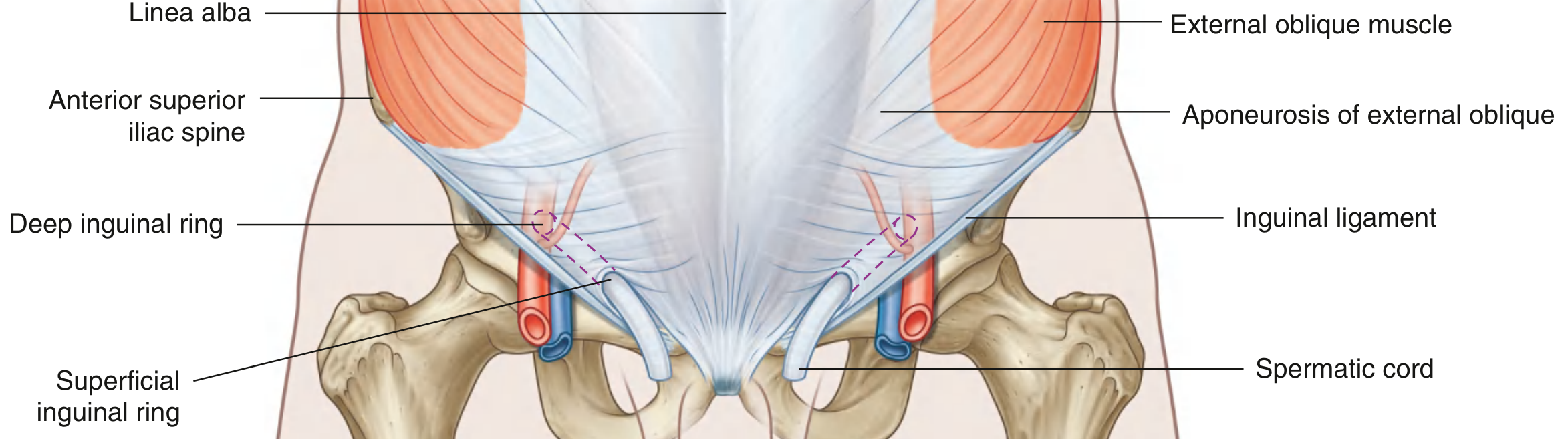
LOCATION
- Lower part of anterior abdominal wall, just above the medial half of the inguinal ligament
- Runs obliquely downward and medially for 4 cm
- Extends from Deep inguinal ring → Superficial inguinal ring
OPENINGS - The Two Rings
Deep (Internal) Inguinal Ring:
- Position: Midpoint of inguinal ligament (midway between ASIS and pubic symphysis), 1.25 cm above the inguinal ligament
- What it is: An opening in the transversalis fascia
- Landmark: Immediately lateral to the inferior epigastric vessels
- Memory: "Deep = lateral, Superficial = medial" (the canal runs obliquely inward)
Superficial (External) Inguinal Ring:
- Position: Just above and lateral to the pubic tubercle
- What it is: A triangular opening in the aponeurosis of external oblique
- Medial crus attaches to pubic symphysis, lateral crus to pubic tubercle
WALLS - "MALT" Mnemonic
| Wall | Structure |
|---|---|
| Anterior | Aponeurosis of External Oblique (whole length) + Internal Oblique (lateral 1/3) |
| Posterior | Transversalis fascia (whole length) + Conjoint tendon (medial 1/3) |
| Roof (Superior) | Arched fibres of Internal oblique + Transversus abdominis |
| Floor (Inferior) | Inguinal ligament + Lacunar ligament (medially) |
- Medial = Conjoint tendon (posterior)
- Anterior = External oblique aponeurosis
- Lateral = Internal oblique + Transversalis fascia
- Top = Internal oblique arch
CONTENTS
| In MALES | In FEMALES |
|---|---|
| Spermatic cord (main content) | Round ligament of uterus (main content) |
| Ilioinguinal nerve | Ilioinguinal nerve |
| Genital branch of genitofemoral nerve | Genital branch of genitofemoral nerve |
Spermatic Cord Contents (3+3+3 rule):
- 3 arteries: Testicular artery + Artery to vas deferens + Cremasteric artery
- 3 nerves: Genital branch of genitofemoral + Sympathetic fibres + Ilioinguinal nerve (outside cord, travels alongside)
- 3 other structures: Vas deferens + Pampiniform plexus of veins + Lymphatics
Coverings of Spermatic Cord (match the layers of abdominal wall):
- External spermatic fascia (from external oblique aponeurosis)
- Cremasteric fascia + Cremaster muscle (from internal oblique)
- Internal spermatic fascia (from transversalis fascia)
MECHANISM OF INTACT INGUINAL CANAL (Most Important for 15 marks!)
- During straining/coughing/lifting: intra-abdominal pressure rises
- Internal oblique and transversus contract → Roof of canal descends (like a shutter)
- Roof meets the floor (inguinal ligament) → Canal is obliterated = closed
- The oblique direction of the canal (inside-outside) means pressure from behind pushes the anterior wall against the posterior wall (like a flap valve)
- Conjoint tendon reinforces the posterior wall medially (guards Hesselbach's triangle)
WHY INDIRECT HERNIA IS MORE COMMON THAN DIRECT:
- Indirect: Goes through DEEP ring → follows the spermatic cord → pushes through SUPERFICIAL ring. Congenital predisposition (patent processus vaginalis).
- Direct: Pushes straight through Hesselbach's triangle (weakness in posterior wall), does NOT go through deep ring.
- Males have 8x more inguinal hernias because the canal is longer and larger.
APPLIED ANATOMY (Clinical Gold)
- Indirect inguinal hernia: Through deep ring → through canal → through superficial ring (controlled by pressure over deep ring)
- Direct inguinal hernia: Through Hesselbach's triangle (not controlled by pressure over deep ring)
- Inguinal orchidopexy: Bring undescended testis down through the inguinal canal to the scrotum
- Ilioinguinal nerve block: Anesthesia for groin surgery
- Laparoscopic hernia repair (TEP/TAPP): Must know all 3 layers and 2 rings
TOPIC 2 - STOMACH ⭐⭐⭐⭐⭐
The Story: "The Food Mixer"
LOCATION
- Lies in the epigastric, umbilical, and left hypochondriac regions
- Extends from the cardiac orifice (T11) to the pylorus (L1)
EXTERNAL FEATURES
Parts (C-Part-B-Py = "Come Play Before Pylorus"):
- Cardia - surrounds cardiac orifice
- Fundus - dome above the cardiac orifice (contains swallowed air - seen on X-ray as "gastric bubble" under left dome of diaphragm)
- Body - the largest part
- Pyloric antrum → Pyloric canal → Pylorus
Two Curvatures:
- Lesser curvature (right/medial border) - shorter; the hepatogastric ligament attaches here; lesser omentum attaches here
- Greater curvature (left/lateral border) - longer; greater omentum attaches here; gives attachment to gastrosplenic ligament
Two Surfaces: Anterior and Posterior
INTERNAL FEATURES
- Rugae (gastric folds): Longitudinal folds of mucosa and submucosa that allow distension
- Gastric pits (foveolae): Where gastric glands open
- Pyloric sphincter: Thickened circular muscle; palpable as a firm ring during surgery; controls gastric emptying
- Magenstrasse (gastric canal): A channel along the lesser curvature that rapidly conducts liquids to the pylorus
- Blood supply determines mucosal zones
RELATIONS
Anterior surface:
- Diaphragm (left part)
- Anterior abdominal wall (lower part of body/antrum - this is why stomach can be reached without displacing bowel in gastrostomy)
- Left lobe of liver (upper part)
Posterior surface (= the "stomach bed"):
- Pancreas (most important - connected via lesser sac)
- Left Kidney
- Left Adrenal
- Spleen
- Splenic artery (runs along upper border of pancreas)
- Transverse mesocolon/colon
- Diaphragm (left part)
VASCULAR SUPPLY - The "CROW" System
| Artery | Source | Supplies |
|---|---|---|
| Left gastric A. | Directly from coeliac trunk | Lesser curvature (left side) |
| Right gastric A. | From hepatic artery proper | Lesser curvature (right side) |
| Right gastro-omental (gastroepiploic) A. | From gastroduodenal artery (from hepatic) | Greater curvature (right side) |
| Left gastro-omental A. | From splenic artery | Greater curvature (left side) |
| Short gastric arteries (x5-7) | From splenic artery | Fundus |
Memory: Lesser curvature = Left + Right GASTRIC arteries. Greater curvature = Left + Right GASTRO-OMENTAL arteries.
VENOUS DRAINAGE
- Left and right gastric veins → Portal vein
- Left gastro-omental and short gastric veins → Splenic vein → Portal vein
- Right gastro-omental → Superior mesenteric vein → Portal vein
- All drain into the PORTAL SYSTEM (important in portal hypertension)
NERVE SUPPLY
- Parasympathetic (vagus): Anterior vagal trunk (L vagus) + Posterior vagal trunk (R vagus) enter at oesophageal hiatus
- Sympathetic: T6-T10 via coeliac plexus; causes DECREASED motility and secretion; carries pain fibres (referred to epigastrium)
LYMPHATIC DRAINAGE
- 4 groups: All drain to the coeliac nodes ultimately
- Lesser curvature → Left gastric nodes → Coeliac nodes
- Right greater curvature → Right gastro-omental nodes → Pyloric nodes → Coeliac
- Left greater curvature → Pancreaticosplenic nodes → Coeliac
- Fundus → Left gastric + Pancreaticosplenic nodes
APPLIED ANATOMY
- Peptic ulcer: 90% duodenal (DU > GU). DU = anterior wall perforation → peritonitis (Anterior perforates, Posterior erodes), Posterior DU = erodes gastroduodenal artery → haematemesis
- Gastric cancer: Spreads along lymphatics; Virchow's node (left supraclavicular) = distant spread via thoracic duct
- Gastrectomy: Billroth I (partial), Billroth II (subtotal) - know complications
- Vagotomy for PUD: Highly selective vagotomy divides only branches to acid-secreting fundus/body
TOPIC 3 - LIVER ⭐⭐⭐⭐⭐
The Story: "The Body's Chemical Factory"
LOCATION
- Right hypochondriac + Epigastric + Small part of left hypochondriac regions
- Upper surface: Under the diaphragm (right dome)
- Surface marking: Right side - from 5th rib in midclavicular line; Left side - to 5th intercostal space in midclavicular line
EXTERNAL FEATURES
Two Surfaces:
- Diaphragmatic surface (superior + anterior + posterior): Smooth, convex, covered by peritoneum except bare area (where liver is stuck directly to diaphragm, no peritoneum)
- Visceral surface (inferior/posterior): Has H-shaped arrangement of fissures and fossae that divides liver into lobes
The H-shaped Pattern on Visceral Surface:
Left limb of H: Right limb of H:
- Left sagittal fissure - Fossa for gallbladder (right, anterior)
(Falciform + Round lig. - Groove for IVC (right, posterior)
anteriorly + Ligamentum
venosum posteriorly)
Crossbar of H = PORTA HEPATIS (liver hilum)
Contains: Portal vein (posterior), Hepatic artery proper (left), Bile duct (right)
Memory: "Portal Bill" = Portal vein (back), Bile duct (right front), arterY (left front)
LOBES:
LIGAMENTS
- Falciform ligament: Connects liver to anterior abdominal wall; free edge = ligamentum teres (round ligament) = obliterated left umbilical vein; separates left and right lobes anteriorly
- Coronary ligament: Attaches posterior diaphragmatic surface to the diaphragm; its edges enclose the bare area (no peritoneum here)
- Left and right triangular ligaments: Lateral extremities of coronary ligament
- Lesser omentum: Hepatogastric + Hepatoduodenal ligaments (right free edge = epiploic foramen/Winslow's foramen boundary)
RELATIONS
- Above: Diaphragm (which separates liver from right lung, heart, pericardium)
- Below (visceral surface):
- Right lobe: Right kidney + right adrenal, hepatic flexure of colon, duodenum (second part)
- Left lobe: Stomach, oesophagus
- Quadrate lobe: Pylorus
- Caudate lobe: IVC (grooved), lesser omentum (separates it from stomach)
VASCULAR SUPPLY - The "Double Inlet"
| Source | Volume | Contents | Enters via |
|---|---|---|---|
| Portal vein | 75% of blood | Nutrient-rich blood from gut | Porta hepatis (posterior) |
| Hepatic artery proper | 25% of blood | Oxygenated blood | Porta hepatis (left side of bile duct) |
APPLIED ANATOMY
- Liver biopsy: Right intercostal route (9th-10th ICS midaxillary line) during expiration
- Hepatomegaly: Palpable below right costal margin; moves with respiration (attached to diaphragm)
- Porta hepatis: "Portal Bill" - in hepatoduodenal ligament - left = artery, right = bile duct, behind = portal vein
- Pringle's manoeuvre: Compress hepatoduodenal ligament (porta hepatis) to control haemorrhage
- Cirrhosis → Portal hypertension: Increased resistance in portal vein → varices + splenomegaly + ascites
TOPIC 4 - PORTAL VEIN ⭐⭐⭐
The Story: "The Nutrient Highway to the Liver"
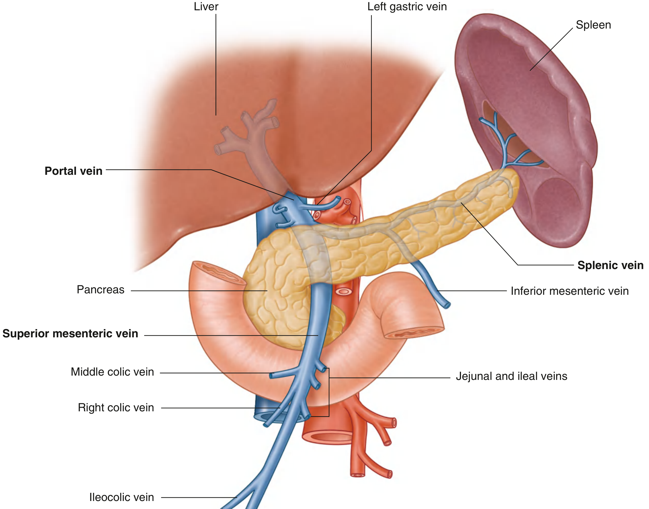
FORMATION
- Portal vein = Superior Mesenteric Vein (SMV) + Splenic Vein
- Formed BEHIND THE NECK OF THE PANCREAS at the level of L2
- Length: ~8 cm
TRIBUTARIES (Direct tributaries to portal vein itself):
- Right and Left gastric veins (drain lesser curvature + lower oesophagus)
- Cystic veins (from gallbladder)
- Para-umbilical veins (connect to anterior abdominal wall veins - dilate in portal hypertension = Caput medusae)
- Inferior mesenteric vein (usually into splenic vein)
- All gut veins (jejunal, ileal, right/left colic, middle colic, ileocolic)
COURSE AND RELATIONS
- Formed at L2 (neck of pancreas)
- Ascends behind the first part of duodenum
- Enters the free edge (right margin) of the lesser omentum
- In lesser omentum: Portal vein is POSTERIOR, Bile duct is to the RIGHT, Hepatic artery is to the LEFT (memory: "Portal-Bill-Art" back to front)
- Enters the liver at the porta hepatis - divides into right and left branches
PORTOSYSTEMIC ANASTOMOSES - THE MOST IMPORTANT PART
| Site | Portal tributaries | Systemic veins | Clinical result |
|---|---|---|---|
| Lower oesophagus (Most important!) | Left gastric vein (portal) | Azygos vein (systemic) | Oesophageal varices → haematemesis |
| Umbilicus | Para-umbilical veins (portal) | Superficial epigastric veins | Caput medusae (dilated veins radiating from umbilicus) |
| Anal canal (lower rectum) | Superior rectal vein (portal) | Middle/inferior rectal veins | Anorectal varices (NOT haemorrhoids - different thing!) |
| Retroperitoneum | Colic/renal veins (portal) | Retroperitoneal systemic veins | Retroperitoneal varices |
| Bare area of liver | Small hepatic tributaries | Diaphragmatic veins | Collaterals in cirrhosis |
- Oesophagus (left gastric ↔ azygos)
- Umbilicus (para-umbilical ↔ superficial epigastric)
- Rectum/Anal canal (superior rectal ↔ middle/inferior rectal)
- Area (bare area of liver)
APPLIED ANATOMY
- Portal hypertension: Cirrhosis (90% cause) → back pressure → all 4 portosystemic anastomoses dilate
- Oesophageal varices (most dangerous) → rupture → massive haematemesis → 30% mortality/bleed
- Splenomegaly (spleen enlarges due to backpressure) → hypersplenism → pancytopenia
- Ascites = portal hypertension + hypoalbuminaemia + secondary hyperaldosteronism
- TIPS (Transjugular Intrahepatic Portosystemic Shunt): Radiological shunt to decompress portal system
TOPIC 5 - PANCREAS ⭐⭐⭐
The Story: "The Hidden Gland Behind Everything"
LOCATION
- Retroperitoneal (secondarily retroperitoneal), lies at L1-L2 level
- Head: In the C-loop of duodenum (to the right of the vertebral column)
- Neck: Overlies the portal vein (SMV + Splenic vein join here!)
- Body: Crosses L1, lies behind lesser sac (stomach in front)
- Tail: Reaches the hilum of spleen (held in splenorenal ligament)
PARTS AND RELATIONS
HEAD (in C-loop of duodenum):
- Anterior: Transverse colon, stomach (via lesser sac)
- Posterior: IVC, right renal vessels, bile duct (grooves the posterior head)
- Medial: Portal vein (formed here at neck)
- The uncinate process hooks around the superior mesenteric vessels
NECK:
- Anterior: Pylorus of stomach
- Posterior: Portal vein formed here (SMV + Splenic vein union)
BODY:
- Anterior: Lesser sac (stomach beyond it)
- Posterior: Aorta, SMA origin, left kidney and adrenal, splenic vein (runs in a groove on the posterior surface)
- Superior border: Splenic artery (tortuous, runs along upper border)
- Inferior border: Transverse mesocolon attaches here
TAIL:
- In splenorenal ligament → contacts spleen
- Splenic artery + vein here
DUCTS
- Main pancreatic duct (of Wirsung): Runs the full length, joins the common bile duct at the ampulla of Vater → drains into 2nd part of duodenum at the major duodenal papilla
- Accessory pancreatic duct (of Santorini): Drains upper head → minor duodenal papilla (2cm above major papilla)
BLOOD SUPPLY
- Head: Superior pancreaticoduodenal artery (from gastroduodenal/hepatic) + Inferior pancreaticoduodenal artery (from SMA) → This anastomosis is clinically important - the SMA/hepatic artery arcades around the head
- Body and tail: Splenic artery (multiple pancreatic branches)
APPLIED ANATOMY
- Acute pancreatitis: Gallstones (block ampulla of Vater) + Alcohol. Amylase/lipase rises. "GET SMASHED" causes. Grey-Turner sign (flank bruising), Cullen's sign (periumbilical bruising) = retroperitoneal haemorrhage
- Carcinoma of head of pancreas: Obstructs bile duct → painless progressive jaundice (Courvoisier's law: palpable non-tender gallbladder = carcinoma not stones). "Head of pancreas carcinoma is the cancer surgeon dreads most" (usually inoperable by detection)
- Whipple's procedure (pancreaticoduodenectomy): For head carcinoma - removes head + duodenum + gallbladder + part of stomach
- Diabetes mellitus: Body/tail carcinoma → destroys islets of Langerhans
TOPIC 6 - KIDNEY ⭐⭐⭐⭐⭐
The Story: "The Body's Filter Plant - Deep in the Back"
LOCATION
- Retroperitoneal organs, on the posterior abdominal wall
- Right: T12-L3 (lower - displaced by liver)
- Left: T11-L2 (higher)
- Each kidney is ~11 cm long, 6 cm wide, 3 cm thick, weighing ~150g
EXTERNAL FEATURES
- Hilum: Medial border - where vessels, ureter, lymphatics enter/leave
- Order of structures at hilum (front to back): Renal vein → Renal artery → Ureter (pelvis)
- Memory: "VAU" - Vein, Artery, Ureter front to back (the ureter is the most posterior)
- Coverings (inside out): Fibrous capsule → Perinephric fat → Renal fascia (Gerota's fascia) → Paranephric fat → Peritoneum (anterior only)
INTERNAL FEATURES
- Cortex: Outer layer - contains glomeruli and convoluted tubules (where filtration happens)
- Medulla: Inner layer - renal pyramids (8-18) → tips = renal papillae → drain into minor calices → major calices → renal pelvis → ureter
- Columns of Bertin: Cortical tissue between pyramids
- Sinus: The fatty space inside the kidney containing the collecting system and vessels
RELATIONS
ANTERIOR RELATIONS (Most asked):
- Suprarenal gland (superiomedial)
- Liver (upper 2/3 - the big neighbour that pushes it down)
- 2nd part of duodenum (medial, no peritoneum between - directly applied)
- Right colic (hepatic) flexure (lower pole)
- Jejunum (small portion)
- Suprarenal gland (superomedial)
- Spleen (upper lateral)
- Stomach (upper medial)
- Tail of pancreas + Splenic vessels (directly applied, crosses the hilum anteriorly)
- Left colic (splenic) flexure (lower pole)
- Jejunum
POSTERIOR RELATIONS (same for BOTH kidneys):
Why diaphragm matters posteriorly: In renal surgery → if you open too high (above 12th rib) → you can enter the pleural cavity = pneumothorax
VASCULAR SUPPLY
- Renal arteries: Branch from aorta at L1 (just below SMA). Right renal artery is LONGER (crosses behind IVC, right renal vein, and head of pancreas)
- Renal veins: Left renal vein is LONGER (crosses anterior to aorta, receives left gonadal vein and left suprarenal vein before entering IVC)
- Segmental arteries: 5 segments (superior, anterosuperior, anteroinferior, inferior, posterior) - end arteries, no anastomosis = infarction if blocked
APPLIED ANATOMY
- Renal calculi: Form in calices/pelvis. Pain (renal colic) radiates from loin to groin (follows ureter and ilioinguinal nerve distribution). "Loin to Groin" = ureteric colic
- Nephrectomy: Posterior approach - 12th rib removal common. Beware subcostal nerve (T12) injury → skin anaesthesia of anterolateral abdominal wall
- Renal transplant: Placed in the ILIAC FOSSA (extraperitoneal), renal artery anastomosed to internal/external iliac artery; renal vein to external iliac vein; ureter to bladder
- Horseshoe kidney: Lower poles fused across midline, inferior mesenteric artery passes between = tethered at IMA
- Renal tumour (Wilms' = nephroblastoma): Most common solid abdominal tumour in children
TOPIC 7 - URINARY BLADDER ⭐⭐⭐
The Story: "The Expandable Bag in the Pelvis"
LOCATION
- In the anterior part of the pelvis, behind the pubic symphysis
- When empty: entirely in pelvis; When full: rises into abdominal cavity
PARTS
- Apex: Points forward toward pubic symphysis; connected to anterior abdominal wall by median umbilical ligament (= obliterated urachus)
- Base (fundus): Posterior surface - triangular; faces backward and downward
- Body: Between apex and base
- Neck: Most inferior and fixed part; where urethra begins
INTERNAL FEATURES
- Trigone: A triangular smooth area on the internal surface of the base
- 3 angles: Two ureteric orifices (posterolateral) + Internal urethral orifice (anterior inferior)
- The mucosa over the trigone is SMOOTH and firmly adherent (unlike rest of bladder which is folded in when empty)
- Interureteric fold/bar = raised ridge connecting the two ureteric openings
- Clinical: The trigone is where bladder tumours and bladder stones commonly occur
RELATIONS
MALE:
- Superior: Loops of small intestine + sigmoid colon
- Posterior: Rectovesical pouch (peritoneum-lined) → then Seminal vesicles + Ampullae of vas deferens + Rectum
- Inferior: Prostate gland (directly below neck of bladder)
- Anterior: Retropubic space (cave of Retzius, filled with fat) + Pubic symphysis
FEMALE:
- Superior: Uterus (anteflexed uterus lies on top of bladder)
- Posterior: Vesicouterine pouch → Cervix → Upper vagina (directly related - important in pelvic surgery)
- Inferior: Pelvic floor (levator ani)
VASCULAR SUPPLY
- Superior vesical artery (from superior vesical = patent part of umbilical artery): Supplies dome
- Inferior vesical artery (males only, from internal iliac): Supplies base, fundus, seminal vesicles, prostate
- Vaginal artery (females) = equivalent to inferior vesical
NERVE SUPPLY
- Parasympathetic (S2-S4) = pelvic splanchnic nerves = nervi erigentes: MOTOR to detrusor muscle (contraction → voiding) + INHIBITORY to internal sphincter
- Sympathetic (L1-L2) via hypogastric plexus: Inhibitory to detrusor + MOTOR to internal sphincter (prevents ejaculation into bladder)
- Pudendal nerve (S2-S4): Somatic motor to external sphincter (voluntary control)
"Parasympathetic = PEE" (S2-S4 → bladder contracts → urine flows) "Sympathetic = STOP" (L1-L2 → bladder stores, sphincter closes)
APPLIED ANATOMY
- Suprapubic cystostomy: Catheter inserted above pubic symphysis when urethra blocked; safe because bladder rises above pubic symphysis when full (no peritoneum in the way anteriorly)
- Bladder cancer: Most common is TCC (transitional cell carcinoma); painless haematuria = until proven otherwise bladder/renal tumour
- Vesicoureteric junction (VUJ): Where ureter enters bladder wall obliquely; acts as a valve; site where renal stones commonly get stuck (other sites: PUJ, pelvic brim)
TOPIC 8 - UTERUS ⭐⭐⭐⭐⭐
The Story: "The House That Holds the Baby"
LOCATION
- Pelvic cavity, between bladder (anterior) and rectum (posterior)
- Normal position: Anteverted + Anteflexed
- Anteversion: Angle between long axis of uterus and vagina (~90°, forward leaning)
- Anteflexion: Angle between body and cervix (~125°, forward bending at internal os)
EXTERNAL FEATURES
- Fundus: Rounded top (above the fallopian tube openings)
- Body: Main mass
- Isthmus: Short constriction between body and cervix (~1 cm)
- Cervix: Lower cylindrical part
- Supravaginal part (above vagina)
- Vaginal part (portio vaginalis - surrounded by vaginal fornices)
INTERNAL FEATURES
- Uterine cavity: Triangular, flattened antero-posteriorly
- Endometrium: Inner lining - undergoes cyclical changes (menstrual cycle)
- Myometrium: Thick smooth muscle (3 layers: outer longitudinal, middle oblique/spiral, inner longitudinal) - spiral arrangement helps expulsion of baby
- Internal os: Junction of body and cervix (important in incompetent cervix)
- External os: Opening of cervix into vagina
- Nulliparous: circular
- Multiparous: transverse slit
RELATIONS
| Direction | Relation |
|---|---|
| Anterior | Vesicouterine pouch → Bladder (body rests on bladder when anteflexed) |
| Posterior | Recto-uterine pouch (Pouch of Douglas - deepest part of peritoneal cavity in female) → Rectum |
| Lateral | Broad ligament; Uterine artery (crosses over ureter - CRITICALLY IMPORTANT); Ureter (2 cm lateral to cervix) |
| Superior | Loops of small intestine (on fundus) |
| Inferior (cervix) | Vagina |
THE MOST IMPORTANT CLINICAL RELATION: The ureter passes 2 cm lateral to the cervix, running UNDER the uterine artery ("water under the bridge"). During hysterectomy, the ureter is at risk when the surgeon ligates the uterine artery. This is the #1 surgical complication in hysterectomy.Memory: "Ureter is water. Uterine artery is bridge. Water passes UNDER the bridge."
SUPPORTS OF THE UTERUS (What holds it in place)
Primary (most important) Supports:
- Transverse (cardinal/Mackenrodt's) ligament: Most important! Thickening of parametrium from cervix to lateral pelvic wall. Prevents uterus prolapsing
- Uterosacral ligament: From cervix backward to sacrum; keeps cervix in position
- Pubocervical ligament: From cervix forward to pubis
Secondary Supports:
- Pelvic floor (levator ani): The "hammock" - most important passive support
- Broad ligament: Wing of peritoneum on each side; contains uterine tube (in free edge), ovary, round ligament, uterine vessels and ureter
Non-Supports (do NOT support against prolapse):
- Round ligament: Goes through inguinal canal to labia majora; maintains anteversion but NOT against prolapse
- Ovarian ligament: Connects ovary to uterus
VASCULAR SUPPLY
- Uterine artery (from internal iliac artery): Main supply. Runs in base of broad ligament, crosses ABOVE the ureter 2 cm lateral to cervix
- Ovarian artery (from aorta at L2): Supplies fundus and tubes via ovarian ligament
- Anastomosis between both is extensive
NERVE SUPPLY
- Sympathetic (T10-L1): Via hypogastric plexus - pain from uterine body referred to T10 dermatome (umbilicus) = labour pains felt around navel
- Parasympathetic (S2-S4): Pelvic splanchnic nerves
- Cervix: Pelvic plexus (S2-S4); less pain from cervix (which is why IUD insertion is tolerable)
LYMPHATIC DRAINAGE
- Fundus: Para-aortic nodes (at L2 - same as ovary, because uterine fundus developed from same embryological origin)
- Body: Internal iliac + external iliac nodes
- Cervix: Internal + external iliac nodes + obturator nodes
APPLIED ANATOMY
- Uterine prolapse: Failure of transverse cardinal ligament + pelvic floor → uterus descends into vagina (1st degree = descent, 2nd = cervix at introitus, 3rd = procidentia = whole uterus outside)
- Ectopic pregnancy: Implantation in fallopian tube (most common); rupture → haemoperitoneum collects in Pouch of Douglas → posterior fornix tenderness (cul-de-sac)
- Cervical cancer: Spread to parametrium + pelvic sidewall = parametrial invasion (inoperable). Pap smear screens for this
- Hysterectomy: Risk = ureter injury (2 cm lateral to cervix)
- Endometriosis: Ectopic endometrium - most common site = Pouch of Douglas + ovary
TOPIC 9 - TESTIS ⭐⭐⭐⭐⭐
The Story: "The Organ That Made the Inguinal Canal"
- Testicular blood supply = testicular artery from AORTA at L2 (not from iliac - never forget this!)
- Testicular lymphatics = para-aortic nodes at L2 (not inguinal nodes!)
- Testicular nerve supply = T10 (referred pain to umbilicus, not to scrotum)
LOCATION
- In the scrotum, suspended by the spermatic cord
- Left testis is usually slightly LOWER than the right
- Temperature = 2-3°C BELOW body temperature (needed for spermatogenesis)
EXTERNAL FEATURES
- Tunica vaginalis: Double serous layer (visceral + parietal); derived from processus vaginalis (peritoneum); surrounds the testis anteriorly and laterally; the potential space between layers = site of hydrocele
- Tunica albuginea: Dense fibrous capsule
- Epididymis: Lies on the posterolateral surface; has head (top), body, tail (bottom); the tail continues as the vas deferens
- Mediastinum testis: Posterior thickening of tunica albuginea where vessels and ducts enter
INTERNAL FEATURES
- Seminiferous tubules: ~900 coiled tubules where sperm is produced
- Sertoli cells: "Nurse cells" - blood-testis barrier; support sperm development; secrete inhibin
- Leydig (interstitial) cells: Between tubules; produce testosterone (under LH stimulation)
- Rete testis: Network of channels in mediastinum testis
- Efferent ductules (x15-20): From rete testis → head of epididymis → epididymis → vas deferens
VASCULAR SUPPLY
- Testicular artery: From AORTA at L2 (directly, not from internal iliac!) - this reflects testicular origin near kidney
- Cremasteric artery: From inferior epigastric
- Artery to vas deferens: From inferior vesical artery
- Pampiniform plexus of veins: Numerous veins that surround the testicular artery like a "net" in the spermatic cord; converge to form testicular vein
- Right testicular vein → directly into IVC
- Left testicular vein → into LEFT RENAL VEIN (not directly to IVC)
- Memory: "Left is longer and drains into left renal vein" - this is why left-sided varicocele (90%) is more common
APPLIED ANATOMY
- Varicocele: Dilated pampiniform plexus; "bag of worms" on left side; left-sided because left testicular vein drains into left renal vein at 90° angle (higher pressure). Can cause subfertility (heated blood backflows, raises testicular temp)
- Testicular torsion: Twisting of spermatic cord; "bell-clapper" deformity (high insertion of tunica vaginalis). Presents with sudden severe scrotal pain. Emergency surgery within 6 hours or testis lost
- Testicular tumours: Lymph drainage to para-aortic nodes (NOT inguinal). If scrotal skin invaded → inguinal nodes. Germ cell tumours (seminoma = most common, teratoma) = peak 20-35 years
- Cryptorchidism (undescended testis): 20x increased risk of testicular cancer; reduced fertility; treat by age 1-2 years with orchidopexy
TOPIC 10 - RECTUM ⭐⭐⭐⭐⭐
The Story: "The Final Stretch Before Exit"
LOCATION
- Begins at the rectosigmoid junction (S3 level), follows the sacral curvature
- Ends at the anorectal junction (where it bends sharply forward = anorectal flexure/perineal flexure)
- 12-15 cm long
EXTERNAL FEATURES
- No mesentery (retroperitoneal lower 2/3; extraperitoneal)
- No haustrations, taeniae coli, or appendices epiploicae (these disappear at rectosigmoid junction)
- 3 lateral flexures (curves): Right, Left, Right (like an S-shape when viewed from front)
- Peritoneal coverage:
- Upper 1/3: Peritoneum covers front AND sides
- Middle 1/3: Peritoneum covers front only (here the peritoneum reflects → Pouch of Douglas in females / Rectovesical pouch in males)
- Lower 1/3: No peritoneum (extraperitoneal)
INTERNAL FEATURES
- 3 transverse rectal folds (Houston's valves): Correspond to the 3 lateral flexures
- Upper fold: on left wall
- Middle fold (most prominent): on right wall - corresponds to level of anterior peritoneal reflection
- Lower fold: on left wall
- Ampulla: Dilated lower part where faeces are stored
- Mucosa: Columnar epithelium (like colon)
RELATIONS
Anterior:
Posterior:
Lateral:
- Upper third: Peritoneum (pararectal fossae)
- Lower two-thirds: Levator ani (pelvic floor), Coccygeus, Pelvic fascia, Pelvic lymph nodes
VASCULAR SUPPLY
- Superior rectal artery (from inferior mesenteric artery): Main supply; anastomoses with middle rectal at portosystemic junction
- Middle rectal artery (from internal iliac): Middle and lower rectum
- Inferior rectal artery (from pudendal artery from internal iliac): Lower rectum/anal canal
- The anastomosis between superior (portal system) and middle/inferior (systemic) = portosystemic anastomosis site
APPLIED ANATOMY
- Rectal examination (PR): Can feel prostate anteriorly (males), cervix anteriorly (females), sacrum posteriorly, ischiorectal fossa laterally
- Rectal carcinoma: Spread along lymphatics → superior rectal nodes → IMA nodes. Below peritoneal reflection → lateral pelvic lymph nodes → internal iliac. Circumferential resection margin matters
- Abdominoperineal resection (APR) vs Anterior resection: Low tumour (<5 cm from anal verge) = APR (permanent colostomy). High tumour = anterior resection (anastomosis)
- Hartmann's operation: Resect sigmoid/rectum, close rectal stump, end colostomy (for obstructed/perforated cancer)
TOPIC 11 - ANAL CANAL ⭐⭐⭐⭐⭐
The Story: "The Exit Gate with Two Different Zones"
LOCATION
- From the anorectal junction (where the perineal flexure is) to the anal verge (where skin begins)
- 4 cm long, passes through the pelvic floor and perineum
- Surrounded by two sphincters
ZONES AND THE PECTINATE (DENTATE) LINE
| Feature | ABOVE Pectinate Line | BELOW Pectinate Line |
|---|---|---|
| Embryology | Endoderm (hindgut) | Ectoderm (proctodeum) |
| Epithelium | Columnar (glandular) | Stratified squamous |
| Venous drainage | Superior rectal vein → Portal system | Middle/inferior rectal veins → Systemic |
| Lymphatics | Internal iliac nodes | Superficial inguinal nodes |
| Nerve supply (sensory) | Autonomic (visceral - insensitive to pain, only sensitive to distension) | Somatic (inferior rectal nerve - sensitive to pain, temperature, touch) |
| Arterial supply | Superior rectal artery | Inferior rectal artery |
"The Pectinate Line divides the PAINLESS zone (above) from the PAINFUL zone (below)"
- Haemorrhoids ABOVE the line = internal haemorrhoids = PAINLESS (but bleed bright red)
- Haemorrhoids BELOW the line = external haemorrhoids = PAINFUL
- Injection sclerotherapy only works ABOVE the line (no pain sensation)
INTERNAL FEATURES
- Anal columns (of Morgagni): 8-10 longitudinal folds of mucosa in the upper anal canal; each column contains a terminal branch of the superior rectal artery + vein (= the internal haemorrhoidal cushions)
- Anal valves: Crescentic folds connecting the bases of anal columns
- Anal sinuses: Pockets between anal valves and wall (anal glands open here → source of perianal abscesses and fistulae)
- Transitional zone (ATZ): 6-12 mm zone above pectinate line; has mixed epithelium; important in surgery (where to cut in ileoanal pouches)
SPHINCTERS
Internal Anal Sphincter (IAS):
- Involuntary smooth muscle
- Continuation of circular muscle of rectum
- Always contracted (maintains 85% of resting anal tone)
- Nerve: Sympathetic (hypogastric plexus - CONTRACTS sphincter = holds faeces) + Parasympathetic (RELAXES for defecation)
- Rectoanal inhibitory reflex (RAIR): When rectum distends → internal sphincter RELAXES (Auerbach's plexus). Absent in Hirschsprung's disease
External Anal Sphincter (EAS):
- Voluntary skeletal muscle
- Continuous with puborectalis (part of levator ani)
- Nerve: Inferior rectal branch of pudendal nerve (S2-S3) + Perineal branch of S4
- Puborectalis: Creates the anorectal angle (~90°) = most important factor in maintaining continence
ANORECTAL ANGLE
APPLIED ANATOMY
- Haemorrhoids: 3 primary positions - 3, 7, 11 o'clock (patient in lithotomy). Internal = above pectinate line = painless bleed. External = below = painful
- Fistula-in-ano: Goodsall's rule: anterior external opening → straight tract to pectinate line; posterior external opening → curved tract to posterior midline. Treat with fistulotomy/fistulectomy (preserving sphincters)
- Anal carcinoma: SCC (squamous = BELOW pectinate line → inguinal node spread); Adenocarcinoma (ABOVE → internal iliac nodes). HIV-associated SCC is rising
- Hirschsprung's disease: Absence of ganglion cells (Auerbach's plexus) → no RAIR → internal sphincter cannot relax → bowel obstruction in neonate
- Perianal abscess/fistula: Infected anal glands (at pectinate line) → abscess in intersphincteric plane → tracks to ischiorectal fossa
MASTER STORY RECAP - How Everything Links:
- Coeliac axis (T12): Stomach, liver, spleen, pancreas (upper abdominal organs)
- SMA (L1): Small intestine, right colon, pancreatic head
- IMA (L3): Left colon, sigmoid, upper rectum
- Internal iliac: Bladder, uterus, rectum (pelvic organs)
- Aorta direct (L2): Kidneys, testis/ovary, suprarenal (retroperitoneal organs)
Create a visual concept map linking all 15 abdominal organs with their vascular/lymphatic drainage hierarchy
A detailed medical anatomy concept map poster showing 15 abdominal and pelvic organs with their vascular and lymphatic drainage hierarchy. Clean white background with color-coded sections. TOP SECTION - ARTERIAL SUPPLY TREE: Title: "ABDOMINAL ORGAN VASCULAR & LYMPHATIC HIERARCHY" Left column labeled "ARTERIAL SUPPLY" with a large red downward tree: - AORTA at top center - Branch 1: COELIAC AXIS (T12) in red box → splits to: Stomach, Liver, Spleen, Pancreas (head), Gallbladder - Branch 2: SMA (L1) in orange box → splits to: Small intestine, Right colon, Pancreas head, Duodenum - Branch 3: IMA (L3) in yellow box → splits to: Left colon, Sigmoid, Upper Rectum - Branch 4: INTERNAL ILIAC in pink box → splits to: Bladder, Uterus, Rectum (lower), Anal canal - Branch 5: DIRECT FROM AORTA (L2) in purple box → splits to: Kidney (renal artery), Testis/Ovary (gonadal artery), Suprarenal - Branch 6: INFERIOR EPIGASTRIC (from external iliac) in blue box → Inguinal canal structures RIGHT COLUMN - VENOUS DRAINAGE: "PORTAL SYSTEM (blue)" box containing: Stomach veins, Intestinal veins, Splenic vein, Pancreatic veins, Inferior mesenteric → ALL arrows pointing to PORTAL VEIN → LIVER → HEPATIC VEINS → IVC "SYSTEMIC" box: Kidneys → Renal veins → IVC; Testis left → Left renal vein → IVC; Testis right → direct to IVC MIDDLE SECTION - ORGAN MAP arranged anatomically in body silhouette outline: Row 1 (upper abdomen): Liver | Stomach | Spleen Row 2: Gallbladder | Pancreas | Left kidney | Right kidney Row 3: Duodenum | Portal vein | Suprarenal Row 4 (pelvis): Bladder | Uterus | Testis Row 5 (lower): Rectum | Anal canal | Inguinal canal Each organ in a rounded colored box connected by arrows to its arterial source and lymphatic destination. BOTTOM SECTION - LYMPHATIC DRAINAGE TABLE: Title: "LYMPHATIC DRAINAGE - FOLLOW THE ARTERY BACK" 4 columns: Column 1 "Organ" | Column 2 "Primary nodes" | Column 3 "Secondary nodes" | Column 4 "Final" - Stomach → Left gastric/coeliac nodes → Para-aortic → Thoracic duct - Liver → Hepatic nodes → Coeliac → Para-aortic - Kidney → Para-aortic (L1-L2) → → Thoracic duct - Testis → Para-aortic (L2) → → Thoracic duct (NOT inguinal!) - Uterus fundus → Para-aortic | body → iliac | cervix → obturator/iliac - Bladder → Internal + external iliac - Rectum upper → IMA nodes → para-aortic | lower → internal iliac - Anal canal above pectinate → internal iliac | below pectinate → INGUINAL KEY RULE BOX at bottom right: "GOLDEN RULE: Lymphatics follow the artery backwards to its origin. Testis drains to PARA-AORTIC not inguinal (because testicular artery comes from aorta at L2)" Color coding: Red = arteries, Blue = veins, Green = lymphatics, Yellow = organ boxes, Orange = portal system Clean medical textbook style, clear labels, professional anatomy poster layout
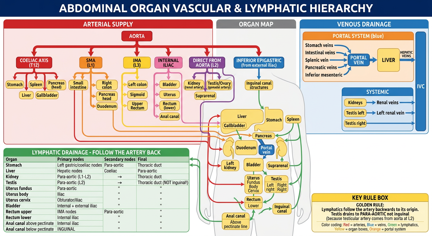
HOW TO READ THIS MAP
THE ONE GOLDEN RULE
"Lymphatics always follow the artery BACKWARDS to its origin." This single rule lets you derive lymphatic drainage for ANY organ without memorizing separately.
ARTERIAL SUPPLY - The 5 Sources
| Source | Level | Organs |
|---|---|---|
| Coeliac axis | T12 | Stomach, Liver, Spleen, Pancreas (body/tail), Gallbladder |
| SMA | L1 | Small intestine, right colon, pancreas head, duodenum |
| IMA | L3 | Left colon, sigmoid, upper rectum |
| Internal iliac | Pelvis | Bladder, Uterus, lower rectum, anal canal |
| Aorta direct | L2 | Kidneys (renal artery), Testis/Ovary (gonadal artery) |
VENOUS DRAINAGE - Portal vs Systemic Split
- Kidneys → Renal veins → IVC
- Right testis → IVC directly
- Left testis → Left renal vein → IVC (this is why left varicocele is more common - higher resistance angle)
LYMPHATIC DRAINAGE - Quick Reference
| Organ | Drains to | Key Exam Point |
|---|---|---|
| Stomach, Liver, Pancreas, Spleen | Coeliac → Para-aortic | Virchow's node = distant spread |
| Kidney | Para-aortic (L1-L2) | Follows renal artery origin |
| Testis | Para-aortic (L2) - NOT inguinal! | #1 exam trap - artery comes from aorta at L2 |
| Uterus fundus | Para-aortic | Follows gonadal/ovarian artery |
| Uterus body | Internal + External iliac | Middle territory |
| Cervix | Internal iliac + Obturator | Parametrial spread = inoperable cancer |
| Bladder | Internal + External iliac | |
| Rectum (upper) | IMA nodes → Para-aortic | Above peritoneal reflection |
| Rectum (lower) + Anal canal above pectinate | Internal iliac | Below peritoneal reflection |
| Anal canal BELOW pectinate line | Superficial inguinal | #2 exam trap - ectodermal origin = skin drainage |
THE 3 EXAM TRAPS TO NEVER MISS
-
Testis drains to para-aortic nodes (L2), NOT inguinal - testicular artery comes from the aorta, not the iliac. If scrotal skin is invaded by tumour, THEN inguinal nodes are involved.
-
Anal canal above pectinate = internal iliac; below pectinate = inguinal - the pectinate line is the embryological boundary where everything changes.
-
Left testicular vein → Left renal vein (NOT directly into IVC like the right) - this explains why left varicocele is 90% of cases.
Make a downloadable PDF study guide linking all abdominal topics to exam formats
mkdir -p /home/daytona/workspace/abdomen-study-guide && ls /home/daytona/workspace/
~/abdomen-study-guide/generate_pdf.py
from reportlab.lib.pagesizes import A4
from reportlab.lib import colors
from reportlab.lib.styles import getSampleStyleSheet, ParagraphStyle
from reportlab.lib.units import cm, mm
from reportlab.platypus import (
SimpleDocTemplate, Paragraph, Spacer, Table, TableStyle,
HRFlowable, PageBreak, KeepTogether
)
from reportlab.lib.enums import TA_CENTER, TA_LEFT, TA_JUSTIFY
from reportlab.platypus import BaseDocTemplate, Frame, PageTemplate
OUTPUT = "/home/daytona/workspace/abdomen-study-guide/Abdomen_Pelvis_Gold_Medal_Study_Guide.pdf"
# ── Colour palette ──────────────────────────────────────────────────────────
DARK_BLUE = colors.HexColor("#1a3a5c")
MID_BLUE = colors.HexColor("#2e6da4")
LIGHT_BLUE = colors.HexColor("#d6e8f7")
GOLD = colors.HexColor("#c8960c")
GOLD_LIGHT = colors.HexColor("#fff5d6")
RED = colors.HexColor("#b30000")
RED_LIGHT = colors.HexColor("#fde8e8")
GREEN = colors.HexColor("#1a6b2a")
GREEN_LIGHT = colors.HexColor("#e6f4ea")
PURPLE = colors.HexColor("#5b1a8c")
PURPLE_LIGHT= colors.HexColor("#f0e8f8")
ORANGE = colors.HexColor("#c45000")
ORANGE_LIGHT= colors.HexColor("#fff0e6")
GREY_LIGHT = colors.HexColor("#f5f5f5")
GREY_MID = colors.HexColor("#cccccc")
WHITE = colors.white
BLACK = colors.black
# ── Styles ───────────────────────────────────────────────────────────────────
styles = getSampleStyleSheet()
def S(name, **kw):
return ParagraphStyle(name, **kw)
cover_title = S("CoverTitle", fontSize=28, textColor=WHITE, alignment=TA_CENTER,
fontName="Helvetica-Bold", spaceAfter=8, leading=34)
cover_sub = S("CoverSub", fontSize=14, textColor=GOLD, alignment=TA_CENTER,
fontName="Helvetica-Bold", spaceAfter=6, leading=18)
cover_body = S("CoverBody", fontSize=11, textColor=WHITE, alignment=TA_CENTER,
fontName="Helvetica", spaceAfter=4, leading=16)
h1 = S("H1", fontSize=16, textColor=WHITE, fontName="Helvetica-Bold",
spaceAfter=4, spaceBefore=6, leading=20)
h2 = S("H2", fontSize=13, textColor=DARK_BLUE, fontName="Helvetica-Bold",
spaceAfter=3, spaceBefore=8, leading=16)
h3 = S("H3", fontSize=11, textColor=MID_BLUE, fontName="Helvetica-Bold",
spaceAfter=2, spaceBefore=5, leading=14)
body= S("Body", fontSize=9, textColor=BLACK, fontName="Helvetica",
spaceAfter=3, leading=13, alignment=TA_JUSTIFY)
bullet=S("Bullet",fontSize=9, textColor=BLACK, fontName="Helvetica",
spaceAfter=2, leading=13, leftIndent=12, bulletIndent=2)
small=S("Small",fontSize=8, textColor=colors.HexColor("#444444"),
fontName="Helvetica", spaceAfter=2, leading=11)
tip = S("Tip", fontSize=9, textColor=GREEN, fontName="Helvetica-Bold",
spaceAfter=2, leading=13, leftIndent=6)
warn = S("Warn",fontSize=9, textColor=RED, fontName="Helvetica-Bold",
spaceAfter=2, leading=13, leftIndent=6)
gold_rule=S("GoldRule",fontSize=10,textColor=GOLD,fontName="Helvetica-Bold",
spaceAfter=3,leading=14,leftIndent=4)
toc_item=S("TocItem",fontSize=10,textColor=DARK_BLUE,fontName="Helvetica",
spaceAfter=2,leading=14,leftIndent=12)
# ── Helper builders ───────────────────────────────────────────────────────────
def section_header(text, bg=DARK_BLUE, fg=WHITE):
data = [[Paragraph(text, h1)]]
t = Table(data, colWidths=[17*cm])
t.setStyle(TableStyle([
("BACKGROUND", (0,0), (-1,-1), bg),
("TOPPADDING", (0,0), (-1,-1), 6),
("BOTTOMPADDING",(0,0),(-1,-1), 6),
("LEFTPADDING", (0,0), (-1,-1), 10),
("ROUNDEDCORNERS",[4]),
]))
return t
def subsection_header(text, bg=LIGHT_BLUE):
data = [[Paragraph(text, h2)]]
t = Table(data, colWidths=[17*cm])
t.setStyle(TableStyle([
("BACKGROUND",(0,0),(-1,-1), bg),
("TOPPADDING",(0,0),(-1,-1),4),
("BOTTOMPADDING",(0,0),(-1,-1),4),
("LEFTPADDING",(0,0),(-1,-1),8),
("LINEBELOW",(0,0),(-1,-1),1.5,MID_BLUE),
]))
return t
def colored_box(content_rows, bg=GOLD_LIGHT, border=GOLD):
"""content_rows: list of Paragraph objects wrapped in single-cell rows"""
data = [[p] for p in content_rows]
t = Table(data, colWidths=[17*cm])
t.setStyle(TableStyle([
("BACKGROUND",(0,0),(-1,-1),bg),
("BOX",(0,0),(-1,-1),1.2,border),
("LEFTPADDING",(0,0),(-1,-1),8),
("RIGHTPADDING",(0,0),(-1,-1),8),
("TOPPADDING",(0,0),(0,0),6),
("BOTTOMPADDING",(0,-1),(-1,-1),6),
("TOPPADDING",(0,1),(-1,-1),2),
("BOTTOMPADDING",(0,0),(-1,-2),2),
]))
return t
def two_col_table(headers, rows, col1=8.5*cm, col2=8.5*cm,
hdr_bg=DARK_BLUE, hdr_fg=WHITE, alt_bg=GREY_LIGHT):
all_rows = [[Paragraph(h, S("th",fontSize=9,textColor=hdr_fg,
fontName="Helvetica-Bold",leading=12))
for h in headers]] + \
[[Paragraph(str(c), small) for c in r] for r in rows]
t = Table(all_rows, colWidths=[col1, col2])
ts = [
("BACKGROUND",(0,0),(-1,0),hdr_bg),
("TEXTCOLOR",(0,0),(-1,0),hdr_fg),
("FONTNAME",(0,0),(-1,0),"Helvetica-Bold"),
("FONTSIZE",(0,0),(-1,0),9),
("TOPPADDING",(0,0),(-1,-1),4),
("BOTTOMPADDING",(0,0),(-1,-1),4),
("LEFTPADDING",(0,0),(-1,-1),6),
("GRID",(0,0),(-1,-1),0.5,GREY_MID),
("ROWBACKGROUNDS",(0,1),(-1,-1),[WHITE, alt_bg]),
]
t.setStyle(TableStyle(ts))
return t
def three_col_table(headers, rows, widths=None, hdr_bg=DARK_BLUE):
widths = widths or [5*cm, 6*cm, 6*cm]
hdr_fg = WHITE
all_rows = [[Paragraph(h, S("th3",fontSize=9,textColor=hdr_fg,
fontName="Helvetica-Bold",leading=12))
for h in headers]] + \
[[Paragraph(str(c), small) for c in r] for r in rows]
t = Table(all_rows, colWidths=widths)
t.setStyle(TableStyle([
("BACKGROUND",(0,0),(-1,0),hdr_bg),
("GRID",(0,0),(-1,-1),0.5,GREY_MID),
("ROWBACKGROUNDS",(0,1),(-1,-1),[WHITE,GREY_LIGHT]),
("TOPPADDING",(0,0),(-1,-1),4),
("BOTTOMPADDING",(0,0),(-1,-1),4),
("LEFTPADDING",(0,0),(-1,-1),5),
]))
return t
def four_col_table(headers, rows, widths=None, hdr_bg=DARK_BLUE):
widths = widths or [4*cm,4.5*cm,4.5*cm,4*cm]
hdr_fg = WHITE
all_rows = [[Paragraph(h, S("th4",fontSize=9,textColor=hdr_fg,
fontName="Helvetica-Bold",leading=12))
for h in headers]] + \
[[Paragraph(str(c), small) for c in r] for r in rows]
t = Table(all_rows, colWidths=widths)
t.setStyle(TableStyle([
("BACKGROUND",(0,0),(-1,0),hdr_bg),
("GRID",(0,0),(-1,-1),0.5,GREY_MID),
("ROWBACKGROUNDS",(0,1),(-1,-1),[WHITE,GREY_LIGHT]),
("TOPPADDING",(0,0),(-1,-1),3),
("BOTTOMPADDING",(0,0),(-1,-1),3),
("LEFTPADDING",(0,0),(-1,-1),4),
]))
return t
def sp(n=4): return Spacer(1, n)
def hr(c=GREY_MID): return HRFlowable(width="100%", thickness=0.8, color=c, spaceAfter=4, spaceBefore=4)
# ════════════════════════════════════════════════════════════════════════════
# BUILD STORY
# ════════════════════════════════════════════════════════════════════════════
story = []
# ── PAGE 1: COVER ────────────────────────────────────────────────────────────
cover_bg_data = [
[Paragraph("", cover_title)], # spacer row
]
# Build cover as a big coloured table
cover_rows = [
[Paragraph("ABDOMEN & PELVIS", cover_title)],
[Paragraph("GOLD MEDAL ANATOMY STUDY GUIDE", cover_sub)],
[Paragraph("", cover_body)],
[Paragraph("TM's Anatomy QBank — LAQ Format | 15-Mark Questions", cover_body)],
[Paragraph("", cover_body)],
[Paragraph("11 Key Organs • Story-Linked Learning • Exam-Ready Answers", cover_body)],
[Paragraph("", cover_body)],
[Paragraph("Vascular Hierarchy • Lymphatic Drainage • Applied Anatomy", cover_body)],
[Paragraph("", cover_body)],
[Paragraph("", cover_body)],
[Paragraph("Structured for Vishram Singh Pattern", S("cs2",fontSize=10,textColor=GOLD,
fontName="Helvetica-BoldOblique",alignment=TA_CENTER,leading=14))],
]
cover_table = Table(cover_rows, colWidths=[17*cm])
cover_table.setStyle(TableStyle([
("BACKGROUND",(0,0),(-1,-1),DARK_BLUE),
("TOPPADDING",(0,0),(-1,-1),6),
("BOTTOMPADDING",(0,0),(-1,-1),6),
("LEFTPADDING",(0,0),(-1,-1),20),
("RIGHTPADDING",(0,0),(-1,-1),20),
("TOPPADDING",(0,0),(0,0),60),
("BOTTOMPADDING",(0,-1),(-1,-1),60),
]))
story.append(cover_table)
story.append(PageBreak())
# ── PAGE 2: TABLE OF CONTENTS ─────────────────────────────────────────────
story.append(section_header("📋 TABLE OF CONTENTS"))
story.append(sp(6))
toc_data = [
("1.", "The Master Story — How All Organs Connect", "Pg 3"),
("2.", "The Universal 15-Mark Format (LEIR VNLA)", "Pg 3"),
("3.", "Topic 1 — Inguinal Canal", "Pg 4"),
("4.", "Topic 2 — Stomach", "Pg 5"),
("5.", "Topic 3 — Liver", "Pg 6"),
("6.", "Topic 4 — Portal Vein & Portosystemic Anastomoses", "Pg 7"),
("7.", "Topic 5 — Pancreas", "Pg 8"),
("8.", "Topic 6 — Kidney", "Pg 9"),
("9.", "Topic 7 — Urinary Bladder", "Pg 10"),
("10.","Topic 8 — Uterus", "Pg 11"),
("11.","Topic 9 — Testis", "Pg 12"),
("12.","Topic 10 — Rectum", "Pg 13"),
("13.","Topic 11 — Anal Canal", "Pg 14"),
("14.","Master Vascular Hierarchy Table", "Pg 15"),
("15.","Master Lymphatic Drainage Table", "Pg 15"),
("16.","Top 30 Exam Facts — Quick Revision", "Pg 16"),
("17.","3 Exam Traps — Never Lose Marks", "Pg 16"),
]
for num, title, pg in toc_data:
row_data = [[
Paragraph(num, S("tn",fontSize=10,textColor=GOLD,fontName="Helvetica-Bold",leading=14)),
Paragraph(title, toc_item),
Paragraph(pg, S("tp",fontSize=10,textColor=MID_BLUE,fontName="Helvetica",
leading=14,alignment=TA_CENTER)),
]]
t = Table(row_data, colWidths=[1*cm, 13.5*cm, 2.5*cm])
t.setStyle(TableStyle([
("TOPPADDING",(0,0),(-1,-1),2),("BOTTOMPADDING",(0,0),(-1,-1),2),
("LEFTPADDING",(0,0),(-1,-1),4),
("LINEBELOW",(0,0),(-1,-1),0.3,GREY_MID),
]))
story.append(t)
story.append(PageBreak())
# ── PAGE 3: MASTER STORY + FORMAT ────────────────────────────────────────────
story.append(section_header("🔗 THE MASTER STORY — How All Organs Connect"))
story.append(sp(6))
story.append(colored_box([
Paragraph("THE FOOD JOURNEY STORY", S("FS",fontSize=11,textColor=GOLD,
fontName="Helvetica-Bold",leading=16)),
Paragraph(
"Food enters <b>Stomach</b> (T2) → digested using <b>Bile from Liver</b> (T3) and "
"<b>Enzymes from Pancreas</b> (T5) → nutrients absorbed → carried to Liver via "
"<b>Portal Vein</b> (T4) → <b>Kidneys</b> (T6) filter blood → urine stored in "
"<b>Bladder</b> (T7) → the <b>Uterus</b> (T8) sits between bladder and rectum in "
"females → the <b>Testis</b> (T9) descended from near kidney (L2) through the "
"<b>Inguinal Canal</b> (T1) → waste exits via <b>Rectum</b> (T10) and "
"<b>Anal Canal</b> (T11).",
body),
], bg=GOLD_LIGHT, border=GOLD))
story.append(sp(8))
story.append(subsection_header("📝 THE UNIVERSAL 15-MARK FORMAT — LEIR VNLA"))
story.append(sp(4))
leir_rows = [
("L", "Location", "Region, vertebral level, peritoneal status (retro/intra)"),
("E", "External Features", "Shape, size, weight, parts, surfaces, borders, curvatures"),
("I", "Internal Features", "Mucosal lining, sphincters, ducts, internal architecture"),
("R", "Relations", "Anterior, posterior, superior, inferior, lateral — all sides"),
("V", "Vascular Supply", "Artery (named source) + Vein (portal or systemic)"),
("N", "Nervous Supply", "Sympathetic (level) + Parasympathetic (nerve name) + somatic"),
("L", "Lymphatic Drainage", "Primary nodes → Secondary nodes → Final destination"),
("A", "Applied Anatomy", "3–5 clinical points: surgery, pathology, investigations"),
]
leir_table = four_col_table(
["Letter","Heading","What to Write",""],
[(r[0],r[1],r[2],"") for r in leir_rows],
widths=[1.2*cm, 3.8*cm, 10*cm, 2*cm],
hdr_bg=MID_BLUE
)
story.append(leir_table)
story.append(sp(6))
story.append(colored_box([
Paragraph("⭐ GOLDEN RULE FOR EVERY ORGAN:",
S("GR",fontSize=10,textColor=RED,fontName="Helvetica-Bold",leading=14)),
Paragraph(
"Lymphatics always follow the artery <b>BACKWARDS</b> to its origin. "
"If the artery comes from the aorta at L2 (kidney, testis) → lymph goes to "
"<b>para-aortic nodes at L2</b>. If artery from coeliac → lymph to <b>coeliac nodes</b>. "
"If artery from internal iliac → lymph to <b>internal iliac nodes</b>.", body),
], bg=RED_LIGHT, border=RED))
story.append(PageBreak())
# ════════════════════════════════════════════════════════════════════════════
# ORGAN PAGES (compact but complete)
# ════════════════════════════════════════════════════════════════════════════
# Helper to build a compact organ page
def organ_page(title, star, color_bg, color_border,
location, ext_feat, int_feat, relations_rows,
artery, vein, nerve, lymph, applied_points, memory=""):
items = []
# Title banner
banner_data = [[
Paragraph(f"{title} {star}",
S("OT",fontSize=15,textColor=WHITE,fontName="Helvetica-Bold",leading=20)),
]]
bt = Table(banner_data, colWidths=[17*cm])
bt.setStyle(TableStyle([
("BACKGROUND",(0,0),(-1,-1),color_border),
("TOPPADDING",(0,0),(-1,-1),7),("BOTTOMPADDING",(0,0),(-1,-1),7),
("LEFTPADDING",(0,0),(-1,-1),12),
]))
items.append(bt)
items.append(sp(5))
# Two-column top row: Location + External Features
loc_data = [[Paragraph("📍 LOCATION", h3)],[Paragraph(location, body)]]
ext_data = [[Paragraph("🔷 EXTERNAL FEATURES", h3)],[Paragraph(ext_feat, body)]]
top_t = Table([[
Table(loc_data, colWidths=[8*cm]),
Table(ext_data, colWidths=[8.5*cm]),
]], colWidths=[8.2*cm, 8.8*cm])
top_t.setStyle(TableStyle([
("VALIGN",(0,0),(-1,-1),"TOP"),
("LEFTPADDING",(0,0),(-1,-1),0),
("RIGHTPADDING",(0,0),(-1,-1),0),
]))
items.append(top_t)
items.append(sp(4))
# Internal Features
items.append(subsection_header("🔬 INTERNAL FEATURES", bg=color_bg))
items.append(Paragraph(int_feat, body))
items.append(sp(4))
# Relations table
items.append(subsection_header("🗺 RELATIONS", bg=color_bg))
rel_table = Table(
[[Paragraph("<b>Direction</b>",small), Paragraph("<b>Structure</b>",small)]] +
[[Paragraph(r[0],small), Paragraph(r[1],body)] for r in relations_rows],
colWidths=[4*cm, 13*cm]
)
rel_table.setStyle(TableStyle([
("BACKGROUND",(0,0),(-1,0),color_border),
("TEXTCOLOR",(0,0),(-1,0),WHITE),
("FONTNAME",(0,0),(-1,0),"Helvetica-Bold"),
("GRID",(0,0),(-1,-1),0.4,GREY_MID),
("ROWBACKGROUNDS",(0,1),(-1,-1),[WHITE,color_bg]),
("TOPPADDING",(0,0),(-1,-1),3),("BOTTOMPADDING",(0,0),(-1,-1),3),
("LEFTPADDING",(0,0),(-1,-1),5),
]))
items.append(rel_table)
items.append(sp(4))
# Vascular + Nerve + Lymph in 3 columns
vasc = [[Paragraph("🩸 VASCULAR SUPPLY",h3)],[Paragraph(artery,body)],
[Paragraph(vein, S("vb",fontSize=9,textColor=MID_BLUE,fontName="Helvetica",
leading=12,spaceAfter=2))]]
nerv = [[Paragraph("⚡ NERVE SUPPLY", h3)],[Paragraph(nerve, body)]]
lymp = [[Paragraph("🟢 LYMPHATICS", h3)],[Paragraph(lymph, body)]]
vnl_t = Table([[
Table(vasc, colWidths=[6*cm]),
Table(nerv, colWidths=[5.2*cm]),
Table(lymp, colWidths=[5.5*cm]),
]], colWidths=[6.2*cm, 5.4*cm, 5.7*cm])
vnl_t.setStyle(TableStyle([
("VALIGN",(0,0),(-1,-1),"TOP"),
("LEFTPADDING",(0,0),(-1,-1),0),
("RIGHTPADDING",(0,0),(-1,-1),2),
]))
items.append(vnl_t)
items.append(sp(4))
# Applied Anatomy
items.append(subsection_header("🏥 APPLIED ANATOMY (Clinical Points)", bg=RED_LIGHT))
for i, pt in enumerate(applied_points, 1):
items.append(Paragraph(f"<b>{i}.</b> {pt}", bullet))
items.append(sp(3))
# Memory trick
if memory:
items.append(colored_box([
Paragraph(f"💡 MEMORY TRICK: {memory}",
S("MT",fontSize=9,textColor=PURPLE,fontName="Helvetica-Bold",leading=13))
], bg=PURPLE_LIGHT, border=PURPLE))
items.append(PageBreak())
return items
# ── TOPIC 1: INGUINAL CANAL ─────────────────────────────────────────────────
story += organ_page(
"TOPIC 1 — INGUINAL CANAL", "⭐⭐⭐⭐⭐",
LIGHT_BLUE, DARK_BLUE,
location="Lower anterior abdominal wall, just above medial half of inguinal ligament. "
"4 cm oblique passage from deep inguinal ring (lateral) to superficial inguinal "
"ring (medial).",
ext_feat="Deep ring: opening in transversalis fascia, midpoint of inguinal ligament, "
"lateral to inferior epigastric vessels. Superficial ring: triangular gap in "
"external oblique aponeurosis, above pubic tubercle.",
int_feat="<b>WALLS (MALT):</b> Anterior = External oblique aponeurosis (whole) + Internal "
"oblique (lateral 1/3). Posterior = Transversalis fascia (whole) + Conjoint "
"tendon (medial 1/3). Roof = Arched fibres of internal oblique + Transversus. "
"Floor = Inguinal ligament + Lacunar ligament (medially).",
relations_rows=[
("Anterior", "External oblique aponeurosis throughout"),
("Posterior", "Transversalis fascia + Conjoint tendon medially"),
("Floor", "Inguinal ligament; lacunar ligament medially"),
("Roof", "Arched fibres of internal oblique + transversus abdominis"),
],
artery="Testicular artery (from aorta at L2) within spermatic cord. "
"Cremasteric artery (from inferior epigastric). "
"Artery to vas deferens (from inferior vesical).",
vein="Pampiniform plexus → Testicular vein → Right: IVC directly. "
"Left: Left renal vein → IVC.",
nerve="Ilioinguinal nerve (L1) — travels inside canal, exits at superficial ring. "
"Genital branch of genitofemoral nerve (L1/L2) — within spermatic cord.",
lymph="From testis: Para-aortic nodes (L2) — NOT inguinal nodes! "
"From scrotal skin: Superficial inguinal nodes.",
applied_points=[
"Indirect inguinal hernia: through deep ring → along canal → superficial ring. "
"Controlled by pressure over deep ring (midinguinal point).",
"Direct inguinal hernia: through Hesselbach's triangle (posterior wall weakness). "
"NOT controlled by pressure over deep ring.",
"Males 8× more hernias than females — longer canal, larger rings.",
"Orchidopexy: Undescended testis brought down through canal to scrotum.",
"Laparoscopic repair (TEP/TAPP): Must know all 3 fascial layers.",
],
memory="'MALT': Medial=conjoint, Anterior=ext.oblique, Lateral=int.oblique, "
"Top=int.oblique arch. Deep ring is LATERAL to inferior epigastric vessels."
)
# ── TOPIC 2: STOMACH ─────────────────────────────────────────────────────────
story += organ_page(
"TOPIC 2 — STOMACH", "⭐⭐⭐⭐⭐",
GREEN_LIGHT, GREEN,
location="Epigastric, umbilical, left hypochondriac regions. Extends from cardiac "
"orifice (T11) to pylorus (L1). J-shaped distensible organ.",
ext_feat="Parts: Cardia, Fundus (above cardiac orifice), Body, Pyloric antrum → "
"Pyloric canal → Pylorus (sphincter). Two curvatures: Lesser (right, shorter, "
"lesser omentum attaches) and Greater (left, longer, greater omentum attaches).",
int_feat="Rugae: longitudinal folds allowing distension. Gastric pits (foveolae): "
"openings of gastric glands. Pyloric sphincter: thickened circular muscle. "
"Magenstrasse: canal along lesser curvature for liquids. "
"Mucosa: simple columnar with gastric glands (fundic, cardiac, pyloric types).",
relations_rows=[
("Anterior surface", "Left lobe of liver (upper), Diaphragm (left), Anterior abdominal wall (lower)"),
("Posterior surface (Gastric Bed)", "'Please Let Lady Spiders Stop To Dance': Pancreas, Left Kidney, Left Adrenal, Spleen, Splenic artery, Transverse mesocolon, Diaphragm"),
("Superior", "Diaphragm; oesophagus enters at cardiac orifice"),
("Inferior", "Transverse colon and mesocolon"),
],
artery="Lesser curvature: Left gastric (direct from coeliac) + Right gastric (from "
"hepatic artery). Greater curvature: Right gastro-omental (from gastroduodenal) "
"+ Left gastro-omental (from splenic). Fundus: Short gastric arteries (from splenic).",
vein="All drain into PORTAL SYSTEM. Left/right gastric → portal vein directly. "
"Left gastro-omental + short gastric → splenic vein. "
"Right gastro-omental → superior mesenteric vein.",
nerve="Parasympathetic: Anterior vagal trunk (L vagus) + Posterior vagal trunk "
"(R vagus) through oesophageal hiatus. "
"Sympathetic: T6–T10 via greater splanchnic nerve → coeliac plexus. "
"Pain from stomach referred to epigastrium (T6-T9).",
lymph="4 groups all drain to coeliac nodes ultimately. "
"Lesser curvature → left gastric nodes. Pyloric region → pyloric nodes. "
"Greater curvature → gastro-omental nodes. Fundus → pancreaticosplenic nodes.",
applied_points=[
"Peptic ulcer: Anterior duodenal ulcer perforates → peritonitis. "
"Posterior DU erodes gastroduodenal artery → haematemesis.",
"Gastric cancer: Spreads via lymphatics. Virchow's node (left supraclavicular) = "
"advanced disease via thoracic duct.",
"Vagotomy for PUD: Highly selective vagotomy spares hepatic and coeliac branches.",
"Gastrostomy: Lower anterior surface of stomach directly contacts anterior "
"abdominal wall — accessible without displacing bowel.",
"Referred pain: Gastric pain felt in epigastrium (T6–T9 dermatome).",
],
memory="Gastric bed: 'Please Let Lady Spiders Stop To Dance' = Pancreas, Left Kidney, "
"Left Adrenal, Spleen, Splenic artery, Transverse mesocolon, Diaphragm."
)
# ── TOPIC 3: LIVER ───────────────────────────────────────────────────────────
story += organ_page(
"TOPIC 3 — LIVER", "⭐⭐⭐⭐⭐",
ORANGE_LIGHT, ORANGE,
location="Right hypochondriac + epigastric + small part of left hypochondriac. "
"Largest abdominal organ (~1500g). Under right dome of diaphragm.",
ext_feat="Diaphragmatic surface: convex, smooth, covered by peritoneum except bare area. "
"Visceral surface: H-shaped arrangement — left limb (falciform/ligamentum venosum), "
"right limb (gallbladder fossa/IVC groove), crossbar = PORTA HEPATIS. "
"Lobes: Right (largest), Left, Caudate (posterior), Quadrate (anterior).",
int_feat="Portal triad: portal vein + hepatic artery branch + bile ductule in each "
"portal tract. Hepatic lobule: hexagonal functional unit. Central vein: "
"drains each lobule to hepatic veins. Sinusoids: between hepatocyte plates. "
"Kupffer cells: macrophages in sinusoids. Spaces of Disse: between hepatocytes "
"and sinusoids — site of fibrosis in cirrhosis.",
relations_rows=[
("Superior/Anterior", "Diaphragm (separates from right lung, pericardium, heart)"),
("Right lobe visceral", "Right kidney + adrenal, hepatic flexure, 2nd part duodenum"),
("Left lobe visceral", "Stomach, oesophagus"),
("Quadrate lobe", "Pylorus of stomach"),
("Caudate lobe", "IVC (grooved posteriorly), lesser omentum anteriorly"),
("Porta hepatis", "Portal vein (posterior), Hepatic artery (left), Bile duct (right)"),
],
artery="DUAL SUPPLY: (1) Hepatic artery proper (25% volume, oxygenated) — from coeliac "
"axis → common hepatic → hepatic artery proper. (2) Portal vein (75% volume, "
"nutrient-rich) — from SMV + splenic vein. Both enter at porta hepatis.",
vein="Right, middle, left hepatic veins → IVC just below diaphragm. "
"No portal hypertension builds here (drains directly to IVC).",
nerve="Sympathetic: T7–T10 via coeliac plexus (hepatic plexus around hepatic artery). "
"Parasympathetic: Vagus (anterior vagal trunk → hepatic branch of lesser omentum). "
"Liver parenchyma insensitive to pain; Glisson's capsule is sensitive.",
lymph="Superficial: subdiaphragmatic nodes → mediastinal nodes. "
"Deep: hepatic nodes (porta hepatis) → coeliac nodes → para-aortic → cisterna chyli.",
applied_points=[
"Pringle's manoeuvre: Compress hepatoduodenal ligament (porta hepatis) between "
"finger and thumb to control haemorrhage during liver surgery.",
"Liver biopsy: Right 9th–10th ICS midaxillary line in full expiration.",
"Porta hepatis = 'Portal Bill': Portal vein (posterior), Bile duct (right), "
"artery (left). Mnemonic: 'Portal Bill Art'.",
"Couinaud's 8 segments: Functional surgical anatomy. Rex-Cantlie line (IVC to "
"gallbladder fossa) divides right and left functional lobes — no surface marking.",
"Cirrhosis: Fibrosis → portal hypertension → varices + splenomegaly + ascites.",
],
memory="At porta hepatis front to back: Bile duct (right) + Artery (left) + Portal vein "
"(behind). 'BAP' — Bile, Artery, Portal (right to left, front to back)."
)
# ── TOPIC 4: PORTAL VEIN ─────────────────────────────────────────────────────
story += organ_page(
"TOPIC 4 — PORTAL VEIN", "⭐⭐⭐",
LIGHT_BLUE, MID_BLUE,
location="Formed behind neck of pancreas at L2. Ascends in free edge of lesser "
"omentum to porta hepatis. Length ~8 cm. No valves.",
ext_feat="Formed by union of Superior Mesenteric Vein (SMV) + Splenic Vein behind "
"neck of pancreas at L2. Passes behind 1st part of duodenum. Enters lesser "
"omentum — in right free edge (epiploic foramen boundary).",
int_feat="In lesser omentum (front to back): Bile duct (right) → Hepatic artery "
"(left) → Portal vein (behind). The portal vein is the most posterior of the "
"three. Divides into right and left branches at porta hepatis.",
relations_rows=[
("Formation (L2)", "Behind neck of pancreas: SMV + Splenic vein unite"),
("Behind", "1st part of duodenum; head of pancreas"),
("In lesser omentum", "Posterior to bile duct (right) and hepatic artery (left)"),
("Anterior", "Omental foramen (Winslow) — can compress here with finger"),
("At porta hepatis", "Divides into right and left hepatic branches"),
],
artery="Receives: Right + left gastric veins (lesser curvature + oesophagus). "
"Para-umbilical veins (round ligament — dilate in portal HTN = caput medusae). "
"Cystic vein (gallbladder). Plus SMV (gut) and splenic vein (spleen + pancreas).",
vein="Delivers blood to liver sinusoids → central veins → hepatic veins → IVC. "
"Normal portal pressure: 5–10 mmHg. Hypertension when >12 mmHg.",
nerve="Autonomic fibres from coeliac plexus follow the portal vein. No direct "
"innervation of portal vein wall (it is thin-walled, no smooth muscle).",
lymph="Hepatic (portal) lymph nodes at porta hepatis → coeliac nodes → para-aortic → "
"cisterna chyli → thoracic duct.",
applied_points=[
"PORTOSYSTEMIC ANASTOMOSES (OEURA): Oesophagus (left gastric ↔ azygos = "
"oesophageal varices), Umbilicus (para-umbilical ↔ epigastric = caput medusae), "
"Rectum (superior ↔ middle/inferior rectal = anorectal varices), "
"Retroperitoneum, Bare Area of liver.",
"Oesophageal varices rupture = massive haematemesis, 30% mortality per bleed.",
"TIPS procedure: Radiological stent between portal vein and hepatic vein — "
"decompress portal system without surgery.",
"Splenomegaly in portal hypertension → hypersplenism → pancytopenia.",
"Pringle's manoeuvre stops portal AND hepatic arterial flow simultaneously.",
],
memory="PORTOSYSTEMIC SITES — 'OEURA': Oesophagus, Umbilicus, Rectum, "
"Retroperitoneum, Area (bare area). All dilate in portal hypertension."
)
# ── TOPIC 5: PANCREAS ────────────────────────────────────────────────────────
story += organ_page(
"TOPIC 5 — PANCREAS", "⭐⭐⭐",
GREEN_LIGHT, GREEN,
location="Secondarily retroperitoneal, at L1–L2. Head in C-loop of duodenum, "
"tail touches hilum of spleen. Dual function: exocrine (acini → digestive "
"enzymes) + endocrine (islets of Langerhans → insulin/glucagon).",
ext_feat="Parts: Head (in duodenal C-loop) + Uncinate process (hooks behind SMV) + "
"Neck (over portal vein formation) + Body (crosses L1) + Tail (splenorenal "
"ligament, contacts spleen). Length ~15 cm.",
int_feat="Main pancreatic duct (Wirsung): runs full length → joins common bile duct → "
"ampulla of Vater → major duodenal papilla (2nd part duodenum). "
"Accessory duct (Santorini): upper head → minor papilla (2 cm above major). "
"Islets of Langerhans: Alpha (glucagon), Beta (insulin), Delta (somatostatin).",
relations_rows=[
("Head anterior", "Transverse colon, gastroduodenal artery, stomach"),
("Head posterior", "IVC, right renal vessels, bile duct (grooves head), portal vein formation"),
("Neck anterior", "Pylorus of stomach"),
("Neck posterior", "Portal vein formed here (SMV + splenic vein)"),
("Body anterior", "Lesser sac, stomach beyond"),
("Body posterior", "Aorta, SMA origin, left kidney + adrenal, splenic vein (in groove)"),
("Body superior", "Splenic artery (tortuous, along upper border)"),
("Tail", "Spleen (in splenorenal ligament with splenic vessels)"),
],
artery="Head: Superior pancreaticoduodenal artery (from gastroduodenal/hepatic) + "
"Inferior pancreaticoduodenal artery (from SMA) — important anastomosis. "
"Body + Tail: Multiple branches from splenic artery.",
vein="Pancreatic veins → splenic vein (body/tail) and SMV (head). "
"All ultimately drain into the PORTAL SYSTEM.",
nerve="Sympathetic: T6–T10 via coeliac plexus → pain fibres (epigastric pain). "
"Parasympathetic: Vagus → stimulates enzyme and insulin secretion.",
lymph="Pancreaticoduodenal nodes → coeliac nodes + superior mesenteric nodes → "
"para-aortic nodes → cisterna chyli.",
applied_points=[
"Acute pancreatitis: Gallstones (block ampulla) + Alcohol. Cullen's sign "
"(periumbilical bruising) + Grey-Turner (flank bruising) = retroperitoneal bleed.",
"Carcinoma of head: Obstructs bile duct → painless progressive jaundice. "
"Courvoisier's law: palpable non-tender gallbladder = carcinoma (not stones).",
"Whipple's operation (pancreaticoduodenectomy): For head carcinoma — removes "
"head + duodenum + gallbladder + distal stomach.",
"Splenic vein runs in groove on posterior body → thrombosis in pancreatitis "
"→ left-sided (sinistral) portal hypertension → gastric varices.",
"Neck of pancreas lies directly over portal vein formation — key surgical danger zone.",
],
memory="'FISH': Head (in duodenal C-loop), body (crosses aorta/SMA), tail (touches Spleen). "
"Neck covers portal vein formation (SMV + splenic vein meet here at L2)."
)
# ── TOPIC 6: KIDNEY ──────────────────────────────────────────────────────────
story += organ_page(
"TOPIC 6 — KIDNEY", "⭐⭐⭐⭐⭐",
LIGHT_BLUE, MID_BLUE,
location="Retroperitoneal, posterior abdominal wall. Right: T12–L3 (lower, pushed by "
"liver). Left: T11–L2 (higher). Each ~11×6×3 cm, ~150g.",
ext_feat="Hilum on medial border — structures (front to back): Renal vein, Renal artery, "
"Ureter (VAU). Coverings: Fibrous capsule → Perinephric fat → Gerota's fascia "
"→ Paranephric fat → Peritoneum (anterior only).",
int_feat="Cortex (outer): glomeruli + convoluted tubules. Medulla: 8–18 renal pyramids "
"with papillae draining → minor calices → major calices → renal pelvis → ureter. "
"Columns of Bertin between pyramids. Sinus: fat + collecting system + vessels.",
relations_rows=[
("Right kidney — Anterior", "Suprarenal gland, Liver (upper 2/3), 2nd part duodenum (directly applied, no peritoneum), Hepatic flexure, Jejunum"),
("Left kidney — Anterior", "Suprarenal gland, Spleen, Stomach, Tail of pancreas + splenic vessels, Splenic flexure, Jejunum"),
("Both — Posterior", "Diaphragm (upper), Psoas major (medial), Quadratus lumborum (lateral), Transversus abdominis"),
("Posterior nerves", "Subcostal nerve T12, Iliohypogastric L1, Ilioinguinal L1"),
],
artery="Renal arteries from aorta at L1 (just below SMA). Right renal artery longer — "
"crosses behind IVC, right renal vein, head of pancreas. "
"5 segmental arteries (end arteries — no anastomosis = infarction if blocked).",
vein="Right renal vein → IVC directly (short). Left renal vein longer — "
"crosses anterior to aorta, receives left gonadal vein + left suprarenal vein → IVC.",
nerve="Sympathetic: T10–L1 via renal plexus (from coeliac + aorticorenal ganglia). "
"Pain from kidney referred to T10 dermatome (loin to groin = ureteric colic). "
"No parasympathetic to kidneys.",
lymph="Para-aortic (lateral aortic) nodes at L1–L2 — follows renal artery back to aorta. "
"→ cisterna chyli → thoracic duct.",
applied_points=[
"Renal colic: Stone in ureter → pain radiates loin to groin (T10-L1 + ilioinguinal "
"nerve distribution). 3 narrowings: PUJ, pelvic brim (crosses iliac vessels), VUJ.",
"Nephrectomy: Posterior approach — 12th rib removal. Beware subcostal nerve (T12) "
"→ skin anaesthesia of anterolateral abdominal wall.",
"Renal transplant: In iliac fossa (extraperitoneal). Renal artery → internal/external "
"iliac artery. Renal vein → external iliac vein. Ureter → bladder.",
"Horseshoe kidney: Lower poles fused across midline, held by IMA → 'tethered'. "
"Cannot ascend normally during development.",
"Left renal vein longer → left testicular/ovarian vein drains into it at 90° "
"→ left varicocele more common than right.",
],
memory="Hilum structures front to back: 'VAU' — Vein, Artery, Ureter. "
"Right kidney anterior: 'Suprarenal-Liver-Duodenum-Colon'. "
"Left kidney anterior: 'Suprarenal-Spleen-Stomach-Pancreas-Colon'."
)
# ── TOPIC 7: URINARY BLADDER ─────────────────────────────────────────────────
story += organ_page(
"TOPIC 7 — URINARY BLADDER", "⭐⭐⭐",
ORANGE_LIGHT, ORANGE,
location="Anterior pelvis, behind pubic symphysis. Empty: entirely pelvic. "
"Full: rises into abdomen above pubic symphysis.",
ext_feat="Apex (→ median umbilical ligament = obliterated urachus), Base/Fundus "
"(posterior, triangular), Body, Neck (most fixed, connects to urethra). "
"Capacity: 500 mL functionally, 1000+ mL before rupture.",
int_feat="Trigone: Smooth triangular area on internal base. 3 angles = 2 ureteric "
"orifices (posterolateral) + 1 internal urethral orifice (antero-inferior). "
"Interureteric bar: ridge connecting ureteric openings. Detrusor muscle: "
"3 layers of smooth muscle; inner + outer longitudinal, middle circular. "
"Rugae: folds in body (absent over trigone).",
relations_rows=[
("Male — Superior", "Loops of small intestine, sigmoid colon"),
("Male — Posterior", "Rectovesical pouch → Seminal vesicles + vas ampullae → Rectum"),
("Male — Inferior", "Prostate gland (directly below neck)"),
("Female — Superior", "Uterus (lies on top when anteflexed)"),
("Female — Posterior", "Vesicouterine pouch → Cervix → Upper vagina"),
("Both — Anterior", "Retropubic space (Cave of Retzius, fatty) → Pubic symphysis"),
],
artery="Superior vesical artery (from patent umbilical artery branch of internal iliac): "
"supplies dome. Inferior vesical artery (males, from internal iliac): base, "
"fundus, seminal vesicles, prostate. Vaginal artery (females) = equivalent.",
vein="Vesical venous plexus → internal iliac veins → common iliac → IVC.",
nerve="Parasympathetic S2–S4 (pelvic splanchnic = nervi erigentes): Motor to detrusor "
"(contraction = voiding). Sympathetic L1–L2 (hypogastric plexus): Inhibits detrusor "
"+ contracts internal sphincter (storage). Pudendal nerve S2–S4: somatic motor "
"to external sphincter (voluntary).",
lymph="Internal iliac + external iliac nodes → common iliac → para-aortic nodes.",
applied_points=[
"Suprapubic cystostomy: Insert above pubic symphysis when full — safe because "
"bladder rises above pubis and no peritoneum anteriorly (Cave of Retzius).",
"Bladder carcinoma (TCC): Painless haematuria until proven otherwise. "
"Transitional cell carcinoma = most common. Staging: T1 (lamina propria), "
"T2 (muscle), T3 (perivesical fat), T4 (adjacent organs).",
"VUJ obstruction: Ureter enters bladder obliquely (anti-reflux flap valve mechanism). "
"Stone commonly stuck here (3rd narrowing of ureter).",
"Cystocele: Bladder bulges into anterior vaginal wall due to pelvic floor weakness.",
"Parasympathetic = 'PEE' (S2-S4 detrusor contracts). Sympathetic = 'STOP' (L1-L2).",
],
memory="Parasympathetic = PEE (S2-S4). Sympathetic = STOP (L1-L2). "
"Suprapubic tap is SAFE because bladder rises above pubis when full."
)
# ── TOPIC 8: UTERUS ──────────────────────────────────────────────────────────
story += organ_page(
"TOPIC 8 — UTERUS", "⭐⭐⭐⭐⭐",
PURPLE_LIGHT, PURPLE,
location="Pelvic cavity between bladder (anterior) and rectum (posterior). "
"Normal position: Anteverted (~90° angle to vagina) + Anteflexed (~125° bend "
"at internal os). Retroversion in 20% of women (normal variant).",
ext_feat="Parts: Fundus (above fallopian tube openings), Body, Isthmus (1 cm), Cervix "
"(supravaginal + vaginal parts). Covered by peritoneum except anteriorly below "
"isthmus (extraperitoneal cervix). Weight ~60g non-pregnant.",
int_feat="Endometrium: columnar epithelium, undergoes cyclical changes. "
"Myometrium: 3 smooth muscle layers (outer longitudinal, middle oblique/spiral, "
"inner longitudinal). External os: circular (nulliparous), transverse slit "
"(multiparous). Internal os: site of incompetent cervix (cervical stitch here).",
relations_rows=[
("Anterior", "Vesicouterine pouch → Bladder (body rests on bladder)"),
("Posterior", "Recto-uterine pouch (Pouch of Douglas, deepest part of peritoneal cavity) → Rectum"),
("Lateral", "Broad ligament; UTERINE ARTERY (crosses ABOVE ureter 2cm lateral to cervix)"),
("Superior (fundus)", "Loops of small intestine"),
("Inferior (cervix)", "Vaginal fornices surround cervix; vagina below"),
],
artery="Uterine artery (main) from internal iliac — runs in base of broad ligament, "
"CROSSES ABOVE URETER 2 cm lateral to cervix ('water under the bridge'). "
"Ovarian artery (from aorta at L2): supplies fundus + fallopian tubes. "
"Extensive anastomosis between both.",
vein="Uterine venous plexus → uterine veins → internal iliac veins → IVC.",
nerve="Sympathetic T10–L1 via hypogastric plexus: pain from uterine body referred "
"to T10 (umbilical level) — labour pains felt at umbilicus. "
"Parasympathetic S2–S4: pelvic splanchnic nerves. "
"Cervix: pelvic plexus (less sensitive — IUD insertion tolerable).",
lymph="Fundus → para-aortic nodes (L2) [follows ovarian/round ligament artery]. "
"Body → internal + external iliac nodes. "
"Cervix → internal iliac + external iliac + obturator nodes.",
applied_points=[
"CRITICAL: Ureter passes 2 cm lateral to cervix UNDER the uterine artery "
"('water under the bridge'). At risk in hysterectomy when uterine artery ligated.",
"Uterine prolapse: Failure of transverse (cardinal/Mackenrodt's) ligament + pelvic "
"floor. Grades: 1st (descent), 2nd (cervix at introitus), 3rd (procidentia).",
"Ectopic pregnancy: Usually in fallopian tube → rupture → blood in Pouch of "
"Douglas → posterior fornix tenderness on examination.",
"Cervical cancer: Parametrial spread makes it inoperable (Stage IIB+). Pap smear "
"detects pre-cancerous CIN changes.",
"Endometriosis: Ectopic endometrium; most common sites = Pouch of Douglas + ovaries.",
],
memory="'Water under the bridge': Uterine artery (bridge) crosses ABOVE ureter (water). "
"Cervix lymphatics → obturator/internal iliac nodes. "
"Fundus lymphatics → para-aortic (like ovary — both drain to aorta level)."
)
# ── TOPIC 9: TESTIS ──────────────────────────────────────────────────────────
story += organ_page(
"TOPIC 9 — TESTIS", "⭐⭐⭐⭐⭐",
GREEN_LIGHT, GREEN,
location="Scrotum, suspended by spermatic cord. Left usually lower. Temperature "
"2–3°C below body temp (required for spermatogenesis). Descended from "
"near L2 (why blood supply and lymphatics go to L2 level).",
ext_feat="Tunica vaginalis (double serous coat, derived from peritoneum). "
"Tunica albuginea (fibrous capsule). Epididymis on posterolateral surface "
"(head, body, tail → vas deferens). Mediastinum testis: posterior thickening "
"where vessels + ducts enter.",
int_feat="Seminiferous tubules (~900): sperm production. Sertoli cells: blood-testis "
"barrier, support sperm, secrete inhibin (inhibits FSH). "
"Leydig cells (interstitial): testosterone (under LH stimulation). "
"Rete testis → efferent ductules (×15) → head of epididymis → vas deferens.",
relations_rows=[
("Anteriorly", "Tunica vaginalis (visceral layer directly, parietal layer separated by potential space = hydrocele site)"),
("Posterolaterally", "Epididymis (head above, body, tail below)"),
("Medially", "Scrotal septum"),
("Above", "Spermatic cord with all its contents"),
],
artery="Testicular artery: from AORTA at L2 (directly — not from iliac!). "
"This reflects testicular origin near kidney at L2. "
"Cremasteric artery (from inferior epigastric). "
"Artery to vas (from inferior vesical artery).",
vein="Pampiniform plexus (venous net around testicular artery in spermatic cord) → "
"Testicular vein. Right: → IVC directly. Left: → LEFT RENAL VEIN → IVC. "
"(Left is longer, drains at 90° angle = higher pressure = left varicocele more common).",
nerve="Sympathetic T10 via testicular plexus (follows testicular artery from aorta). "
"Pain referred to T10 dermatome = umbilicus. Cremasteric reflex: L1-L2 "
"(femoral branch of genitofemoral nerve afferent, genitofemoral efferent).",
lymph="Para-aortic nodes (lateral aortic nodes at L2) — NOT inguinal nodes! "
"If scrotal SKIN invaded by tumour → THEN inguinal nodes involved. "
"→ para-aortic → cisterna chyli → thoracic duct.",
applied_points=[
"EXAM TRAP: Testicular lymph drains to PARA-AORTIC nodes (L2), NOT inguinal nodes. "
"Only scrotal skin drainage goes to inguinal nodes.",
"Varicocele: Dilated pampiniform plexus ('bag of worms'). 90% left-sided — "
"left testicular vein drains into left renal vein at 90° angle (higher resistance).",
"Testicular torsion: 'Bell-clapper' deformity (high tunica vaginalis insertion). "
"Emergency surgery within 6 hours. Cremasteric reflex ABSENT (unlike epididymitis).",
"Testicular tumours: Germ cell (seminoma, teratoma) = peak 20–35 years. "
"Painless lump. Spread to para-aortic nodes first.",
"Cryptorchidism: 20× increased cancer risk. Treat with orchidopexy by age 1–2 years.",
],
memory="Testicular artery from AORTA at L2 → lymph to PARA-AORTIC at L2. "
"Left varicocele (90%) because left testicular vein drains into left renal vein "
"at 90° = higher resistance = backpressure = pampiniform plexus dilates."
)
# ── TOPIC 10: RECTUM ─────────────────────────────────────────────────────────
story += organ_page(
"TOPIC 10 — RECTUM", "⭐⭐⭐⭐⭐",
RED_LIGHT, RED,
location="Rectosigmoid junction at S3 to anorectal junction (perineal flexure). "
"12–15 cm long. Follows sacral curvature. No mesentery.",
ext_feat="No taeniae coli, haustrations, or appendices epiploicae. 3 lateral flexures "
"(R-L-R when viewed anteriorly). Peritoneal coverage: Upper 1/3 = front + sides; "
"Middle 1/3 = front only (peritoneal reflection here = Pouch of Douglas in "
"females / rectovesical pouch in males); Lower 1/3 = no peritoneum (extraperitoneal).",
int_feat="3 Houston's valves (transverse rectal folds): upper (left), middle (right, most "
"prominent = at peritoneal reflection level), lower (left). "
"Ampulla: dilated lower rectum for faecal storage. "
"Mucosa: columnar epithelium (like colon) above anorectal junction.",
relations_rows=[
("Male — Anterior", "Rectovesical pouch → Seminal vesicles + vas ampullae → Prostate → Membranous urethra (top to bottom)"),
("Female — Anterior", "Recto-uterine pouch (Pouch of Douglas) → Cervix → Posterior vaginal wall"),
("Both — Posterior", "Sacrum, Coccyx → Piriformis, Coccygeus → Median sacral artery → Sympathetic trunks"),
("Lateral", "Upper: peritoneum (pararectal fossae). Lower: Levator ani, pelvic fascia, pelvic lymph nodes"),
],
artery="Superior rectal artery (main): from IMA — portal system territory. "
"Middle rectal artery: from internal iliac — systemic territory. "
"Inferior rectal artery: from pudendal artery (internal iliac). "
"→ Portosystemic anastomosis between superior and middle/inferior rectal vessels.",
vein="Superior rectal vein → IMV → portal system. "
"Middle + inferior rectal veins → internal iliac → systemic. "
"Anastomosis here = anorectal portosystemic site.",
nerve="Sympathetic L1–L2 via hypogastric plexus: inhibits motility, controls internal "
"anal sphincter. Parasympathetic S2–S4 (pelvic splanchnic): stimulates peristalsis, "
"inhibits sphincter (defecation). Somatic: pudendal nerve for external sphincter.",
lymph="Upper rectum → superior rectal nodes → IMA nodes → para-aortic. "
"Lower rectum (below peritoneal reflection) → internal iliac nodes. "
"Lymphatics do NOT cross the peritoneal reflection.",
applied_points=[
"PR (per rectal) examination: Feel prostate (male) / cervix (female) anteriorly, "
"sacrum posteriorly, ischiorectal fossa laterally.",
"Rectal carcinoma below peritoneal reflection: Spread laterally to pelvic wall "
"→ circumferential resection margin (CRM) determines prognosis.",
"APR (abdominoperineal resection) vs anterior resection: Low tumour (<5 cm from "
"anal verge) = APR + permanent colostomy.",
"Middle Houston's valve corresponds to peritoneal reflection level — key in "
"rigid sigmoidoscopy (15 cm from anal verge = peritoneal cavity).",
"Lateral lymph node dissection: For tumours below peritoneal reflection to clear "
"internal iliac nodes — reduces local recurrence.",
],
memory="Anterior to rectum in males (top-bottom): 'Seminal vessels Visit Prostate's "
"Membrane' = Seminal vesicles, Vas, Prostate, Membranous urethra."
)
# ── TOPIC 11: ANAL CANAL ─────────────────────────────────────────────────────
story += organ_page(
"TOPIC 11 — ANAL CANAL", "⭐⭐⭐⭐⭐",
ORANGE_LIGHT, ORANGE,
location="From anorectal junction (perineal flexure) to anal verge. 4 cm long. "
"Passes through pelvic floor (levator ani) and perineum.",
ext_feat="Two sphincters: Internal anal sphincter (IAS — smooth muscle, involuntary, "
"provides 85% resting tone) and External anal sphincter (EAS — skeletal "
"muscle, voluntary, 3 parts). Puborectalis creates anorectal angle (~90°) "
"= most important continence mechanism.",
int_feat="Pectinate (dentate) line: MOST IMPORTANT LANDMARK — divides upper (endoderm, "
"columnar, insensitive) from lower (ectoderm, squamous, sensitive). "
"Anal columns of Morgagni (8–10): contain terminal superior rectal artery + vein "
"(= internal haemorrhoidal cushions at 3, 7, 11 o'clock). "
"Anal valves at bases of columns. Anal sinuses: pockets behind valves "
"(anal glands open here → source of fistulae and abscesses).",
relations_rows=[
("Above pectinate line", "Columnar epithelium, portal venous drainage (superior rectal → IMV), autonomic nerve supply (insensitive to pain), lymph → internal iliac"),
("Below pectinate line", "Stratified squamous, systemic venous drainage (middle/inferior rectal), somatic nerve supply (inferior rectal branch of pudendal = PAINFUL), lymph → inguinal"),
("Lateral", "Ischiorectal (ischioanal) fossa — fat-filled space allowing distension"),
("Posterior", "Anococcygeal body (ligament)"),
],
artery="Above pectinate: Superior rectal artery (from IMA, portal). "
"Below pectinate: Inferior rectal artery (from pudendal, from internal iliac, systemic). "
"Middle rectal: supplies sphincters.",
vein="Above pectinate: Superior rectal vein → IMV → PORTAL SYSTEM. "
"Below pectinate: Inferior rectal vein → pudendal vein → internal iliac → SYSTEMIC. "
"→ Portosystemic anastomosis at this level.",
nerve="Above pectinate: Autonomic (visceral) — insensitive to pain, only sensitive to "
"stretch/distension. Below pectinate: Inferior rectal nerve (branch of pudendal, "
"S2–S3) — sensitive to pain, temperature, touch.",
lymph="Above pectinate line → internal iliac nodes. "
"Below pectinate line → superficial inguinal nodes. "
"(Ectodermal origin = skin drainage = inguinal nodes.)",
applied_points=[
"Haemorrhoids: Internal = above pectinate = painless bleeding (autonomic nerve). "
"External = below pectinate = painful (somatic pudendal nerve). "
"Primary positions: 3, 7, 11 o'clock (patient in lithotomy).",
"Anal carcinoma: Squamous cell carcinoma (below pectinate) → inguinal node spread. "
"Adenocarcinoma (above pectinate) → internal iliac nodes.",
"Fistula-in-ano: Goodsall's rule: anterior external opening → straight tract to "
"pectinate line; posterior opening → curved tract to posterior midline.",
"Hirschsprung's disease: Absent Auerbach's plexus → no RAIR → IAS cannot relax "
"→ neonatal bowel obstruction. Segment always includes internal sphincter.",
"ANORECTAL ANGLE (90°): Maintained by puborectalis — most important continence "
"factor. Division of puborectalis = faecal incontinence.",
],
memory="PECTINATE LINE RULE: 'Above = Inside (portal, autonomic, internal iliac lymph, "
"columnar). Below = Outside (systemic, somatic, inguinal lymph, squamous)'. "
"Everything flips at this one line!"
)
# ── MASTER TABLES PAGE ────────────────────────────────────────────────────────
story.append(section_header("📊 MASTER VASCULAR & LYMPHATIC HIERARCHY TABLES"))
story.append(sp(6))
story.append(subsection_header("🩸 ARTERIAL SUPPLY — ALL 11 ORGANS", bg=RED_LIGHT))
story.append(sp(3))
art_rows = [
("Stomach","Coeliac → L.gastric, R.gastric, gastro-omental, short gastric","T12"),
("Liver","Coeliac → Hepatic artery proper (25%) + Portal vein (75%)","T12"),
("Pancreas","Coeliac + SMA → pancreaticoduodenal arteries; Splenic (body/tail)","T12/L1"),
("Portal vein","Formed by SMV + Splenic vein (NO arterial supply)","L2"),
("Kidney","Direct from Aorta → Renal artery (5 segmental = end arteries)","L1"),
("Bladder","Internal iliac → Superior vesical + Inferior vesical","Pelvis"),
("Uterus","Internal iliac → Uterine artery (crosses ABOVE ureter 2cm lat to cervix)","Pelvis"),
("Testis","DIRECT from Aorta → Testicular artery","L2"),
("Rectum","IMA → Superior rectal; Internal iliac → Middle + Inferior rectal","L3/Pelvis"),
("Anal canal","IMA → Superior rectal (above PC); Pudendal → Inferior rectal (below PC)","L3/Pelvis"),
("Inguinal canal","Inf. epigastric (Cremasteric); Aorta (Testicular); Inf. vesical (Artery to vas)","L2"),
]
story.append(four_col_table(
["Organ","Arterial Supply","Level",""],
[(r[0],r[1],r[2],"") for r in art_rows],
widths=[3.5*cm, 9.5*cm, 2.5*cm, 1.5*cm],
hdr_bg=RED
))
story.append(sp(8))
story.append(subsection_header("🟢 LYMPHATIC DRAINAGE — ALL 11 ORGANS", bg=GREEN_LIGHT))
story.append(sp(3))
lymp_rows = [
("Stomach","Coeliac nodes","Para-aortic","Thoracic duct"),
("Liver","Hepatic (portal) nodes → Coeliac","Para-aortic","Thoracic duct"),
("Pancreas","Pancreaticoduodenal nodes","Coeliac + SMA nodes","Para-aortic"),
("Portal vein","Hepatic nodes at porta","Coeliac","Para-aortic"),
("Kidney","Para-aortic (L1–L2)","—","Cisterna chyli"),
("Bladder","Internal + External iliac","Common iliac","Para-aortic"),
("Uterus — Fundus","Para-aortic (L2)","—","Thoracic duct"),
("Uterus — Body","Int + Ext iliac","Common iliac","Para-aortic"),
("Uterus — Cervix","Int iliac + Obturator","Common iliac","Para-aortic"),
("Testis ⚠️","PARA-AORTIC (L2) — NOT inguinal!","—","Thoracic duct"),
("Rectum — upper","Superior rectal → IMA nodes","Para-aortic","Cisterna chyli"),
("Rectum — lower","Internal iliac nodes","Common iliac","Para-aortic"),
("Anal — above PC","Internal iliac","Common iliac","Para-aortic"),
("Anal — below PC ⚠️","Superficial INGUINAL","External iliac","Para-aortic"),
]
story.append(four_col_table(
["Organ/Part","Primary Nodes","Secondary Nodes","Final"],
lymp_rows,
widths=[4.5*cm, 5*cm, 4*cm, 3.5*cm],
hdr_bg=GREEN
))
story.append(PageBreak())
# ── QUICK REVISION PAGE ────────────────────────────────────────────────────────
story.append(section_header("⚡ TOP 30 EXAM FACTS — Quick Revision"))
story.append(sp(5))
facts = [
("1","Portal vein","Formed behind NECK OF PANCREAS at L2 by SMV + Splenic vein"),
("2","Hilum of kidney","Front-to-back: VEIN → ARTERY → URETER (VAU)"),
("3","Testis lymph","Para-aortic nodes (L2) — NEVER inguinal (unless scrotal skin involved)"),
("4","Left varicocele","Left testicular vein → Left renal vein at 90° = high resistance = 90% left"),
("5","Uterine artery","Crosses ABOVE ureter 2 cm lateral to cervix — injured in hysterectomy"),
("6","Anal canal above PC","Autonomic nerves = PAINLESS; portal venous drainage; internal iliac lymph"),
("7","Anal canal below PC","Pudendal nerve = PAINFUL; systemic venous drainage; INGUINAL lymph"),
("8","Portosystemic sites","OEURA: Oesophagus, Umbilicus, Rectum, Retroperitoneum, Area (bare)"),
("9","Liver blood supply","Portal vein = 75% volume; Hepatic artery = 25% volume"),
("10","Porta hepatis","Portal vein (behind), Bile duct (right), Artery (left) — 'BAP'"),
("11","Pancreatic neck","Portal vein formed directly behind it — danger in surgery"),
("12","Gastric bed","PLEASE LET LADY SPIDERS STOP TO DANCE (7 structures)"),
("13","Inguinal canal","Deep ring = lateral to inf. epigastric vessels (in transversalis fascia)"),
("14","Bladder nerve","Parasympathetic = PEE (S2-S4). Sympathetic = STOP (L1-L2)"),
("15","Suprapubic cystostomy","Safe because full bladder rises above pubic symphysis"),
("16","Courvoisier's law","Palpable non-tender gallbladder = carcinoma of pancreatic head"),
("17","Haemorrhoid positions","3, 7, 11 o'clock in lithotomy (correspond to end branches of superior rectal artery)"),
("18","Rectal peritoneum","Upper 1/3 = front+sides; Middle 1/3 = front only; Lower 1/3 = none"),
("19","Renal transplant","Placed in iliac fossa; renal artery → iliac artery; NOT in retroperitoneal position"),
("20","Right kidney lower","Because liver pushes it down (right kidney at T12-L3, left at T11-L2)"),
("21","Goodsall's rule","Anterior fistula-in-ano: straight; Posterior: curved to posterior midline"),
("22","Anorectal angle","90°, maintained by puborectalis — most important continence factor"),
("23","Uterus fundus lymph","Para-aortic (not iliac) — fundus drains with ovarian vessels to L2"),
("24","SMA origin","L1 — supplies small intestine + right colon (jejunum to 2/3 transverse colon)"),
("25","IMA origin","L3 — supplies left colon + sigmoid + upper rectum"),
("26","Renal colic levels","3 narrow sites: PUJ, crossing iliac vessels at pelvic brim, VUJ"),
("27","Pancreatic cancer","Head → painless jaundice. Body/tail → diabetes mellitus"),
("28","Ectopic pregnancy","Blood collects in Pouch of Douglas → posterior fornix tenderness"),
("29","Pectinate line","Everything changes here — embryological boundary of endoderm/ectoderm"),
("30","Lymph rule","Lymphatics FOLLOW THE ARTERY back to its origin — derive all drainage from this"),
]
facts_table = Table(
[[Paragraph("<b>#</b>",small),
Paragraph("<b>Topic</b>",small),
Paragraph("<b>Key Fact</b>",small)]] +
[[Paragraph(f[0], S("fn",fontSize=9,textColor=GOLD,fontName="Helvetica-Bold",leading=12)),
Paragraph(f[1], S("ft",fontSize=9,textColor=DARK_BLUE,fontName="Helvetica-Bold",leading=12)),
Paragraph(f[2], small)] for f in facts],
colWidths=[0.8*cm, 3.8*cm, 12.4*cm]
)
facts_table.setStyle(TableStyle([
("BACKGROUND",(0,0),(-1,0),DARK_BLUE),
("TEXTCOLOR",(0,0),(-1,0),WHITE),
("FONTNAME",(0,0),(-1,0),"Helvetica-Bold"),
("GRID",(0,0),(-1,-1),0.4,GREY_MID),
("ROWBACKGROUNDS",(0,1),(-1,-1),[WHITE,GREY_LIGHT]),
("TOPPADDING",(0,0),(-1,-1),3),
("BOTTOMPADDING",(0,0),(-1,-1),3),
("LEFTPADDING",(0,0),(-1,-1),4),
("FONTSIZE",(0,1),(-1,-1),8.5),
]))
story.append(facts_table)
story.append(sp(8))
story.append(section_header("⚠️ THE 3 EXAM TRAPS — Never Lose Marks", bg=RED))
story.append(sp(5))
traps = [
("TRAP 1", "TESTIS LYMPH → PARA-AORTIC, NOT INGUINAL",
"The testis originated near the kidney (L2). As it descended through the inguinal canal, "
"it TOOK ITS BLOOD SUPPLY AND LYMPHATICS with it from the aorta at L2. Therefore: testicular "
"lymphatics = para-aortic nodes at L2. Inguinal nodes are only involved if the SCROTAL SKIN "
"(not the testis itself) is invaded by tumour."),
("TRAP 2", "BELOW PECTINATE LINE → INGUINAL NODES (not internal iliac)",
"The lower anal canal (below pectinate line) is derived from ectoderm = skin = perineal skin. "
"All skin of the perineum drains to SUPERFICIAL INGUINAL nodes. Above the pectinate line = "
"endoderm = gut = drains to internal iliac nodes (like all pelvic viscera). Anal carcinoma "
"BELOW the line = inguinal node involvement; ABOVE the line = internal iliac nodes."),
("TRAP 3", "UTERINE ARTERY CROSSES ABOVE URETER (not below)",
"The uterine artery is the 'bridge'. The ureter is 'water'. WATER PASSES UNDER THE BRIDGE. "
"The ureter runs 2 cm lateral to the cervix UNDER the uterine artery. During hysterectomy, "
"when the surgeon clamps and cuts the uterine artery, the ureter is at risk of being cut or "
"kinked. This is the #1 complication of hysterectomy. Always remember: artery above, ureter below."),
]
for trap in traps:
trap_data = [
[Paragraph(trap[0], S("TL",fontSize=11,textColor=WHITE,fontName="Helvetica-Bold",leading=14)),
Paragraph(trap[1], S("TT",fontSize=11,textColor=GOLD, fontName="Helvetica-Bold",leading=14))],
[Paragraph("", small), Paragraph(trap[2], body)],
]
tt = Table(trap_data, colWidths=[2.5*cm, 14.5*cm])
tt.setStyle(TableStyle([
("BACKGROUND",(0,0),(0,-1), RED),
("BACKGROUND",(1,0),(1,0), RED),
("BACKGROUND",(1,1),(1,1), RED_LIGHT),
("TOPPADDING",(0,0),(-1,-1),5),
("BOTTOMPADDING",(0,0),(-1,-1),5),
("LEFTPADDING",(0,0),(-1,-1),8),
("BOX",(0,0),(-1,-1),1.5,RED),
("VALIGN",(0,0),(-1,-1),"TOP"),
]))
story.append(tt)
story.append(sp(6))
# ── BUILD PDF ─────────────────────────────────────────────────────────────────
doc = SimpleDocTemplate(
OUTPUT,
pagesize=A4,
rightMargin=2*cm, leftMargin=2*cm,
topMargin=1.8*cm, bottomMargin=1.8*cm,
title="Abdomen & Pelvis Gold Medal Study Guide",
author="Orris Medical Education",
subject="Anatomy LAQ 15-Mark Answers",
)
doc.build(story)
print(f"PDF created: {OUTPUT}")
cd /home/daytona/workspace/abdomen-study-guide && python generate_pdf.py 2>&1