

This is a given case, now Read it and later I will give you what explanation I have prepared
- Patient: 24-year-old female
- Chief complaint: Rashes over the trunk, most prominent on the back
- History:
- ~1 month ago: a single round rash on the chest that resolved spontaneously in ~1 week (no treatment)
- Several days after that rash disappeared: multiple small rashes appeared on the back
- Itching occurs only when hot/sweating
- Applied antifungal cream for 3 weeks with no improvement
- Image findings: Multiple scattered, oval-to-round, salmon-pink/erythematous macules/patches distributed over the back, following the skin cleavage lines (Christmas tree pattern)
- Herald patch (the initial solitary round chest lesion that self-resolved)
- Subsequent smaller secondary lesions on the trunk
- Distribution along cleavage lines
- Self-limiting course
- No response to antifungal treatment
This is my presentation
python3 -c " import pdfplumber with pdfplumber.open('/tmp/workspace/attachments/e7ada6c6-c05c-496f-bda3-e596e84c7f40/DERMA CASE GROUP 2.pdf') as pdf: print(f'Pages: {len(pdf.pages)}') for i, page in enumerate(pdf.pages): print(f'\n--- PAGE {i+1} ---') text = page.extract_text() if text: print(text) else: print('[No text extracted - may be image-based]') "
Feedback: Dermatology Case Presentation - Group 2 (Pityriasis Rosea)
Overall Impression
What Your Group Did Well
Lesion Description (Slide 6)
- Excellent. You covered morphology, arrangement, distribution, and evolution - all four key descriptors.
- Correctly identified the Christmas Tree pattern along Langer's cleavage lines.
- The collarette scale detail is precise and shows good textbook knowledge.
Primary Diagnosis (Slide 7)
- Clean, concise definition hitting all the hallmarks.
- Correctly attributed etiology to HHV-6/HHV-7 and NOT to fungi - this directly connects to why antifungal cream failed.
- Good epidemiology mention (age 10-35, slight female predominance, seasonal pattern).
Pathogenesis (Slide 8)
- The flowchart is simple and accurate. For a case presentation, this level is appropriate.
- Suggestion: You could add one extra step - mention that HHV-6/7 reactivation from a latent state (not primary infection) is the proposed mechanism. This shows deeper understanding.
Clinical Features (Slides 9-10)
- Well-organized comparison between herald patch and secondary eruption.
- Histopathology inclusion on Slide 10 is a nice touch, and accurately listed.
Why This Diagnosis (Slide 11)
- Excellent checklist format. All 7 findings directly tied back to the case.
- The "no response to antifungal" point is particularly strong - it actively rules OUT tinea and rules IN pityriasis rosea.
Differential Diagnoses (Slides 12-16)
- This is the strongest section of your presentation.
- You used a RULE IN / RULE OUT format for 5 differentials - this is thorough and clinically mature.
- Tinea corporis, secondary syphilis, guttate psoriasis, nummular eczema, and pityriasis versicolor are all appropriate choices.
- Secondary syphilis inclusion is impressive - many students forget this classic mimic.
- For pityriasis versicolor, you correctly noted KOH would be negative for "spaghetti and meatballs" - excellent detail.
Lab Investigations (Slides 17-20)
- Good selection: KOH prep, VDRL/RPR, skin biopsy, CBC, HIV test, and HHV PCR.
- Each lab includes purpose, expected result, and rationale - well done.
- Including HIV testing for atypical/severe presentations shows clinical thinking beyond the textbook.
Treatment (Slides 21-22)
- Good coverage of the treatment ladder: reassurance → topical steroids → antihistamines → acyclovir.
- Mechanism of action for each drug class is a nice addition.
- You listed side effects and drug interactions - thorough.
Areas for Improvement
1. Acyclovir Dosing - Minor Accuracy Issue
2. Phototherapy Mentioned But Not Explained
- Expand it (narrowband UVB, used in persistent/severe cases), or
- Remove it to avoid an incomplete point that examiners might probe.
3. Pathogenesis Could Be Slightly Deeper
- HHV-6/7 reactivation occurs in the skin keratinocytes
- Triggers a CD4+ T-cell mediated immune response
- This produces the characteristic spongiosis (intercellular edema) seen on histology
4. Prognosis - Missing One Point
5. References
- Add the Drago et al. study on HHV-6/7 and acyclovir (this is the main evidence base for your pathogenesis and treatment claims)
- Consider citing a review article for completeness
Summary Scorecard
| Section | Score | Comment |
|---|---|---|
| Case presentation | ✅ Excellent | Clear and concise |
| Lesion description | ✅ Excellent | All 4 domains covered |
| Primary diagnosis | ✅ Excellent | Accurate and complete |
| Pathogenesis | ✅ Good | Could be one step deeper |
| Clinical features | ✅ Excellent | Well-organized |
| Differential diagnosis | ✅ Excellent | 5 differentials, RULE IN/OUT format |
| Lab investigations | ✅ Excellent | Purpose + result + rationale |
| Treatment | ✅ Good | Phototherapy needs expansion |
| Prognosis | ✅ Good | One point missing |
| References | ⚠️ Adequate | Could add primary literature |
Bottom Line
Now help me understand all the slides and explain all the points from which the doctor can ask me questions
pityriasis rosea herald patch Christmas tree distribution back
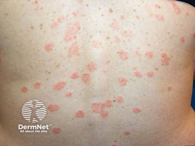
This image depicts a clinical dermatology photograph of the back (posterior trunk) showing multiple erythematous, oval to round plaques characteristic of pityriasis rosea. Acquired in routine outpatient photography, the lesions are salmon-pink to rose-colored, 0.5–2 cm in diameter, with fine, collarette-like scaling at the outer margin and a smooth central surface. The distribution is trunk-predominant with clustering along the spinal regions, consistent with the classic Christmas-tree pattern along skin cleavage lines. The inflammatory plaques may appear slightly raised and may coalesce in areas of friction. No confluent involvement of the face or mucosa is evident. In pityriasis rosea, the eruption often follows a herald patch, though this image focuses on the secondary eruption phase. The differential diagnosis at this stage includes tinea corporis, guttate psoriasis, secondary syphilis, and eczema; however, the sharp, circular to oval morphology, peripheral scaling, and trunk-centric distribution support pityriasis rosea. The clinical significance rests in recognizing its typical self-limited course, usually resolving within several weeks to months and requiring supportive care for itch. This image is relevant for educational demonstrations of PR skin manifestations, photographic recognition in dermatology training, and comparative studies of pityriasis rosea presentations across phototypes. Potential clinical uses include triage, patient education, and dermatologic image search for pattern recognition.
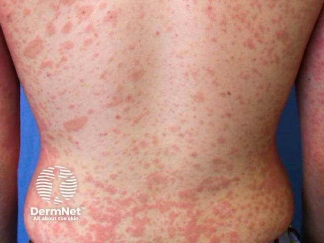
Clinical photograph of the back demonstrating pityriasis rosea-like eruption. High-resolution color image capturing multiple oval annular plaques with salmon-pink hue and fine collarette scaling along the trunk. The long axis of each lesion is oriented approximately parallel to the ribs, creating a pattern that follows the thoracic dermatomal distribution on the dorsal chest and flanks. Lesions are predominantly superficial, ill-defined at the periphery, with central clearing and variable sizes ranging from several millimeters to a centimeter. The overall appearance is erythematous, mildly pruritic, and arranged in a trunk-centered, zigzag or Christmas tree distribution, typical of pityriasis rosea. Differential considerations include tinea corporis, pityriasis versicolor, guttate psoriasis, and nummular eczema. Clinically, this presentation supports the diagnosis of pityriasis rosea when accompanied by a herald patch or secondary eruption; histopathology is not provided. The image is intended for education, differential diagnosis training, and radiological-free dermatology research; useful for teaching pattern recognition, dermography, and dermatoepidemiology. The photograph emphasizes trunk involvement with characteristic collarette scaling, aiding clinicians in distinguishing PR from mimickers and in counseling patients about the self-limited course (usually 6-8 weeks) and symptomatic management options. No systemic symptoms are evident; findings support a benign, self-limited cutaneous eruption in most cases for clinicians.
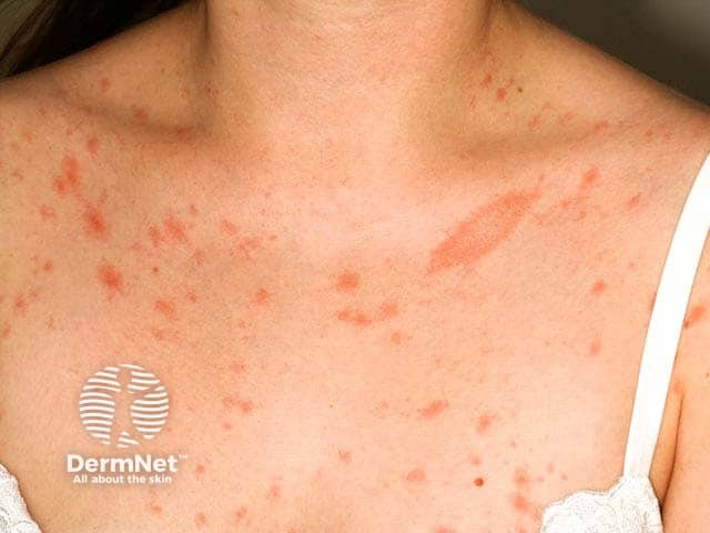
Clinical dermatology photograph of the upper back and posterior neck demonstrates classic pityriasis rosea with a herald patch. The main lesion is a solitary, oval, salmon‑pink plaque with fine, collarette scaling along the outer margin. The lesion size is larger than subsequent papules and appears slightly raised with less surface involvement centrally. Surrounding the trunk shows numerous smaller erythematous papules and plaques distributed in a crest-like or zigzag pattern consistent with secondary eruption along Langer's lines, sometimes described as a Christmas tree distribution on the torso. Coloration ranges from pink to coral; scaling is peripheral and secondary lesions may be pruritic. The overall clinical impression is pityriasis rosea in an early or evolving stage, with herald patch indicating onset often preceding generalized eruption by days to weeks. Differential considerations include tinea corporis and secondary syphilis, which can resemble patchy erythema and collarette scaling; correlate clinically and with serology if indicated. The image highlights typical dermatologic features such as circinate patches, mild edema, and intact epidermis without crusting or vesiculation. This presentation is self-limited, benign, and usually resolves within 6–8 weeks with symptomatic care. Reassurance and avoidance of irritants are appropriate management, with follow-up to ensure resolution. If lesions persist or patient reports systemic symptoms, consider dermoscopic evaluation and confirmatory testing as indicated. Early diagnosis guides management decisions.
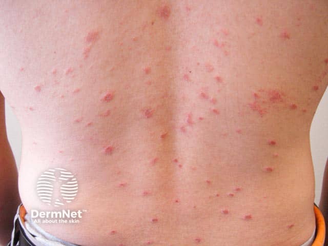
This is a high-resolution clinical photograph of the dorsal trunk showing a mildly symptomatic pityriasis rosea eruption. The image captures numerous discrete erythematous to pink papules and plaques on the back, many displaying fine overlying scale concentrated at the periphery (collarette scale). Lesions are small to medium in size (roughly 3-6 mm) and arranged diffusely across the posterior torso, without mucosal involvement. The distribution is characteristic for pityriasis rosea, with trunk predominance and a tendency to align along cleavage lines, producing a subtle christmas tree pattern in classic cases, though such configuration may be less evident in isolated back photographs. The coloration is uniform, with slight central clearing in some lesions and a flatter, smooth surface in others. No vesiculation, crusting, or purpura is evident. The image does not reveal the herald patch, which may precede or accompany the eruption. This photographic record is intended for dermatology education, differential diagnosis training, and clinical pattern recognition; it supports rapid recognition of PR versus mimickers such as tinea corporis and psoriasis. The image could be used in teaching modules, teledermatology consultations, and AI-based pattern recognition research to improve diagnostic accuracy and triage in skin-of-color presentations when available.
Complete Study Guide: Pityriasis Rosea
Slide-by-Slide Breakdown + Expected Doctor Questions
SLIDE 3-4: THE CASE
The herald patch - a single, round, self-resolving rash on the chest that disappeared in one week, BEFORE the widespread eruption appeared. This sequential pattern is pathognomonic. About 80% of PR cases have a herald patch, and it precedes the secondary eruption by approximately 2 weeks in adults.
Pityriasis rosea-related pruritus is worsened by heat and sweating because elevated skin temperature and sweat lower the itch threshold - this is also called the "hot bath sign" or thermally aggravated pruritus. This is a classic feature of PR and is different from allergic conditions where itching is constant.
Because pityriasis rosea is NOT a fungal infection - it is a viral exanthem, most likely caused by HHV-6/HHV-7 reactivation. Antifungal agents (like clotrimazole, miconazole) only target fungal cell membrane ergosterol - they have no activity against herpesviruses. The failure of antifungal therapy is actually a diagnostic clue that helps confirm PR and rule out tinea corporis and pityriasis versicolor.
SLIDE 6: LESION DESCRIPTION
Langer's lines (also called lines of cleavage or tension lines) are natural orientations of collagen fibers in the dermis. On the trunk/back, they run diagonally downward and outward from the spine, like branches of a Christmas tree. In PR, the oval lesions align their long axis parallel to these lines, producing the Christmas tree pattern. This happens because the virus spreads along dermatomes and perivascular lymphatics that follow these planes.
A collarette scale is a ring of fine, crinkled scale that sits just inside the periphery of each lesion, with the free edge of the scale pointing inward (toward the center of the lesion). It forms because the active inflammatory process starts at the outer edge of the plaque and moves centrally. The center of the lesion heals first while the periphery is still active, lifting the dead skin inward. This is sometimes called the "hanging curtain sign" when the skin is stretched.
Feature Herald Patch Secondary Lesions Size 2-10 cm (large) 0.5-2 cm (smaller) Number Single Multiple Location Trunk (50%), neck, extremities Trunk, proximal limbs Timing Appears 1-2 weeks first Appear after herald patch involutes Distribution Isolated Christmas tree pattern
Salmon color refers to a pink-orange hue caused by mild inflammation with superficial vascular dilation in fair skin. In darker-skinned patients, lesions may appear hyperpigmented or even hypopigmented after resolution. Recognizing this color helps distinguish PR from the silvery-white scale of psoriasis or the brown macules of pityriasis versicolor.
SLIDE 7: PRIMARY DIAGNOSIS - DEFINITION & ETIOLOGY
The name was coined by Gibert in 1860. "Pityriasis" comes from Greek meaning scaly, and "rosea" means pink. Together: pink scales - which accurately describes the appearance of the lesions.
The most accepted theory is reactivation of HHV-6 and/or HHV-7 (human herpesviruses). These viruses are acquired in early childhood (nearly universal by age 3) and remain latent in mononuclear cells (T-lymphocytes and monocytes). Reactivation - possibly triggered by stress, fever, or immunosuppression - leads to viremia and then the skin eruption. Key evidence:
- Watanabe et al. (2002) demonstrated active HHV-6/HHV-7 replication in skin lesion mononuclear cells
- Drago et al. found HHV-6/7 in serum of PR patients including those who miscarried
- HHV-7 is detected more often than HHV-6 in most series
Because it is caused by reactivation of a latent virus that nearly everyone already carries - not a new primary infection being transmitted person to person. The virus is not being actively shed in respiratory secretions or skin contact. Clustering of cases in households/dormitories has been reported occasionally, but true horizontal transmission is not established.
This is a common "trap" question. Drug-induced PR-like rashes can be caused by: captopril, imatinib, interferon, lisinopril, adalimumab, lamotrigine, rituximab, lithium, barbiturates, gold, arsenic, and notably the BCG vaccine. Always ask about recent medications/vaccinations to exclude a drug eruption masquerading as PR.
SLIDE 8: PATHOGENESIS
- Latent HHV-6/7 resides in skin keratinocytes and circulating CD4+ T-lymphocytes/monocytes from childhood infection
- A trigger (febrile illness, stress, immunosuppression) causes viral reactivation and viremia
- The virus seeds the skin via hematogenous spread, particularly to keratinocytes
- The immune system mounts a CD4+ T-cell mediated response against virally infected cells
- This inflammatory response causes spongiosis (intercellular edema of the epidermis) and superficial perivascular lymphocytic infiltrate in the dermis
- The result is the characteristic papulosquamous eruption
- Once viral antigen is cleared, the immune response resolves and lesions heal spontaneously within 6-8 weeks
The herald patch represents the site of initial viral seeding - essentially the first focus where the virus reaches the skin after viremia begins. The secondary eruption represents subsequent widespread hematogenous dissemination to the entire skin surface. The 1-2 week delay corresponds to the time needed for viremia to spread broadly before the immune response contains it.
SLIDE 9-10: CLINICAL FEATURES & HISTOPATHOLOGY
Approximately 80% of cases have an identifiable herald patch. It is absent in about 20%, making those cases harder to diagnose. It can also appear simultaneously with the secondary eruption, or rarely be the only manifestation.
Prodromal/concurrent symptoms occur in 5-69% of patients and include:
- Fatigue and malaise
- Headache
- Nausea
- Mild upper respiratory symptoms (sore throat, runny nose)
- Arthralgias These symptoms reflect the underlying viral reactivation/viremia.
Spongiosis is intercellular edema within the epidermis - fluid accumulates between keratinocytes, stretching the desmosomes and giving a "spongy" appearance under the microscope. It is the hallmark of eczematous/inflammatory reactions. In PR it is mild and helps distinguish it from psoriasis (which shows NO spongiosis but has Munro microabscesses).
Finding What It Indicates Mild spongiosis Epidermal edema from inflammation Focal parakeratosis Retained nuclei in stratum corneum - rapid cell turnover Mild acanthosis Epidermal thickening Extravasated RBCs Leaky vessels - mild inflammation Superficial perivascular lymphocytic infiltrate T-cell mediated immune response around dermal capillaries
No. The histological findings in PR are not specific - they can be seen in other papulosquamous diseases. Biopsy is done only if: (a) the clinical picture is atypical, (b) the eruption persists beyond 12 weeks, or (c) secondary syphilis cannot be excluded. The biopsy helps by excluding other diagnoses rather than confirming PR.
SLIDE 11: WHY THIS DIAGNOSIS
The herald patch - the initial single round lesion on the chest that spontaneously resolved in one week before the widespread eruption appeared. This sequential history is nearly pathognomonic for PR and is not seen in any of the main differential diagnoses.
It actively rules out the two most common fungal mimics:
- Tinea corporis - would respond to azole antifungals within 2-4 weeks
- Pityriasis versicolor - would respond to topical antifungals Three weeks of antifungal with zero improvement strongly supports a non-fungal etiology, pointing toward PR.
SLIDE 12-16: DIFFERENTIAL DIAGNOSES
Feature PR Tinea Corporis Herald patch Yes No Lesion border Collarette scale (inward-pointing) Raised, active, outward-pointing scaly border Central clearing Subtle Pronounced (annular) KOH prep Negative Positive (hyphae) Distribution Christmas tree on trunk Scattered, any site Antifungal response No Yes
Because secondary syphilis is a great mimic and missing it has serious consequences (untreated syphilis progresses to tertiary disease including cardiovascular and neurosyphilis). Syphilis incidence is rising globally. The key distinguishing features of secondary syphilis are: palmoplantar involvement (palms and soles), mucous membrane patches, generalized lymphadenopathy, condyloma lata, and positive VDRL/RPR. PR typically spares palms/soles and has no lymphadenopathy.
The Auspitz sign is pinpoint bleeding that occurs when scale is removed from a psoriatic plaque (due to dilated dermal capillaries extending into thinned suprapapillary epidermis). It is characteristic of psoriasis (including guttate psoriasis). It is absent in PR, which helps differentiate them.
This describes the KOH microscopic appearance of Pityriasis (Tinea) Versicolor - caused by Malassezia furfur. Short hyphae = "spaghetti", clusters of yeast spores = "meatballs". This appearance would be absent in PR (KOH negative), which is a key distinguishing test.
Wickham striae are fine white lines/streaks visible on the surface of flat-topped violaceous papules of Lichen Planus. This is absent in PR. Lichen planus could occasionally be confused with the papular variant of PR.
SLIDE 17-20: LABORATORY INVESTIGATIONS
Potassium hydroxide (KOH) prep is a bedside test for fungal infection:
- Scrape the active border of the lesion onto a glass slide
- Add 10-20% KOH solution + gentle heat (optional)
- KOH dissolves keratin/debris but leaves fungal hyphae intact
- View under microscopy: positive = fungal hyphae (tinea) or "spaghetti and meatballs" (tinea versicolor)
- In PR: expected to be NEGATIVE
VDRL (Venereal Disease Research Laboratory) is a non-treponemal screening test for syphilis. It detects reagin antibodies (anti-cardiolipin/anti-phospholipid) produced by the body in response to treponemal infection. A positive VDRL is then confirmed with a treponemal test (FTA-ABS or TPPA). In PR, VDRL is expected to be negative. False positives can occur in pregnancy, SLE, and other infections.
HIV testing is done because immunosuppression can cause atypical, severe, or persistent PR. In HIV patients, PR may be more widespread, have unusual morphology (vesicular, papular, purpuric), resist treatment, and last longer than 12 weeks. So if the clinical picture is atypical or severe, checking the immune status is appropriate.
Because: (a) most adults are already seropositive for HHV-6/7 from childhood infection, making serology hard to interpret; (b) distinguishing latent vs active infection by PCR requires quantitative methods; (c) PR is a clinical diagnosis and the result does not change management; (d) the test is mainly used in research settings or in transplant patients where HHV-6 reactivation has more serious implications.
SLIDE 21-22: TREATMENT
The cornerstone is reassurance - telling the patient that PR is self-limiting and will resolve in 6-8 weeks (sometimes up to 12 weeks) without any specific treatment. General measures: avoid overheating, wear loose cotton clothing, use gentle soap, avoid hot showers, avoid scratching. Most patients need nothing beyond this.
Mechanism: Topical steroids bind intracellular glucocorticoid receptors → suppress phospholipase A2 → decrease arachidonic acid → reduce prostaglandins and leukotrienes → less inflammation and pruritus. In PR, mild-to-moderate potency steroids are used (e.g., hydrocortisone 1% for face/flexures, triamcinolone 0.1% for trunk). Avoid high-potency steroids for prolonged periods - they cause skin atrophy, striae, telangiectasia, and hypopigmentation.
PR pruritus has a histamine-mediated component. H1 antihistamines block histamine at H1 receptors in skin and CNS → reduce itch sensation. Choices:
- Cetirizine/Loratadine (non-sedating) - daytime use
- Hydroxyzine (sedating) - useful at night because itch is often worse at night and the sedation helps sleep Dry mouth and drowsiness (with hydroxyzine) are the main side effects.
Acyclovir is an antiviral used in moderate-to-severe PR, preferably early in the course. Mechanism: Acyclovir is phosphorylated by viral thymidine kinase to acyclovir triphosphate → inhibits viral DNA polymerase → terminates DNA chain elongation → stops HHV-6/7 replication. Dose: 800 mg orally 5 times daily for 7 days. Evidence: Ganguly (2014) - a randomized, double-blind, placebo-controlled trial - showed acyclovir hastened resolution and reduced severity. It is NOT used routinely in mild cases.
Narrowband UVB (NB-UVB) phototherapy is used in cases of persistent or severe PR that fail to resolve within the typical timeframe. Mechanism: UVB radiation suppresses cutaneous immune responses, induces T-cell apoptosis, and reduces inflammatory cytokines in the skin. It is a second-line option for resistant/persistent cases. A study by Yalkova et al. (2004) supported UVB use in PR.
Hydroxyzine potentiates CNS depression when combined with alcohol, opioids, benzodiazepines, or other sedatives. This is a pharmacodynamic interaction (additive CNS depression), not a pharmacokinetic one. It can also cause QT prolongation at high doses.
SLIDE 23: PROGNOSIS
Recurrence is uncommon - 1.8% to 3.7% in most studies (Fitzpatrick's reports <3%). When recurrence happens, the second episode usually lacks a herald patch, involves fewer lesions, and resolves faster than the first.
Darker-skinned patients are more prone to post-inflammatory hyperpigmentation (PIH) - the healed skin appears darker than normal for weeks to months. This is because melanocytes respond to inflammation by producing excess melanin. PIH is not permanent and fades over time. No permanent scarring occurs in PR.
Yes. PR in early pregnancy (first 15 weeks) is associated with: premature delivery, neonatal hypotonia, and fetal loss (miscarriage). Drago et al. demonstrated HHV-6/7 in the serum of pregnant women with PR who miscarried. This is why - while PR is benign in most people - it needs to be taken seriously in a pregnant patient, and obstetric monitoring is warranted.
BONUS: Curveball Questions the Doctor May Ask
"This condition almost always resolves completely within 6-8 weeks and rarely comes back. Less than 3% of people experience a recurrence. You may notice some darkening of the skin where the rashes were, but that will fade over time and leave no scars."
PR in the first 15 weeks of pregnancy is associated with miscarriage and neonatal complications due to HHV-6/7 viremia crossing the placenta. Management would include OB-GYN referral and closer monitoring. Acyclovir use in pregnancy requires careful risk-benefit assessment.
A rash persisting beyond 12 weeks is called the persistent form of PR. At that point, you should: (a) reconsider the diagnosis - is this actually secondary syphilis, parapsoriasis, or mycosis fungoides? (b) reassess for underlying immunosuppression (HIV); (c) perform skin biopsy for histopathology; (d) consider NB-UVB phototherapy or a short course of acyclovir.
Some patients have atypical PR involving the face, scalp, axillae, groin, or sparing the trunk entirely (inverse pattern). This is more common in children and patients with darker skin. The Christmas tree pattern may be absent. These atypical cases are harder to diagnose and require broader differential workup.


This is the case, go through it
Case Summary - Group 2 (Endocrinology)
Patient Profile
- Name: Ms. A.B.
- Age: 40 years old
- Sex: Female
- Occupation: Office administrator
- Chief Complaint: "Palpitations and weight loss" for 3 months
History of Present Illness
- Progressive palpitations
- Unintentional weight loss of 8 kg despite increased appetite
- Heat intolerance
- Excessive sweating
- Nervousness and irritability
- Easy fatigability
- Increased bowel movements (3-4 times daily, non-bloody, loose but not watery)
- Fine tremors of the hands
- Difficulty sleeping
- Hair thinning
- Recent onset of "eye bulging" noticed by coworkers
- Sensation of neck fullness without pain
- Neck pain
- Recent viral illness
- Iodine or amiodarone intake
- Thyroid medication use
- Postpartum state
Menstrual History
- Oligomenorrhea for the past 4 months
- No prior history of thyroid disease
Past Medical History
- No hypertension, diabetes, or cardiac disease
- No prior thyroid disorder
- No recent pregnancy
Family History
- Mother had a "thyroid problem" treated with radioactive iodine (likely hyperthyroidism)
- No known autoimmune diseases in siblings
Social History
- Non-smoker
- Occasional coffee intake
- No illicit drug use
Review of Systems (Pertinent Positives)
- Weight loss despite increased appetite
- Palpitations
- Heat intolerance
- Anxiety
- Increased bowel frequency
- Oligomenorrhea
- Eye prominence (exophthalmos)
My Clinical Impression
| Clue | Significance |
|---|---|
| Weight loss + increased appetite | Hypermetabolic state from excess T3/T4 |
| Palpitations + fine tremors | Sympathetic overstimulation |
| Heat intolerance + sweating | Elevated basal metabolic rate |
| Eye bulging (exophthalmos/proptosis) | Graves' ophthalmopathy - SPECIFIC to Graves', not other causes of hyperthyroidism |
| Neck fullness (goiter) | Thyroid enlargement due to TSH-receptor antibody stimulation |
| Oligomenorrhea | Hormonal disruption from thyroid excess |
| Family history of hyperthyroidism treated with RAI | Strong autoimmune/genetic predisposition |
| Denies iodine/amiodarone/thyroid meds | Rules out drug-induced hyperthyroidism |
| Denies postpartum state | Rules out postpartum thyroiditis |
| Denies viral illness | Rules out subacute thyroiditis |
This is my presentation, now explain me everything and also cover all the possible points from which doctor will ask questions
python3 -c " import pdfplumber with pdfplumber.open('/tmp/workspace/attachments/e7ada6c6-c05c-496f-bda3-e596e84c7f40/ENDO CASE 2.pdf') as pdf: print(f'Pages: {len(pdf.pages)}') for i, page in enumerate(pdf.pages): print(f'\n--- PAGE {i+1} ---') text = page.extract_text() if text: print(text) else: print('[No text - image-based page]') "
Complete Study Guide: Graves' Disease (Endocrinology Case 2)
Slide-by-Slide Explanation + All Expected Doctor Questions
SLIDES 3-5: PATIENT PROFILE & HISTORY
| Symptom | Mechanism |
|---|---|
| Weight loss + increased appetite | T3/T4 increase basal metabolic rate (BMR) - the body burns more energy than it takes in. Appetite increases to compensate but cannot keep up |
| Palpitations | T3 directly increases heart rate (chronotropy) and force of contraction (inotropy). It also increases sensitivity to catecholamines (adrenaline) |
| Heat intolerance + sweating | Elevated BMR generates excess heat. The body tries to dissipate it through sweat |
| Nervousness, irritability, anxiety | T3 increases adrenergic receptor density and sensitivity in the nervous system |
| Fine hand tremors | Thyroid hormone increases neuromuscular excitability and adrenergic tone |
| Diarrhea/increased bowel movements | Thyroid hormone increases gut motility - but note: movements are loose, NOT watery, NOT bloody - typical of hyperthyroid bowel |
| Insomnia | CNS overstimulation from excess adrenergic tone |
| Hair thinning | Thyroid hormone accelerates hair cycling - follicles enter telogen (shedding) phase too quickly |
| Oligomenorrhea | Excess T3/T4 disrupts the hypothalamic-pituitary-ovarian axis - GnRH pulsatility is disrupted, reducing LH/FSH, leading to irregular cycles |
| Easy fatigability | Despite hypermetabolism, muscles are catabolized for energy - muscle weakness (thyroid myopathy) results |
Most causes of weight loss come with DECREASED appetite (cancer, depression, chronic illness). Weight loss WITH increased appetite means the body is consuming more calories than it can absorb - a hypermetabolic state. This pattern is highly characteristic of hyperthyroidism (and less commonly diabetes).
The mother had a "thyroid problem" treated with radioactive iodine - RAI is used almost exclusively for hyperthyroidism (Graves' or toxic nodular goiter). This suggests a genetic/autoimmune predisposition to Graves' disease. Graves' is known to have HLA associations (HLA-DR3, HLA-B8) and CTLA4 gene polymorphisms, and has increased concordance in monozygotic twins.
Postpartum thyroiditis (PPT) is another common cause of hyperthyroidism in young women - it occurs 1-6 months after delivery due to rebound autoimmune activity. By denying recent pregnancy, PPT is eliminated as a cause without needing further testing.
Iodine excess can cause hyperthyroidism via the Jod-Basedow phenomenon (iodine load triggers autonomous thyroid hormone synthesis in a pre-existing nodular gland). Amiodarone contains ~37% iodine by weight and causes thyrotoxicosis in up to 10% of patients. Excluding these removes drug-induced causes.
SLIDE 6: PRIMARY DIAGNOSIS - GRAVES' DISEASE
Graves' disease is the most common cause of hyperthyroidism worldwide, accounting for ~80% of all cases. It is an organ-specific autoimmune disease in which the body produces IgG autoantibodies - called Thyroid-Stimulating Immunoglobulins (TSI) or TRAb (TSH receptor antibodies) - that bind to and activate the TSH receptor on thyroid follicular cells. This causes:
- Continuous, TSH-independent thyroid hormone production → hyperthyroidism
- Diffuse thyroid enlargement → goiter
- Orbital fibroblast stimulation → ophthalmopathy
- Skin fibroblast stimulation → dermopathy (pretibial myxedema)
- Hyperthyroidism (thyrotoxicosis)
- Diffuse toxic goiter (symmetrically enlarged thyroid)
- Infiltrative ophthalmopathy (Graves' eye disease / exophthalmos)
Some sources add a 4th: pretibial myxedema (dermopathy) and 5th: thyroid acropachy (digital clubbing + periosteal new bone) - though these are less common.
- Peak incidence: 20-40 years (your patient is 40 - right at the upper edge)
- Women affected 5-7 times more than men
- Estimated prevalence: 1.5-2% of women in the US
- Strong autoimmune/genetic background - associated with other autoimmune diseases
SLIDE 7: WHY GRAVES' DISEASE? (Clinical Justification)
- Young-middle-aged female - fits the demographic exactly
- Hypermetabolic symptoms (weight loss, heat intolerance, sweating, palpitations, tremors) - all caused by excess T3/T4
- Eye bulging (exophthalmos/proptosis) - nearly pathognomonic for Graves'; NOT seen in toxic nodular goiter or other causes
- Neck fullness without pain - diffuse painless goiter; pain would suggest subacute thyroiditis
- Oligomenorrhea - reproductive axis disruption from thyrotoxicosis
- No iodine/amiodarone/thyroid meds - drug causes excluded
- No recent viral illness - subacute thyroiditis excluded
- No postpartum state - postpartum thyroiditis excluded
- Family history of hyperthyroidism treated with RAI - autoimmune genetic predisposition
SLIDE 8: WHY DOES EYE BULGING OCCUR? (Graves' Ophthalmopathy Pathogenesis)
- B lymphocytes produce TRAb (TSH receptor antibodies)
- The TSH receptor is expressed not only on thyroid cells but also on orbital fibroblasts (connective tissue cells behind the eye)
- TRAb binds to TSH receptors on orbital fibroblasts → activates them
- CD4+ T cells infiltrate the orbit and secrete cytokines (IL-1, TNF, IFN-γ)
- Activated fibroblasts produce large quantities of glycosaminoglycans (GAGs) - mainly hyaluronic acid
- GAGs are highly hydrophilic - they attract water → orbital edema
- The extraocular muscles swell with edema and GAG deposition → muscle enlargement
- The orbital fat volume also increases
- All of this increases volume behind the eyeball in the bony orbit, pushing the eyeball forward → exophthalmos/proptosis
- Exophthalmos/Proptosis - forward protrusion of eyeball (>20mm is abnormal; measured by Hertel exophthalmometer)
- Lid retraction - upper eyelid pulled up, revealing sclera above iris (Dalrymple sign)
- Lid lag (von Graefe sign) - upper lid lags behind eyeball on downward gaze
- Diplopia - double vision from restricted extraocular muscles
- Eye irritation, redness, tearing - due to exposed conjunctiva/cornea
- Chemosis - conjunctival edema
- In severe cases: corneal ulceration, compressive optic neuropathy → vision loss
Yes - this is called euthyroid Graves' ophthalmopathy (or euthyroid Graves' disease). The TRAb attacks the orbit independently of thyroid status. About 5-10% of Graves' ophthalmopathy patients are euthyroid at diagnosis. This shows that the orbital and thyroid manifestations are driven by the same antibody but can be uncoupled.
A localized skin thickening, usually over the shins (pretibial area), caused by the same mechanism as ophthalmopathy - TRAb stimulates skin fibroblasts to deposit glycosaminoglycans in the dermis. The skin becomes thickened, indurated, and non-pitting, with a scaly appearance. Seen in about 1-5% of Graves' patients. It is almost exclusively seen in Graves' disease.
The rarest manifestation of Graves' disease (0.1-1%). Characterized by digital clubbing + soft tissue swelling of the hands/feet + periosteal new bone formation on X-ray. Often occurs in patients who already have ophthalmopathy and pretibial myxedema (the "triple triad"). It is not related to thyroid function levels.
SLIDE 9: CLINICAL FEATURES
Tachycardia - your slide correctly includes "tachycardia (expected)" even though the patient only reports palpitations. Thyroid hormone causes sinus tachycardia (HR typically 90-120). In severe cases it can precipitate atrial fibrillation (seen in ~10-15% of hyperthyroid patients, especially older patients). This is why an ECG is included in the workup.
A bruit (whooshing sound) heard over the thyroid with a stethoscope, caused by markedly increased blood flow through the hyperactive, enlarged gland. It is characteristic of Graves' disease (diffuse toxic goiter) and can also be seen on Doppler ultrasound as the "thyroid inferno" pattern (massively increased vascularity).
SLIDES 10-14: DIFFERENTIAL DIAGNOSES
Feature Graves' Disease Toxic MNG Age Younger (20-40) Older (>40-50) Thyroid texture Diffuse, smooth enlargement Nodular, irregular TRAb Positive Negative Ophthalmopathy Present (unique to Graves') Absent Scan Diffuse uniform uptake Multiple hot nodules Onset Relatively acute Slowly progressive (years)
Toxic adenoma is a single autonomously functioning thyroid nodule (Plummer disease) that produces excess T3/T4 independent of TSH. Key differences:
- Single palpable nodule (vs. diffuse goiter)
- No ophthalmopathy
- Scan shows one hot nodule with suppression of the rest of the gland
- TRAb negative
Subacute thyroiditis is a viral/post-viral inflammatory condition of the thyroid. Hyperthyroidism occurs because inflammation destroys follicles and releases stored T3/T4 - not because of new synthesis. Key differentiators:
- Painful, tender thyroid (our patient has painless neck fullness - rules it out)
- Often preceded by viral illness (our patient denies this)
- Low RAIU (radioactive iodine uptake) - because follicles are destroyed, not hyperactive
- High ESR, fever, malaise
- Self-limiting - hyperthyroid phase followed by hypothyroid phase, then recovery
Taking exogenous thyroid hormone (T4 or T3 preparations) - either accidentally (over-replacement) or deliberately (for weight loss). Key distinguishing feature: low or undetectable thyroglobulin (because the thyroid gland is suppressed and not making its own hormone). The thyroid is small (not enlarged). Our patient denies thyroid medication use.
Pheo is a catecholamine-secreting tumor that causes: palpitations, sweating, anxiety, hypertension, and weight loss - all of which overlap with hyperthyroidism. Key differentiators:
- No goiter, no exophthalmos in pheo
- No weight gain despite increased appetite (pheo causes weight loss without increased appetite)
- TSH is normal in pheo (not suppressed)
- Pheo: elevated 24-hour urine catecholamines/metanephrines or plasma metanephrines
- Pheo classically causes episodic hypertension with severe headache + sweating + palpitations ("the 5 Ps")
SLIDES 15-16: LABORATORY INVESTIGATIONS
TSH will be very low (<0.01 mIU/L) - often undetectable. Here is why: The TRAb/TSI antibodies continuously stimulate TSH receptors on thyroid cells → thyroid produces massive amounts of T3/T4 → elevated T3/T4 feeds back to the pituitary via negative feedback → pituitary suppresses TSH release. TSH is the most sensitive test; a suppressed TSH is the first abnormality in hyperthyroidism.
- Total T4 includes both protein-bound T4 (inactive, bound to TBG/albumin) and free T4
- Free T4 (FT4) is the biologically active unbound fraction - only ~0.04% of total T4
- Free T4 is preferred in diagnosis because it is not affected by changes in binding proteins (e.g., pregnancy raises TBG and falsely elevates total T4)
- In Graves': FT4 elevated, TSH suppressed
T3 toxicosis is a form of hyperthyroidism where T3 is disproportionately elevated but T4 is normal or only mildly elevated. It is seen in:
- Early Graves' disease
- Toxic nodular goiter
- Iodine deficiency states (thyroid preferentially makes T3) This is why testing both FT3 and FT4 is important - if TSH is low but T4 is normal, check FT3.
TRAb (TSH receptor antibody) or TSI (Thyroid Stimulating Immunoglobulin) - the most specific test because:
- These antibodies are the actual CAUSE of Graves' disease
- Positive in >95% of Graves' patients
- Not found in other causes of hyperthyroidism (toxic nodular goiter, thyroiditis, factitious)
- Also used to monitor response to treatment and predict remission
- Anti-TPO (anti-thyroid peroxidase) antibodies are directed against the enzyme thyroid peroxidase
- Positive in ~70-80% of Graves' patients AND in Hashimoto's thyroiditis
- They are less specific - cannot distinguish Graves' from Hashimoto's
- TRAb is Graves'-specific; anti-TPO is a general marker of thyroid autoimmunity
On color Doppler ultrasound, the hyperactive Graves' thyroid shows massive increase in blood flow throughout the gland, producing a mosaic of color signals resembling flames - called the "thyroid inferno" sign. It also confirms diffuse enlargement (vs. nodular). This helps differentiate Graves' (high flow) from thyroiditis (low/normal flow).
Condition RAIU Graves' disease HIGH and diffuse (entire gland active) Toxic MNG HIGH but patchy (hot nodules + suppressed areas) Toxic Adenoma HIGH in one spot, suppressed rest Subacute thyroiditis VERY LOW (follicles destroyed, no uptake) Factitious hyperthyroidism VERY LOW (exogenous hormone suppresses the gland) This single test separates "production excess" (high uptake) from "release excess/destruction" (low uptake).
That is a copy-paste error. ECG is listed under labs but the "most specific test" description belongs to TRAb (from slide 15). ECG in Graves' is done to detect sinus tachycardia or atrial fibrillation as baseline before starting treatment. Expected ECG finding: sinus tachycardia. Be prepared for this if the doctor notices it.
SLIDES 17-21: TREATMENT
Three options, often used in sequence or combination:
- Beta-blockers - immediate symptom relief (do NOT reduce thyroid hormone, only block its peripheral effects)
- Antithyroid drugs (Methimazole or PTU) - reduce thyroid hormone synthesis
- Definitive therapy - Radioactive Iodine (I-131) or Thyroidectomy
Propranolol blocks β1 and β2 receptors → reduces palpitations, tachycardia, tremors, anxiety. At high doses, propranolol (unlike selective beta-blockers) also inhibits peripheral conversion of T4 → T3 (by inhibiting deiodinase). However, it does NOT reduce thyroid hormone production or address the autoimmune cause - it only controls symptoms while waiting for antithyroid drugs to work (which takes 4-8 weeks).
Methimazole inhibits thyroid peroxidase (TPO) - the enzyme that:
- Oxidizes iodide (I-) to iodine (I2)
- Organifies iodine onto tyrosine residues on thyroglobulin
- Couples iodotyrosines to form T3 and T4 By blocking TPO, methimazole stops new T3/T4 synthesis. Important: it does NOT release pre-formed stored hormone - so there is a delay of 2-6 weeks before levels fall (time for stored hormone to be depleted).
- Methimazole: once-daily dosing (better compliance), faster effect, fewer severe side effects
- PTU: twice or three times daily, associated with severe hepatotoxicity (black box warning for liver failure)
- PTU is preferred ONLY in:
- First trimester of pregnancy (methimazole is teratogenic - causes aplasia cutis, choanal atresia)
- Thyroid storm (PTU also blocks T4→T3 peripheral conversion)
- Methimazole intolerance
Agranulocytosis = severe drop in neutrophils (<500 cells/μL) → life-threatening susceptibility to infection. Occurs in 0.1-0.5% of patients on methimazole or PTU, usually in the first 3 months but can occur at any time. The hallmark symptom is fever + sore throat - this is why you tell every patient: "If you develop fever or sore throat, STOP the medication immediately and go to the ER for a CBC." This is one of the most testable points in pharmacology.
RAI causes massive thyroid cell death → releases large amounts of thyroid antigens including TSH receptor fragments into the bloodstream → this triggers a surge in TRAb antibody production → more TRAb reaches the orbital fibroblasts → worsens the inflammatory orbital process. This is why RAI is contraindicated in moderate-to-severe active Graves' ophthalmopathy. If RAI must be used in a patient with mild eye disease, corticosteroids are given concurrently to dampen the antibody surge.
- Recurrent laryngeal nerve (RLN) injury → hoarseness, voice change (the RLN runs just behind the thyroid in the tracheoesophageal groove)
- Hypocalcemia from hypoparathyroidism → if parathyroid glands are accidentally removed or devascularized → low PTH → low calcium → tetany, numbness, Chvostek's/Trousseau's sign
- Bleeding/hematoma → can compress the airway (emergency)
- Hypothyroidism → permanent in total thyroidectomy → lifelong levothyroxine required
- Thyroid storm (if patient not adequately prepared preoperatively)
The patient MUST be made euthyroid before surgery to prevent thyroid storm intraoperatively. Steps:
- Methimazole for 4-6 weeks until euthyroid
- Lugol's iodine (potassium iodide) for 7-10 days before surgery - iodine reduces thyroid vascularity (Wolff-Chaikoff effect) making surgery safer
- Beta-blocker to control heart rate
SLIDE 22: MANAGEMENT OF GRAVES' OPHTHALMOPATHY
The Clinical Activity Score (CAS) grades orbital inflammation (0-7 points): spontaneous orbital pain, gaze-evoked pain, eyelid swelling, eyelid redness, conjunctival redness, chemosis, caruncle swelling. A CAS ≥3/7 = active disease. Severity is classified by EUGOGO (European Group on Graves' Orbitopathy) as mild, moderate-severe, or sight-threatening.
Smoking is the strongest modifiable risk factor for Graves' ophthalmopathy. Cigarette smoke contains free radicals and antigenic compounds that:
- Increase orbital fibroblast activation
- Increase glycosaminoglycan production
- Worsen hypoxia in retroorbital tissue
- Amplify the autoimmune response Smokers with Graves' disease have 4x greater risk of severe ophthalmopathy. Smoking cessation is the single most impactful lifestyle advice.
Selenium is an essential component of selenoproteins including glutathione peroxidase (an antioxidant). Oxidative stress plays a role in Graves' ophthalmopathy. A European randomized trial (EUGOGO, 2011) showed that selenium supplementation (200 mcg/day for 6 months) significantly improved mild Graves' ophthalmopathy and quality of life compared to placebo. It is recommended for mild, active disease where antioxidant protection is beneficial.
BONUS: CURVEBALL QUESTIONS
Thyroid storm is a life-threatening exacerbation of thyrotoxicosis triggered by physiologic stress (surgery, infection, trauma, childbirth). Features include: hyperpyrexia (>40°C), extreme tachycardia/AF, agitation/psychosis, vomiting, diarrhea, and potentially coma. It is diagnosed clinically using the Burch-Wartofsky Point Scale. Treatment: PTU (blocks synthesis AND T4→T3 conversion) + iodine (after PTU) + corticosteroids + beta-blockers + aggressive supportive care. Our patient is at risk if she undergoes any surgical procedure before being made euthyroid.
Graves' in pregnancy is dangerous for both mother and fetus:
- Use PTU in first trimester (methimazole is teratogenic)
- Switch to methimazole in 2nd/3rd trimester (PTU has hepatotoxicity risk)
- TRAb crosses the placenta → can cause neonatal hyperthyroidism in the newborn
- Target FT4 in the upper-normal range to avoid fetal hypothyroidism
- RAI is absolutely contraindicated in pregnancy
When a large amount of iodine is given suddenly (e.g., Lugol's iodine preoperatively), the thyroid temporarily shuts down thyroid hormone synthesis. This is an autoregulatory phenomenon - excess iodine inhibits TPO. It lasts about 10-14 days before the thyroid "escapes." This escape failure is exploited in surgery prep: use iodine for exactly 7-10 days to reduce vascularity and hormone production, then operate before escape occurs.
The opposite of Wolff-Chaikoff. In a patient with a pre-existing nodular or autonomously functioning thyroid, sudden exposure to a large iodine load (CT contrast, amiodarone, iodinated supplements) provides unlimited substrate for hormone synthesis → hyperthyroidism. This is why iodinated contrast is given carefully in patients with nodular thyroid disease.
After 12-18 months of antithyroid drug therapy:
- Taper and stop methimazole
- Check TRAb levels - if negative or very low → good chance of remission (~50%)
- Monitor TFTs (TSH, FT4) at 4-6 week intervals after stopping
- If levels remain normal for 12 months off medication → remission Relapse occurs in ~50% of patients within 2 years - definitive therapy (RAI or surgery) is then recommended.
Key Numbers to Memorize
| Fact | Value |
|---|---|
| TSH in hyperthyroidism | < 0.01 mIU/L (suppressed) |
| Normal TSH range | 0.4 - 4.0 mIU/L |
| Methimazole dose | 10-30 mg/day |
| PTU dose | 100-150 mg every 8 hours |
| Propranolol dose | 10-40 mg every 6-8 hours |
| Agranulocytosis incidence | 0.1-0.5% of patients |
| Graves' in women vs men | 5-7:1 female predominance |
| Peak incidence age | 20-40 years |
| Relapse rate after ATD | ~50% within 2 years |
| Course of illness to remission | 12-18 months of ATD therapy |