
Warfarin - mechanism of action uses adverse drug reactions and drug interactions
warfarin mechanism of action vitamin K coagulation factors
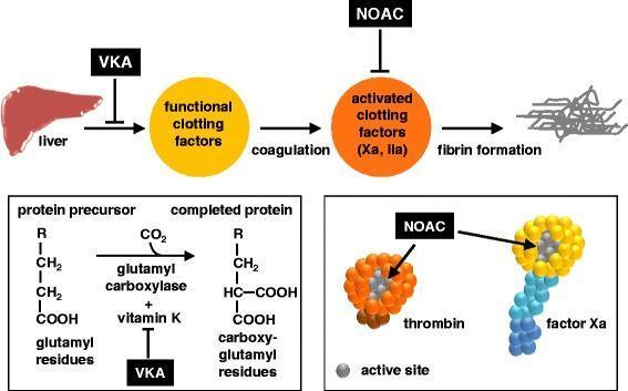
A multi-panel pathophysiology diagram illustrating the mechanism of action of oral anticoagulants on the coagulation cascade. The top panel shows a linear pathway: the liver produces functional clotting factors, which undergo coagulation to become activated clotting factors (Xa and IIa/thrombin), ultimately leading to fibrin formation. Vitamin K antagonists (VKA) are shown inhibiting the liver's production of functional factors, while non-vitamin K antagonist oral anticoagulants (NOACs) inhibit the activated factors. The bottom-left panel details the biochemical mechanism of VKAs, showing they inhibit the glutamyl carboxylase and vitamin K-dependent conversion of glutamyl residues on protein precursors into carboxy-glutamyl residues on completed clotting proteins. The bottom-right panel provides a molecular-level view of NOAC action, depicting thrombin (orange sphere) and factor Xa (yellow head with blue tail) being inhibited at their respective active sites (indicated by grey coloring). This educational graphic highlights the distinction between indirect synthesis inhibition by VKAs and direct enzymatic inhibition by NOACs/DOACs.
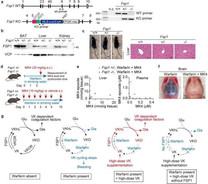
This composite educational graphic illustrates the role of Ferroptosis Suppressor Protein 1 (FSP1) as a warfarin-resistant vitamin K (VK) reductase. Panel (a) shows genomic schematic maps of Fsp1 wild-type and knockout alleles alongside genotyping PCR results. Panel (b) provides an immunoblot for FSP1 expression in adipose tissue, liver, and kidney across genotypes. Panel (c) displays gross morphological and H&E stained liver histology of Fsp1 mice, demonstrating normal architecture. Panel (d) outlines a clinical study design evaluating MK4 (vitamin K2) levels and prothrombin time under high-dose warfarin treatment. Panel (e) uses scatter plots to show the concentrations of MK4 and MK4 epoxide in liver and plasma. Panel (f) presents a clinical photograph comparing mouse brains: the warfarin-only specimen exhibits massive cerebral hemorrhage (dark discoloration), while the warfarin plus MK4 specimen appears normal, illustrating the antidotal effect of MK4. Finally, panel (g) provides pathophysiology flowcharts of the vitamin K cycle, detailing the enzymatic pathways involving VKOR and FSP1, and the mechanism by which high-dose vitamin K bypasses warfarin inhibition to enable carboxylation of coagulation factors.
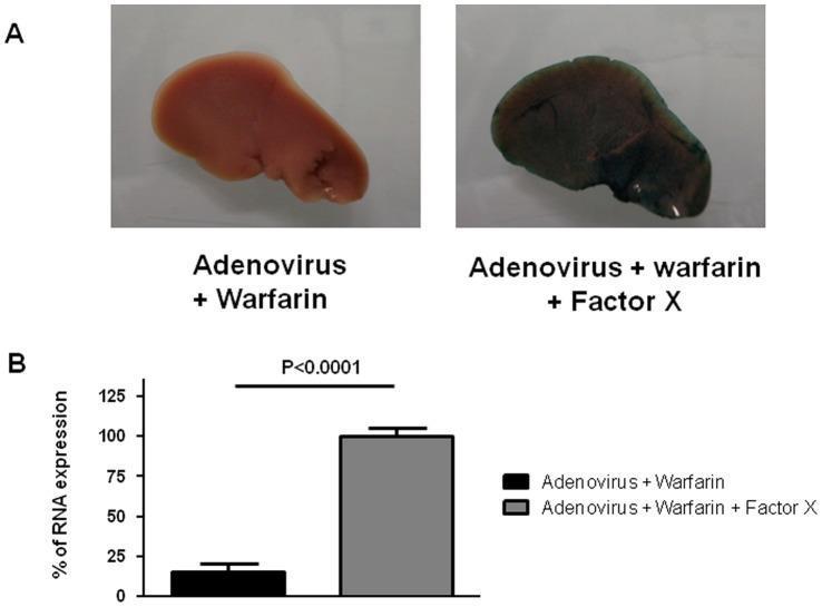
This composite educational image, categorized as Clinical Imaging/Specimen, illustrates the role of blood coagulation Factor X in adenovirus transduction using a mouse model. Panel A presents two gross liver specimens stained for β-galactosidase activity (Lac-Z reporter gene). The specimen labeled 'Adenovirus + Warfarin' appears pale pink/tan, indicating minimal viral transduction due to warfarin-induced depletion of vitamin K-dependent factors. In contrast, the 'Adenovirus + Warfarin + Factor X' specimen shows intense dark blue-green staining across the hepatic surface, demonstrating that exogenous Factor X supplementation restores adenoviral infection. Panel B contains a corresponding bar graph quantifying the percentage of RNA expression in adrenal glands. The 'Adenovirus + Warfarin' group (black bar) shows significantly lower expression compared to the 'Adenovirus + Warfarin + Factor X' group (gray bar), which is set as the 100% reference point. Statistical significance is indicated by a p-value < 0.0001. This figure demonstrates that adenovirus type 5 infection in both liver and adrenal tissues is dependent on Factor X, bypassing traditional CAR receptor-mediated pathways.
Warfarin - Complete Pharmacological Profile
Mechanism of Action
The Vitamin K Cycle
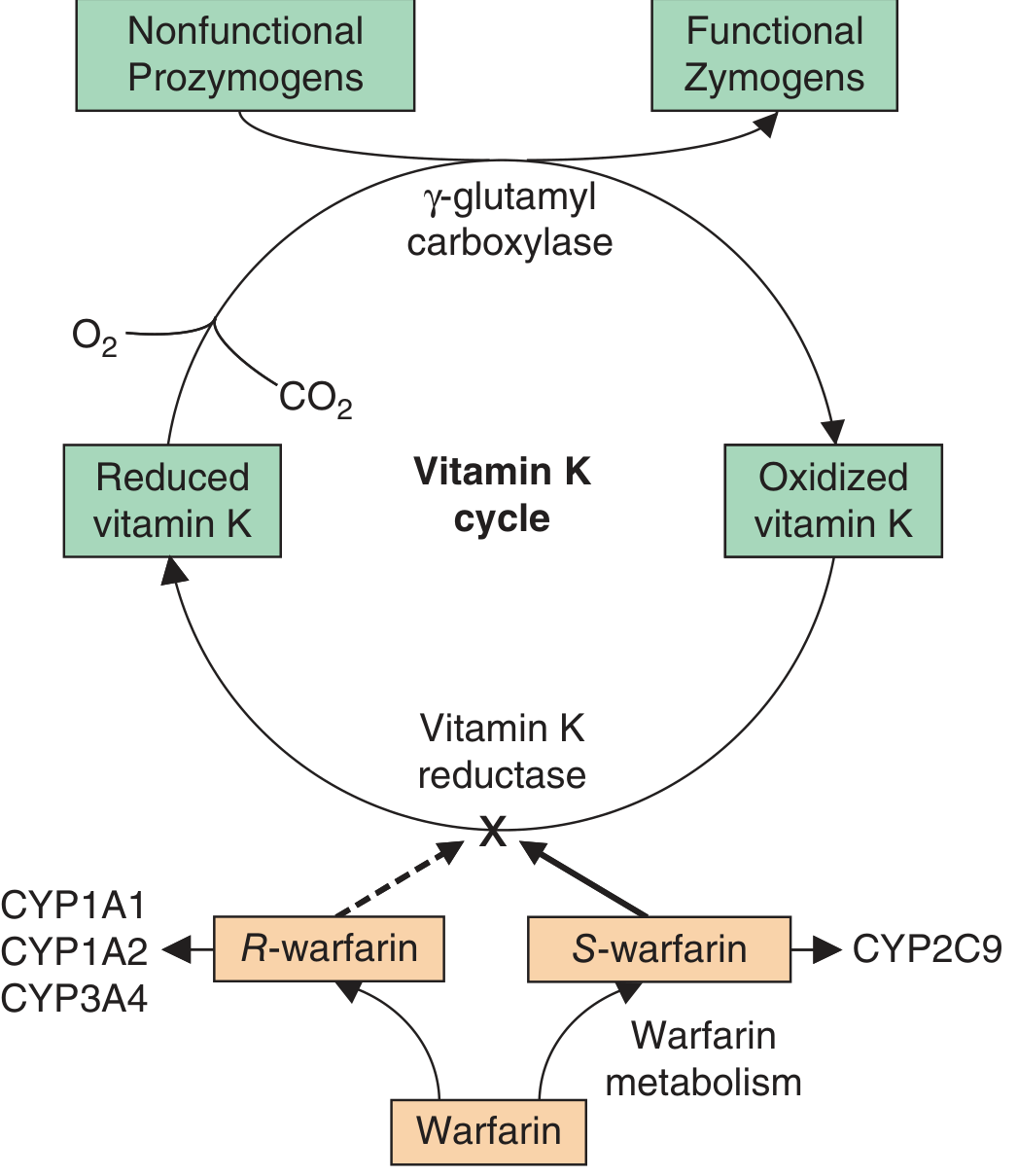
- Vitamin K-dependent clotting factors (II, VII, IX, X) and anticoagulant proteins (C and S) possess glutamic acid residues at their N-termini.
- A posttranslational modification adds a carboxyl group to the gamma-carbon of these residues, generating gamma-carboxyglutamic acid (Gla). This is essential because it permits calcium-dependent binding to anionic phospholipid surfaces.
- This gamma-carboxylation reaction is catalyzed by gamma-glutamyl carboxylase, which requires reduced vitamin K (vitamin K hydroquinone) as a cofactor.
- During carboxylation, vitamin K hydroquinone is oxidized to vitamin K epoxide. The epoxide must then be reduced back to vitamin K by vitamin K epoxide reductase (VKORC1) to keep the cycle going.
- Warfarin inhibits VKORC1, blocking the regeneration of active vitamin K. This depletes reduced vitamin K, impairing gamma-carboxylation, and results in the synthesis of nonfunctional prozymogens - partially carboxylated clotting proteins with little or no biological activity.
Stereochemistry & Pharmacokinetics
- Warfarin is a racemic mixture of R- and S-enantiomers.
- S-warfarin is 4x more potent than R-warfarin.
- S-warfarin is metabolized by CYP2C9; R-warfarin by CYP1A2 and CYP3A4.
- Oral bioavailability is nearly 100%; peak blood levels ~90 minutes after ingestion.
- Plasma half-life: 36-42 hours.
-
97% protein-bound (to albumin).
- Onset of action is delayed (4-5 days for full antithrombotic effect) because it requires depletion of already-circulating active clotting factors. The rate-limiting step is the fall in functional prothrombin (factor II, t½ = 72 hours) and factor X (t½ = 24 hours).
- Because of this delay, patients with active thrombosis need a bridging parenteral anticoagulant (heparin, LMWH, fondaparinux) for at least 5 days.
Genetic Pharmacology
- CYP2C9 variants (*2, *3): Reduce S-warfarin metabolism → lower dose requirements and higher bleeding risk.
- VKORC1 variants: Affect enzyme susceptibility to inhibition → Asians have highest prevalence of sensitive variants; Africans have lowest.
- Braunwald's Heart Disease, 15th Ed.; Harrison's Principles of Internal Medicine, 22nd Ed.
Uses / Indications
| Indication | Target INR |
|---|---|
| DVT / Pulmonary Embolism (treatment & secondary prophylaxis) | 2.0 - 3.0 |
| Atrial fibrillation (stroke prevention) | 2.0 - 3.0 |
| Mechanical prosthetic heart valves (bileaflet) | 2.0 - 3.0 |
| Mechanical prosthetic heart valves (tilting disk, older models) | 2.5 - 3.5 |
| Recurrent systemic thromboembolism | 2.5 - 3.5 |
| Antiphospholipid syndrome with recurrent thrombosis | 2.5 - 3.5 |
- Katzung's Basic and Clinical Pharmacology, 16th Ed.
Adverse Drug Reactions
1. Bleeding (Major ADR)
- The most common and serious complication; risk increases with supratherapeutic INR.
- Can occur at any site: GI tract (most common), genitourinary, intracranial (most feared).
- Risk factors: older age, CYP2C9 variants, high INR, concurrent antiplatelet drugs, falls.
- Management of major bleeding:
- Withhold warfarin.
- Administer Vitamin K1 (phytomenadione) IV or oral - reverses effect within 12-24 hours.
- For life-threatening bleeding: 4-factor prothrombin complex concentrate (PCC) for rapid reversal; Fresh Frozen Plasma (FFP) is an alternative.
2. Warfarin-Induced Skin Necrosis
- Rare but serious complication, typically occurring 3-5 days after initiation.
- Caused by a transient hypercoagulable state due to the short half-life of Protein C (natural anticoagulant) being depleted before the procoagulant factors.
- More common in patients with underlying Protein C or S deficiency.
- Characterized by painful skin lesions progressing to necrosis, typically in fat-rich areas (breast, buttocks, thighs, abdomen).
3. Purple Toe Syndrome
- Rare complication (usually within 3-8 weeks of therapy).
- Caused by cholesterol microemboli released from atheromatous plaques, precipitated by anticoagulation.
- Painful, cyanotic discoloration of toes; can progress to gangrene.
4. Teratogenicity
- Warfarin crosses the placenta and is teratogenic, particularly in the first trimester (weeks 6-12).
- Causes warfarin embryopathy: nasal hypoplasia, stippled epiphyses (chondrodysplasia punctata), limb hypoplasia, optic atrophy.
- Second and third trimester exposure: risk of fetal/neonatal bleeding (including intracranial hemorrhage), CNS abnormalities.
- Warfarin is contraindicated in pregnancy (except in some women with mechanical heart valves where the thrombotic risk outweighs fetal risk).
5. Other Adverse Effects
-
Alopecia (hair loss)
-
Osteoporosis (with long-term use)
-
Fatigue, feeling cold
-
Braunwald's Heart Disease; Harrison's Principles of Internal Medicine, 22nd Ed.
Drug Interactions
Pharmacokinetic Interactions - Increased Effect (↑ INR, ↑ bleeding risk)
| Drug | Mechanism |
|---|---|
| Amiodarone | Inhibits CYP2C9 AND CYP3A4 - inhibits metabolism of both S- and R-warfarin |
| Metronidazole | Stereoselectively inhibits CYP2C9 - inhibits S-warfarin metabolism |
| Fluconazole | Stereoselectively inhibits CYP2C9 - inhibits S-warfarin metabolism |
| Trimethoprim-sulfamethoxazole | Stereoselectively inhibits CYP2C9 - inhibits S-warfarin metabolism |
| Cimetidine | Inhibits both S- and R-warfarin metabolism (non-selective CYP inhibition) |
| Disulfiram | Inhibits both enantiomers |
| Phenylbutazone / Sulfinpyrazone | Inhibit S-warfarin metabolism AND displace albumin-bound warfarin (increase free fraction) - most dangerous combination |
Pharmacokinetic Interactions - Decreased Effect (↓ INR, ↓ efficacy)
| Drug | Mechanism |
|---|---|
| Rifampin | Potent CYP induction - markedly increases warfarin metabolism |
| Barbiturates (phenobarbital) | CYP induction - increases warfarin metabolism |
| Cholestyramine | Binds warfarin in the intestine - reduces absorption and bioavailability |
Pharmacodynamic Interactions - Increased Effect
| Drug/Factor | Mechanism |
|---|---|
| Aspirin (high doses) | Inhibits platelet function (additive bleeding risk) |
| NSAIDs | Platelet dysfunction + GI mucosal damage |
| Heparin, LMWH, direct oral anticoagulants | Additive anticoagulation |
| 3rd-generation cephalosporins | Kill intestinal bacteria producing vitamin K + directly inhibit VKORC1 |
| Hepatic disease | Reduces synthesis of clotting factors; increases warfarin sensitivity |
| Hyperthyroidism | Increases catabolism of clotting factors |
| SSRIs | Inhibit platelet aggregation (additive bleeding) |
Pharmacodynamic Interactions - Decreased Effect
| Drug/Factor | Mechanism |
|---|---|
| Vitamin K (dietary / supplements) | Competitive antagonism - bypasses VKORC1 block |
| Diuretics (chlorthalidone, spironolactone) | Concentrate clotting factors |
| Hypothyroidism | Decreases catabolism of clotting factors |
| Hereditary resistance | VKORC1 mutations that resist warfarin inhibition |
- Katzung's Basic and Clinical Pharmacology, 16th Ed.; Braunwald's Heart Disease; Goodman & Gilman's Pharmacological Basis of Therapeutics
Key Clinical Points
- Reversal agent: Vitamin K1 (oral: slow reversal in 12-24h; IV: faster). For urgent reversal: 4-factor PCC (preferred) or FFP.
- Start with heparin overlap when treating acute VTE - give both for ≥5 days AND until INR is ≥2.0 on two consecutive readings.
- Avoid in pregnancy (teratogen) - use LMWH instead.
- Narrow therapeutic window - monitor INR regularly; many patients benefit from anticoagulation clinics.
- Food interactions - high vitamin K foods (leafy greens: spinach, kale) can reduce efficacy; patients should maintain a consistent vitamin K intake rather than eliminating it.
- Superseeded for many indications by DOACs (direct oral anticoagulants like rivaroxaban, apixaban, dabigatran), which have fewer interactions, no routine monitoring, and predictable pharmacokinetics - but warfarin remains first-line for mechanical heart valves and in patients with antiphospholipid syndrome.