
A 35 year old rowdy take part in a fight. He got a cut in the back of his right arm. Which are the artery and nerves affected. Write the origin and termination of this artery
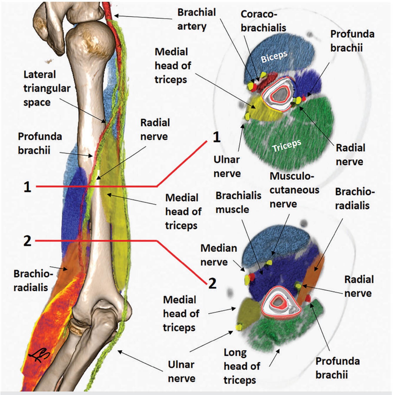
profunda brachii artery radial nerve spiral groove posterior arm anatomy
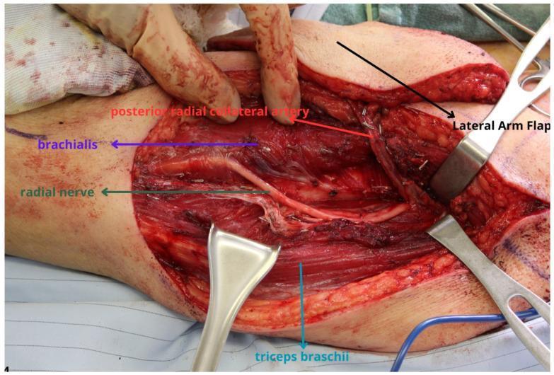
This clinical photograph shows an intraoperative view of a lateral arm flap harvest, highlighting critical neurovascular anatomy of the upper extremity. The surgical field displays the dissection of a fasciocutaneous flap, with a metal retractor reflecting the 'Lateral Arm Flap' to expose the underlying muscular and neurovascular structures. Key landmarks identified by color-coded labels include the radial nerve, which is seen coursing through the spiral groove region between the brachialis muscle (superior/anterior) and the triceps brachii muscle (inferior/posterior). The posterior radial collateral artery, a branch of the profunda brachii and the primary vascular pedicle for this flap, is shown in close proximity to the radial nerve. The image illustrates the surgical plane and the delicate relationship between the radial nerve and the vascular pedicle, emphasizing the clinical importance of nerve protection during flap elevation. This material is intended for advanced surgical education in reconstructive plastic surgery and microsurgery.
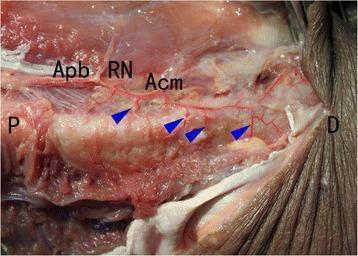
This clinical photograph shows a posterior dissection of a human arm specimen, oriented with proximal (P) and distal (D) labels. The image demonstrates the neurovascular anatomy along the humeral shaft following a Minimally Invasive Plate Osteosynthesis (MIPO) procedure. Key identified structures include the arteria profunda brachii (Apb), the radial nerve (RN), and the arteria collateralis media (Acm). Blue arrowheads highlight several accessory nutrient arteries. These arteries are shown originating from the arteria profunda brachii and entering the mid-distal portion of the humeral shaft posteriorly through the radial groove. The educational focus is the preservation of these critical vascular structures after MIPO, contrasting with potential disruption in more invasive surgical approaches. The image serves as an anatomical reference for orthopedic surgeons and medical students regarding the vascular supply of the humerus and the preservation of biological integrity during fracture fixation.
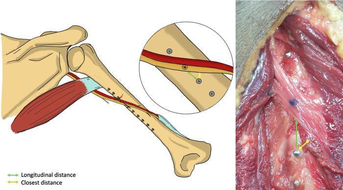
This composite educational graphic includes an anatomical illustration and a corresponding cadaveric clinical photograph focusing on the posterior view of a right humerus during a surgical procedure. The illustration on the left depicts the humeral shaft, shoulder girdle, and the course of the radial nerve (yellow) and profunda brachii artery (red) as they spiral across the posterior aspect of the bone. Several metallic screws are shown penetrating the humeral shaft from an anterolateral plate fixation. A magnified inset highlights the relationship between the screw tips and the neurovascular structures. The clinical photograph on the right shows a deep dissection of the upper arm, revealing the reddish-striated musculature, the humeral bone, and the exposed radial nerve. Both the diagram and the photograph utilize a color-coded measurement system: a green double-headed arrow indicates the longitudinal distance from the screw hole to the neurovascular structure, while a yellow double-headed arrow indicates the closest perpendicular distance. The content demonstrates the risk of iatrogenic neurovascular injury during humeral plating, specifically assessing the proximity of screw placement to vital structures.
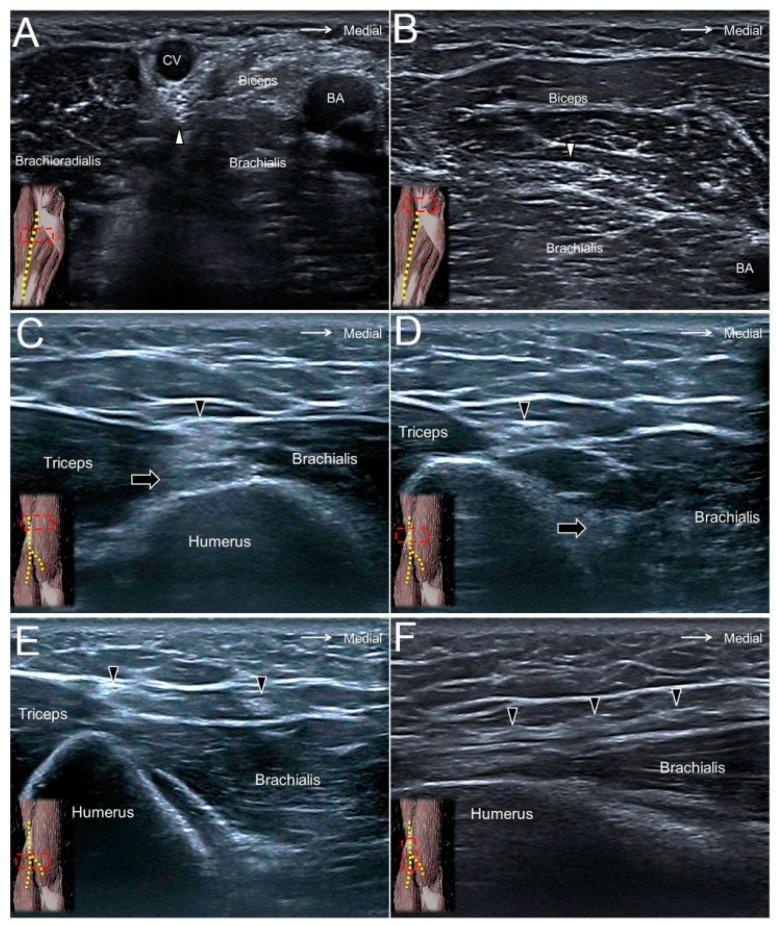
A composite clinical diagnostic image featuring six ultrasound panels (A-F) that demonstrate the sonographic appearance and anatomical landmarks of upper limb cutaneous nerves. Panel A and B (cross-sectional) show the lateral antebrachial cutaneous nerve (white arrowhead) at the elbow crease and distal forearm, illustrating its proximity to the cephalic vein (CV), biceps brachii muscle, and brachial artery (BA). Panels C, D, and E (cross-sectional) visualize the posterior antebrachial cutaneous nerve (black arrowhead) as it exits the spiral groove, descends into the distal forearm, and passes near the lateral epicondyle. These views highlight the nerve's superficial position relative to the triceps brachii muscle, brachialis muscle, and humerus. Panel F provides a long-axis longitudinal view of the posterior antebrachial cutaneous nerve, showing its linear hyperechoic trajectory along the brachialis fascia. Black arrows in C and D indicate the radial nerve's deeper course. Small anatomical orientation diagrams in each panel correlate the sonographic probe position with the arm's surface anatomy.
Structures Affected by a Cut on the Back of the Right Arm
1. Artery Affected: Profunda Brachii Artery (Deep Artery of the Arm)
Origin
Course
Termination
- Radial collateral artery - continues anteriorly, anastomosing with the radial recurrent artery in front of the lateral epicondyle
- Middle collateral artery - passes posteriorly, anastomosing with the interosseous recurrent artery behind the lateral epicondyle
2. Nerve(s) Affected
Primary Nerve: Radial Nerve
| Feature | Detail |
|---|---|
| Origin | Terminal branch of the posterior cord of the brachial plexus (C5, C6, C7, C8, T1) |
| Course in arm | Passes between the long head and between the lateral and medial heads of triceps; winds obliquely from medial to lateral in the radial groove on the posterior surface of humerus |
| Exit from posterior compartment | Pierces the lateral intermuscular septum to enter the anterior compartment, lying between brachialis and brachioradialis |
| Enters forearm | Anterior to the lateral epicondyle, just deep to brachioradialis |
Muscles innervated by radial nerve in the arm (all lost if cut here):
- Triceps brachii (all 3 heads) - C6, C7, C8 - elbow extension
- Anconeus
- Brachioradialis (branch given off as nerve exits posterior compartment)
- Extensor carpi radialis longus
- Lateral part of brachialis
Cutaneous branches (sensory loss):
-
Posterior cutaneous nerve of the arm
-
Inferior lateral cutaneous nerve of the arm
-
Posterior cutaneous nerve of the forearm
-
Ultimately - sensory loss over dorsum of hand and lateral 3.5 fingers
-
Gray's Anatomy for Students, p. 880-881
Clinical Consequences of Injury at This Level
| Finding | Explanation |
|---|---|
| Wrist drop | Paralysis of wrist extensors (ECRL, ECRB, ECU) |
| Finger drop | Loss of extensor digitorum and other finger extensors |
| Inability to extend elbow | If triceps is involved (depends on exact level) |
| Sensory loss | Dorsum of hand, lateral 3.5 fingers |
| Significant haemorrhage | Due to profunda brachii artery injury |