
Orbital extinteration right and frozen biopsy
orbital exenteration surgery technique
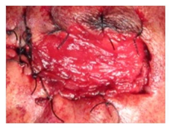
This clinical intraoperative photograph displays a reconstructive surgical procedure following orbital exenteration for squamous cell carcinoma. The image shows a pedicled temporalis muscle flap that has been medially transposed to fill the left orbital cavity. The muscle flap is characterized by its bright red, vascularized, and striated texture. It is secured into the defect using multiple black non-absorbable sutures, which are visible around the perimeter of the flap, anchoring it to the skin of the supraorbital region, the bridge of the nose, and the maxillary buttress. This surgical technique is utilized to provide vascularized bulk for the obliteration of the orbital socket and to provide a base for subsequent split-thickness skin grafting. The photograph serves as an educational example of maxillofacial reconstructive surgery and the management of extensive periorbital malignancies.
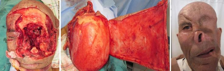
A three-panel clinical progression series demonstrating facial reconstructive surgery for a Type IV orbital-facial defect. The left panel shows the intraoperative state following an orbital exenteration and extensive tumor resection, resulting in a large, raw surgical cavity involving the left mid-face, forehead, and cheek with exposed deep tissue and irregular margins. The center panel illustrates the surgical technique, showing a large Converse scalping flap being prepared and mobilized for transposition; the flap consists of a substantial portion of the scalp and forehead skin. The right panel displays the post-operative outcome at a 2-year follow-up. The scalp flap successfully covers the primary defect, although significant facial asymmetry remains. Key features include the absence of the left eye, displacement and distortion of the nose and upper lip, and different skin texture/pigmentation between the donor flap and the native facial skin. This series illustrates complex maxillofacial reconstruction following oncological resection in the head and neck specialty.
Orbital Exenteration
Definition
Indications
- Gross invasion of periorbital fat, extraocular muscles, or optic nerve by tumor (most common: sinonasal carcinoma, maxillary sinus tumors, sebaceous carcinoma, squamous cell carcinoma, melanoma, adenoid cystic carcinoma)
- Invasion through the orbital periosteum (periorbita) into orbital contents - confirmed either clinically or intraoperatively by frozen section
- Severe refractory orbital mucormycosis/zygomycosis not controllable by debridement
- Other modalities (radiotherapy, local excision) have failed to control disease
- Total maxillectomy cases where tumor has breached the orbital floor into contents
Surgical Steps (Total Maxillectomy with Orbital Exenteration)
- Skin incisions from medial to lateral canthi; skin flaps raised off the orbicularis oculi muscles
- Superior orbital periosteum incised; subperiosteal dissection along the orbital roof back to the orbital apex - done circumferentially (except the quadrant continuous with the maxillectomy specimen)
- Lidocaine injection around the superior orbital fissure (SOF) and optic foramen - to prevent autonomic cardiac arrhythmias
- Neurovascular structures of the SOF carefully ligated and divided
- Optic nerve and ophthalmic artery ligated and divided
- Extraocular muscles transected at the orbital apex
- Medial and inferior orbital walls removed en bloc (in sinonasal carcinoma cases)
- Frozen sections obtained to ensure clear margins
Reconstruction Options
| Defect Type | Reconstruction Option |
|---|---|
| Soft tissue only | Free rectus abdominis flap |
| Bone + soft tissue (better cosmesis/function) | Scapula flap or DCIA (deep circumflex iliac artery) flap |
| With dental rehabilitation | Osseointegrated implants + obturator |
| Simple lining | Split-thickness skin graft |
| Large/complex | Converse scalping flap, temporalis muscle flap |
Frozen Section Biopsy - Role in Orbital Exenteration
What It Is
Specific Role at the Orbit
"The orbital periosteum may be involved with tumour while the underlying fat and orbital contents are not, and the use of peri-operative frozen section can determine those patients whose orbits can be safely retained. If frozen section confirms tumour has not penetrated the periosteum it can be resected and repaired without adversely affecting outcome."
In Craniofacial Resection
Frozen Section vs. Mohs Surgery (for Eyelid/Periorbital Tumors)
| Technique | Details | Best Use |
|---|---|---|
| Standard frozen section | Margins of excised specimen examined intraoperatively; if clear, reconstruction same day; if positive, re-excise | Standard periorbital tumors |
| Mohs micrographic surgery | Layered excision; specimens examined frozen; map of margins built layer by layer - maximum tumor removal with minimum normal tissue sacrifice | Sclerosing BCC, SCC, recurrent tumors, medial/lateral canthal tumors, diffuse finger-like extensions |
Clinical Pearls
- Right orbital exenteration specifically refers to complete removal of right orbital contents; "right-sided" involvement is anatomically close to the ethmoid sinus and cribriform plate, making skull base and dural involvement more likely during craniofacial resection
- Frozen section at the optic nerve stump should be obtained to confirm tumor-free margin
- Intraoperative lidocaine at the SOF is mandatory to prevent bradycardia/arrhythmia from traction on the ciliary ganglion
- Orbital involvement - even with exenteration - worsens prognosis significantly in sinonasal malignancy; 5-year absolute local control is ~50% for all sinonasal malignancies combined
- Cummings Otolaryngology Head and Neck Surgery, Ch. 94 (Total Maxillectomy with Orbital Exenteration, p. 1748-1749)
- KJ Lee's Essential Otolaryngology, Craniofacial Resection steps 24-25 (p. 892-893)
- Scott-Brown's Otorhinolaryngology Head & Neck Surgery, Management of the Orbit (pp. 7430-7444)
- Kanski's Clinical Ophthalmology, Surgical Excision - Frozen Section & Mohs (pp. 2585-2588)
Create a surgical reference guide on orbital exenteration for resident training
What format should the surgical reference guide be in? · What is the primary training level of the residents? · Which specialty is this guide for?
orbital exenteration defect reconstruction flap
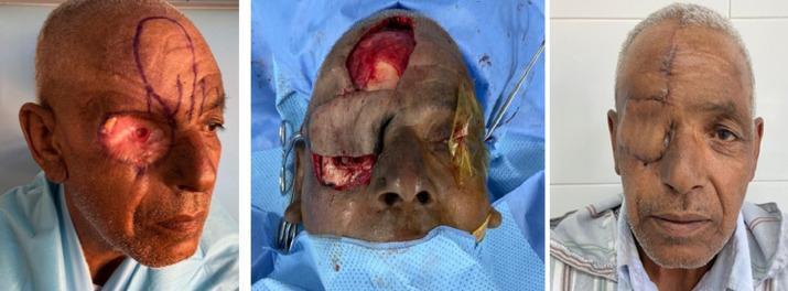
A series of three clinical photographs documenting the stages of orbital exenteration cavity reconstruction using a pedicled paramedian forehead flap. The left image (pre-operative/initial stage) shows a total right orbital exenteration defect with erythematous granulation tissue and surgical ink markings on the forehead delineating the donor flap based on the supratrochlear artery. The center image (intra-operative) illustrates the surgical transposition of the skin flap, which has been rotated 180 degrees to cover the orbital defect, leaving a secondary donor site on the upper forehead. The right image (post-operative) shows the final result at 4 months, demonstrating a well-healed reconstruction with the flap completely obliterating the orbital cavity. There is a visible midline vertical scar on the forehead and peripheral sutures where the flap was integrated into the facial skin. The texture and color of the flap are consistent with the surrounding facial tissue, indicating successful graft take and aesthetic integration.
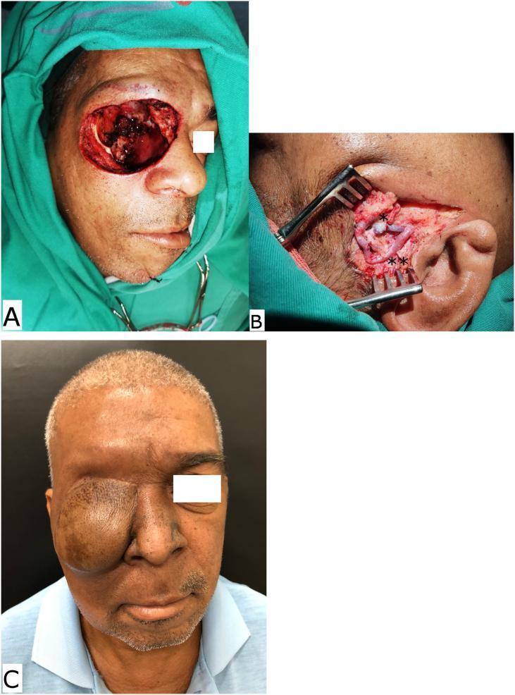
A sequence of clinical photographs illustrating the surgical reconstruction of a midface defect following tumor resection. (A) Intraoperative view of a right orbital exenteration and midface defect after surgical excision of advanced cutaneous squamous cell carcinoma, showing exposed orbital cavity and surrounding facial soft tissue. (B) High-magnification intraoperative view of the microsurgical field in the preauricular region. It demonstrates the microanastomosis of the superficial temporal recipient vessels: a single asterisk (*) marks the venous anastomosis between the superficial temporal vein and the flap vein, while a double asterisk (**) indicates the arterial anastomosis between the superficial temporal artery and the flap artery. (C) Late postoperative photograph of the patient following reconstruction with an anterolateral thigh (ALT) free flap. The flap provides voluminous coverage of the orbital and midface defect, demonstrating successful tissue transfer and wound closure. This series is an educational resource for plastic and reconstructive surgery, highlighting head and neck oncology, microvascular free flap reconstruction, and management of advanced skin malignancies.
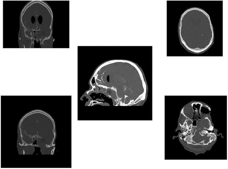
This set of diagnostic images consists of multiple Computed Tomography (CT) cisternogram views (coronal, axial, and sagittal) of the skull and facial bones. The images demonstrate status post-extensive surgical intervention for tumor resection and orbital exenteration. Key findings include a large bony defect in the left anterior cranial fossa and orbital region. The coronal and axial views show the absence of normal left orbital architecture and soft tissue contents, consistent with orbital exenteration. There is evidence of a transorbital and transnasal surgical approach, with disruption of the ethmoid and sphenoid sinuses to accommodate a pedicled nasoseptal flap and dermal fat graft for reconstruction of the skull base. The sagittal view highlights the discontinuity of the frontal bone and sphenoid sinus complex. High-density contrast material is visible within the subarachnoid space and basal cisterns without evidence of active extravasation, confirming a successful repair of a high-flow cerebrospinal fluid (CSF) leak. Bony margins appear irregular, indicating post-operative remodeling and surgical reconstruction.
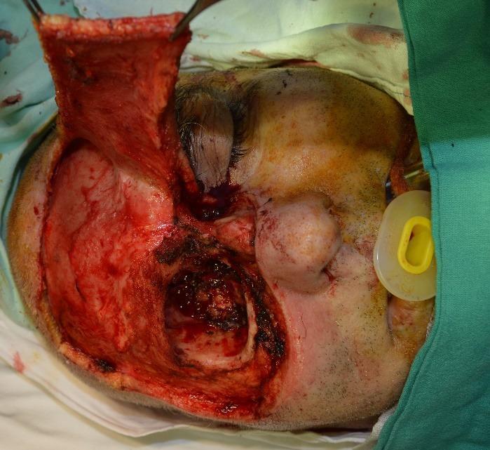
This clinical photograph captures a high-angle intraoperative view of a radical surgical excision and facial reconstruction. The patient is in a supine position, with the right side of the face showing intact skin, a closed eye, and an airway device (nasal cannula with a yellow cushion) in the right nostril. The left side of the face demonstrates a significant surgical defect following a subtotal maxillectomy and orbital exenteration. A large, vascularized frontal myofascial flap has been dissected and is being held superiorly by surgical instruments, ready for rotation to cover the primary defect. The surgical field reveals exposed deep soft tissues, bone margins of the orbital and maxillary regions, and evidence of cauterization and surgical trauma. This image serves as an educational resource for plastic and reconstructive surgery, illustrating the preparation of a regional flap for complex midface reconstruction following oncological resection.
orbital anatomy layers periorbita extraocular muscles
orbital prosthesis after exenteration osseointegrated implant
mkdir -p /tmp/workspace/orbital-exenteration-guide && ls /tmp/workspace/orbital-exenteration-guide/
~/orbital-exenteration-guide/build_guide.py
from docx import Document
from docx.shared import Pt, Inches, RGBColor, Cm
from docx.enum.text import WD_ALIGN_PARAGRAPH
from docx.enum.table import WD_TABLE_ALIGNMENT, WD_ALIGN_VERTICAL
from docx.oxml.ns import qn
from docx.oxml import OxmlElement
import urllib.request
import os
# ── helpers ─────────────────────────────────────────────────────────────────
def set_col_width(table, col_idx, width_inches):
for row in table.rows:
row.cells[col_idx].width = Inches(width_inches)
def shade_cell(cell, fill_hex):
tc = cell._tc
tcPr = tc.get_or_add_tcPr()
shd = OxmlElement('w:shd')
shd.set(qn('w:val'), 'clear')
shd.set(qn('w:color'), 'auto')
shd.set(qn('w:fill'), fill_hex)
tcPr.append(shd)
def add_horizontal_rule(doc):
p = doc.add_paragraph()
p.paragraph_format.space_before = Pt(2)
p.paragraph_format.space_after = Pt(2)
pPr = p._p.get_or_add_pPr()
pBdr = OxmlElement('w:pBdr')
bottom = OxmlElement('w:bottom')
bottom.set(qn('w:val'), 'single')
bottom.set(qn('w:sz'), '6')
bottom.set(qn('w:space'), '1')
bottom.set(qn('w:color'), '2E4A7A')
pBdr.append(bottom)
pPr.append(pBdr)
def add_section_heading(doc, text, level=1):
h = doc.add_heading(text, level=level)
h.paragraph_format.space_before = Pt(14) if level == 1 else Pt(10)
h.paragraph_format.space_after = Pt(4)
for run in h.runs:
run.font.color.rgb = RGBColor(0x2E, 0x4A, 0x7A) if level == 1 else RGBColor(0x1A, 0x6A, 0x5A)
return h
def add_body(doc, text, bold=False, indent=False):
p = doc.add_paragraph()
p.paragraph_format.space_after = Pt(4)
if indent:
p.paragraph_format.left_indent = Inches(0.3)
run = p.add_run(text)
run.font.size = Pt(11)
run.bold = bold
return p
def add_bullet(doc, text, level=0, bold_prefix=None):
p = doc.add_paragraph(style='List Bullet')
p.paragraph_format.space_after = Pt(2)
p.paragraph_format.left_indent = Inches(0.3 + level * 0.25)
if bold_prefix:
run = p.add_run(bold_prefix)
run.bold = True
run.font.size = Pt(11)
rest = p.add_run(text)
rest.font.size = Pt(11)
else:
run = p.add_run(text)
run.font.size = Pt(11)
return p
def add_numbered(doc, text, bold_prefix=None):
p = doc.add_paragraph(style='List Number')
p.paragraph_format.space_after = Pt(3)
p.paragraph_format.left_indent = Inches(0.3)
if bold_prefix:
run = p.add_run(bold_prefix)
run.bold = True
run.font.size = Pt(11)
rest = p.add_run(text)
rest.font.size = Pt(11)
else:
run = p.add_run(text)
run.font.size = Pt(11)
return p
def add_callout_box(doc, title, content, bg_hex="EBF4FA", border_hex="2E4A7A"):
"""Add a highlighted callout/tip box using a 1-cell table."""
tbl = doc.add_table(rows=1, cols=1)
tbl.alignment = WD_TABLE_ALIGNMENT.CENTER
cell = tbl.cell(0, 0)
shade_cell(cell, bg_hex)
# left border accent
tc = cell._tc
tcPr = tc.get_or_add_tcPr()
tcBdr = OxmlElement('w:tcBdr')
left = OxmlElement('w:left')
left.set(qn('w:val'), 'single')
left.set(qn('w:sz'), '24')
left.set(qn('w:space'), '0')
left.set(qn('w:color'), border_hex)
tcBdr.append(left)
tcPr.append(tcBdr)
p = cell.paragraphs[0]
p.paragraph_format.space_before = Pt(4)
p.paragraph_format.space_after = Pt(4)
p.paragraph_format.left_indent = Inches(0.1)
if title:
r = p.add_run(f"{title} ")
r.bold = True
r.font.size = Pt(11)
r.font.color.rgb = RGBColor(0x2E, 0x4A, 0x7A)
r2 = p.add_run(content)
r2.font.size = Pt(11)
doc.add_paragraph() # spacer
return tbl
def download_image(url, path):
try:
req = urllib.request.Request(url, headers={'User-Agent': 'Mozilla/5.0'})
with urllib.request.urlopen(req, timeout=15) as r:
with open(path, 'wb') as f:
f.write(r.read())
return True
except Exception as e:
print(f" Image download failed: {e}")
return False
def add_image_with_caption(doc, img_path, caption, width=5.5):
if os.path.exists(img_path):
p = doc.add_paragraph()
p.alignment = WD_ALIGN_PARAGRAPH.CENTER
run = p.add_run()
run.add_picture(img_path, width=Inches(width))
cap = doc.add_paragraph(caption)
cap.alignment = WD_ALIGN_PARAGRAPH.CENTER
cap.paragraph_format.space_after = Pt(10)
for run in cap.runs:
run.font.size = Pt(9)
run.font.italic = True
run.font.color.rgb = RGBColor(0x55, 0x55, 0x55)
else:
p = doc.add_paragraph(f"[Figure: {caption}]")
p.alignment = WD_ALIGN_PARAGRAPH.CENTER
# ── download images ──────────────────────────────────────────────────────────
IMG_DIR = "/tmp/workspace/orbital-exenteration-guide/images/"
os.makedirs(IMG_DIR, exist_ok=True)
images = {
"recon_temporalis.jpg": "https://cdn.orris.care/cdss_images/pmc_clinical_VQA_6a0dc032870aa6b1103219e3a5a4a6c24c714e33b18b1f06e556e137a144d277.jpg",
"recon_forehead_flap.jpg": "https://cdn.orris.care/cdss_images/pmc_clinical_VQA_2b7fe69f3e969a39bc89d0ad4110c34e00348a479c948158cb589377561403ac.jpg",
"recon_ALT_flap.jpg": "https://cdn.orris.care/cdss_images/pmc_clinical_VQA_39cc81da905aed6eac6a7e8e42eadd186bfc54257d78cb9357b763083ddb7a3c.jpg",
}
downloaded = {}
for fname, url in images.items():
dest = IMG_DIR + fname
ok = download_image(url, dest)
downloaded[fname] = dest if ok else None
print(f" {'OK' if ok else 'FAIL'}: {fname}")
# ── build document ───────────────────────────────────────────────────────────
doc = Document()
# Page margins
for section in doc.sections:
section.top_margin = Cm(2.0)
section.bottom_margin = Cm(2.0)
section.left_margin = Cm(2.5)
section.right_margin = Cm(2.0)
# Default Normal style
style = doc.styles["Normal"]
style.font.name = "Calibri"
style.font.size = Pt(11)
# ── COVER ────────────────────────────────────────────────────────────────────
doc.add_paragraph()
title_p = doc.add_paragraph()
title_p.alignment = WD_ALIGN_PARAGRAPH.CENTER
title_run = title_p.add_run("ORBITAL EXENTERATION")
title_run.bold = True
title_run.font.size = Pt(26)
title_run.font.color.rgb = RGBColor(0x2E, 0x4A, 0x7A)
sub_p = doc.add_paragraph()
sub_p.alignment = WD_ALIGN_PARAGRAPH.CENTER
sub_run = sub_p.add_run("Surgical Reference Guide for Residents")
sub_run.font.size = Pt(16)
sub_run.font.color.rgb = RGBColor(0x1A, 0x6A, 0x5A)
sub_run.bold = True
doc.add_paragraph()
info_p = doc.add_paragraph()
info_p.alignment = WD_ALIGN_PARAGRAPH.CENTER
info_p.add_run("Ophthalmology / Oculoplastics | Head & Neck Surgery / ENT\n").font.size = Pt(11)
info_p.add_run("PGY 1-2 Resident Training | August 2026").font.size = Pt(11)
add_horizontal_rule(doc)
doc.add_paragraph()
# Scope note
add_callout_box(doc,
"Scope:",
"This guide covers the indications, surgical anatomy, step-by-step technique, frozen section use, reconstruction options, complications, and post-operative management of orbital exenteration. "
"It is intended as a quick-reference companion to operative experience, not a substitute for direct supervision.",
bg_hex="FFF8E1", border_hex="F57F17"
)
# ── TABLE OF CONTENTS ────────────────────────────────────────────────────────
add_section_heading(doc, "Contents")
toc_items = [
"1. Overview & Definition",
"2. Anatomy Review",
"3. Indications",
"4. Contraindications & Special Considerations",
"5. Pre-operative Workup",
"6. Types of Exenteration",
"7. Step-by-Step Operative Technique",
"8. Intraoperative Frozen Section Biopsy",
"9. Reconstruction Options",
"10. Complications",
"11. Post-operative Management",
"12. Prognosis",
"13. Key Teaching Points",
"14. References",
]
for item in toc_items:
p = doc.add_paragraph(item, style="List Number")
p.paragraph_format.space_after = Pt(1)
for run in p.runs:
run.font.size = Pt(11)
doc.add_page_break()
# ─────────────────────────────────────────────────────────────────────────────
# SECTION 1: OVERVIEW
# ─────────────────────────────────────────────────────────────────────────────
add_section_heading(doc, "1. Overview & Definition")
add_body(doc,
"Orbital exenteration is the surgical removal of the entire orbital contents — globe, extraocular muscles, "
"orbital fat, lacrimal gland, and typically the eyelids. It is a radical procedure reserved for life-threatening "
"or locally destructive pathology when no organ-preserving option is feasible. It should only be performed "
"with curative intent."
)
add_callout_box(doc,
"Key Principle:",
"Orbital exenteration sacrifices vision and orbital anatomy permanently. The decision must balance oncologic "
"necessity against functional and psychosocial consequences — always involve the patient in a thorough informed consent discussion.",
bg_hex="FDECEA", border_hex="C62828"
)
# ─────────────────────────────────────────────────────────────────────────────
# SECTION 2: ANATOMY
# ─────────────────────────────────────────────────────────────────────────────
add_section_heading(doc, "2. Anatomy Review")
add_body(doc, "Structures removed in exenteration:")
structures = [
("Globe", "containing lens, vitreous, retina, choroid, sclera"),
("Extraocular muscles", "superior, inferior, medial, lateral rectus; superior and inferior oblique"),
("Orbital fat", "fills extraconal and intraconal space"),
("Lacrimal gland", "located in superolateral orbit"),
("Orbital periosteum (periorbita)", "fibrous lining of bony orbit; key frozen section decision layer"),
("Eyelids", "in standard exenteration; may be spared in lid-sparing variant"),
("Optic nerve", "ligated at orbital apex / optic foramen"),
]
for name, desc in structures:
add_bullet(doc, f" {desc}", bold_prefix=name + ":")
doc.add_paragraph()
add_body(doc, "Critical neurovascular structures at the orbital apex:")
apex_items = [
"Superior orbital fissure (SOF): cranial nerves III, IV, V1, VI + superior ophthalmic vein",
"Optic foramen: optic nerve (CN II) + ophthalmic artery",
"Ciliary ganglion (adjacent): vagal stimulation via traction can cause bradycardia/arrhythmia",
]
for item in apex_items:
add_bullet(doc, item)
add_callout_box(doc,
"Resident Pearl:",
"Always inject lidocaine around the SOF and optic foramen before ligation to prevent autonomic-mediated cardiac arrhythmias from traction on the ciliary ganglion.",
bg_hex="E8F5E9", border_hex="2E7D32"
)
# ─────────────────────────────────────────────────────────────────────────────
# SECTION 3: INDICATIONS
# ─────────────────────────────────────────────────────────────────────────────
add_section_heading(doc, "3. Indications")
add_section_heading(doc, "3a. Oncologic Indications", level=2)
add_body(doc, "Exenteration is indicated when tumor invades orbital contents beyond the periorbita:")
ind_table = doc.add_table(rows=1, cols=3)
ind_table.style = 'Table Grid'
ind_table.alignment = WD_TABLE_ALIGNMENT.CENTER
hdr = ind_table.rows[0].cells
for cell, text in zip(hdr, ["Tumor Type", "Scenario Requiring Exenteration", "Specialty"]):
shade_cell(cell, "2E4A7A")
p = cell.paragraphs[0]
r = p.add_run(text)
r.bold = True
r.font.color.rgb = RGBColor(0xFF, 0xFF, 0xFF)
r.font.size = Pt(10)
rows_data = [
("Sinonasal SCC / Adenocarcinoma", "Invasion through periorbita into fat, EOM, or optic nerve", "ENT / H&N"),
("Sebaceous gland carcinoma", "Diffuse pagetoid spread; extensive eyelid/orbital involvement", "Ophthalmology"),
("Squamous cell carcinoma (eyelid/conjunctiva)", "Orbital invasion; failure of prior excision", "Ophthalmology"),
("Adenoid cystic carcinoma (lacrimal gland)", "Perineural spread to apex; recurrence after local excision", "Both"),
("Malignant melanoma (conjunctival/uveal)", "Orbital invasion; other modalities failed", "Ophthalmology"),
("Maxillary sinus carcinoma", "Total maxillectomy with orbital extension through floor", "ENT / H&N"),
("Basal cell carcinoma (sclerosing/recurrent)", "Orbital invasion at medial or lateral canthus", "Ophthalmology"),
("Mucormycosis / Zygomycosis", "Invasive fungal sinusitis with orbital/intracranial extension not controllable by debridement alone", "ENT / Both"),
("Rhabdomyosarcoma", "Recurrence after chemoradiation; life-threatening disease", "Peds / H&N"),
]
for d in rows_data:
row = ind_table.add_row().cells
for cell, text in zip(row, d):
p = cell.paragraphs[0]
r = p.add_run(text)
r.font.size = Pt(10)
doc.add_paragraph()
add_section_heading(doc, "3b. Non-oncologic Indications (Rare)", level=2)
for item in [
"Intractable pain from a blind, phthisical eye (end-stage)",
"Severe orbital infection with necrotizing fasciitis unresponsive to all other management",
"Massive orbital trauma with unsalvageable contents",
]:
add_bullet(doc, item)
# ─────────────────────────────────────────────────────────────────────────────
# SECTION 4: CONTRAINDICATIONS
# ─────────────────────────────────────────────────────────────────────────────
add_section_heading(doc, "4. Contraindications & Special Considerations")
for item in [
"Non-curative intent (exenteration rarely palliative; individualize)",
"Bilateral disease where exenteration would render patient totally blind — multidisciplinary review required",
"Unresectable disease (carotid encasement, bilateral skull base involvement, extensive brain invasion >2 cm)",
"Patient refusal after full informed consent",
"Medical co-morbidities that preclude general anesthesia (relative)",
]:
add_bullet(doc, item)
add_callout_box(doc,
"Special Situation:",
"In invasive mucormycosis, orbital exenteration may be lifesaving if fungal spread is rapid, even without established oncologic criteria being met. "
"Current evidence suggests orbital preservation with aggressive debridement may be sufficient in selected cases — always consult infectious disease + neurosurgery urgently.",
bg_hex="FFF3E0", border_hex="E65100"
)
# ─────────────────────────────────────────────────────────────────────────────
# SECTION 5: PRE-OP WORKUP
# ─────────────────────────────────────────────────────────────────────────────
add_section_heading(doc, "5. Pre-operative Workup")
add_section_heading(doc, "5a. Imaging", level=2)
for item in [
"CT orbits with contrast: bone erosion, periorbita integrity, sinus involvement, skull base",
"MRI orbits (fat-sat + contrast): soft tissue extent, EOM invasion, optic nerve, perineural spread",
"CT chest/abdomen/pelvis or PET-CT: distant metastases staging",
]:
add_bullet(doc, item)
add_section_heading(doc, "5b. Pathologic Confirmation", level=2)
for item in [
"Incisional biopsy of primary tumor prior to exenteration",
"Discuss with pathology for intraoperative frozen section availability",
]:
add_bullet(doc, item)
add_section_heading(doc, "5c. Multidisciplinary Planning", level=2)
for item in [
"Ophthalmology, Head & Neck Surgery, Plastics/Reconstructive Surgery — joint operative planning",
"Radiation oncology: role of adjuvant radiotherapy (adjuvant RT improves local control in positive margins)",
"Anesthesia: plan for significant blood loss; arterial line + large-bore IV access",
"Prosthetics / Maxillofacial team: plan for orbital prosthesis or obturator",
]:
add_bullet(doc, item)
add_section_heading(doc, "5d. Informed Consent Points", level=2)
for item in [
"Permanent loss of vision and the eye",
"Significant facial disfigurement",
"Need for orbital prosthesis or reconstructive surgery",
"Risk of incomplete tumor clearance and need for adjuvant treatment",
"Psychosocial impact; refer to support services pre-operatively",
]:
add_bullet(doc, item)
# ─────────────────────────────────────────────────────────────────────────────
# SECTION 6: TYPES
# ─────────────────────────────────────────────────────────────────────────────
doc.add_page_break()
add_section_heading(doc, "6. Types of Exenteration")
types_table = doc.add_table(rows=1, cols=4)
types_table.style = 'Table Grid'
types_table.alignment = WD_TABLE_ALIGNMENT.CENTER
hdr = types_table.rows[0].cells
for cell, text in zip(hdr, ["Type", "Structures Removed", "Eyelids", "Indication"]):
shade_cell(cell, "1A6A5A")
p = cell.paragraphs[0]
r = p.add_run(text)
r.bold = True
r.font.color.rgb = RGBColor(0xFF, 0xFF, 0xFF)
r.font.size = Pt(10)
types_data = [
("Total (standard)", "Globe, EOM, fat, lacrimal gland, eyelids, conjunctiva, periorbita", "Removed", "Eyelid/skin involvement by tumor"),
("Lid-sparing (subtotal)", "Globe, EOM, fat, lacrimal gland; periorbita resected", "Preserved (lid margins/tarsal plates sacrificed)", "Orbital contents involved but eyelid skin uninvolved; lids cover defect + osseointegrated implants"),
("Extended", "All of above + adjacent bone (orbital walls, roof), sinus structures, maxilla, skull base", "Variable", "Tumor extends into adjacent bony structures"),
("Total maxillectomy + orbital exenteration", "Maxilla en bloc with orbital contents", "Removed", "Maxillary/sinonasal tumor invading orbital floor and contents"),
]
for d in types_data:
row = types_table.add_row().cells
for cell, text in zip(row, d):
p = cell.paragraphs[0]
r = p.add_run(text)
r.font.size = Pt(10)
doc.add_paragraph()
add_callout_box(doc,
"Decision Point:",
"Use intraoperative frozen section of the orbital periosteum to guide whether lid-sparing exenteration is sufficient vs. total exenteration required. "
"If frozen section confirms tumor has NOT penetrated the periosteum, the periosteum can be resected and repaired without adversely affecting oncologic outcome (Scott-Brown's).",
bg_hex="EBF4FA", border_hex="1565C0"
)
# ─────────────────────────────────────────────────────────────────────────────
# SECTION 7: OPERATIVE TECHNIQUE
# ─────────────────────────────────────────────────────────────────────────────
add_section_heading(doc, "7. Step-by-Step Operative Technique")
add_body(doc, "The following describes total orbital exenteration as part of total maxillectomy, the most commonly encountered scenario in combined ENT/ophthalmology practice.")
doc.add_paragraph()
steps = [
("Patient Positioning & Prep",
"Supine, head elevated 15-20 degrees. General anesthesia with endotracheal intubation. "
"Prep face and neck; wide draping to allow access to facial skin, oral cavity, and neck. "
"Arterial line monitoring recommended."),
("Incision Planning",
"Weber-Ferguson incision with subciliary extension for total maxillectomy approach. "
"Skin incisions placed from medial to lateral canthi for isolated orbital exenteration. "
"Mark incisions with surgical pen before injection of epinephrine."),
("Skin Flap Elevation",
"Skin flaps raised off the upper and lower orbicularis oculi muscles. "
"In total exenteration, eyelids are excised en bloc with specimen. "
"In lid-sparing approach, lids are dissected free and preserved."),
("Periorbital Dissection",
"Superior orbital periosteum (periorbita) incised along the orbital rim. "
"Subperiosteal dissection performed circumferentially along the orbital walls — roof, lateral wall, medial wall, floor — "
"back toward the orbital apex. The quadrant continuous with the maxillectomy specimen is left last."),
("Lidocaine Injection at Apex",
"MANDATORY: Inject lidocaine 1% with epinephrine around the superior orbital fissure (SOF) and optic foramen "
"BEFORE ligation. This prevents autonomic-mediated cardiac arrhythmias from traction on the ciliary ganglion."),
("Ligation at the SOF",
"Neurovascular structures at the SOF (CN III, IV, V1, VI, superior ophthalmic vein) are carefully identified, "
"ligated with suture or hemoclips, and divided."),
("Optic Nerve & Ophthalmic Artery",
"Optic nerve identified at the optic foramen. "
"Both the optic nerve and ophthalmic artery are ligated with suture and divided. "
"Send optic nerve stump for frozen section to confirm clear margin."),
("Extraocular Muscle Transection",
"All six extraocular muscles divided at the orbital apex using scissors or electrocautery."),
("Specimen Removal",
"Remaining soft tissue attachments at the posterior orbit divided with heavy Mayo scissors. "
"In total maxillectomy cases, the entire specimen (maxilla + orbital contents) is removed en bloc. "
"Pass specimen immediately to pathology for orientation, frozen sections, and permanent sections."),
("Margin Assessment",
"Obtain multiple frozen section biopsies from: anterior orbital fat margin, periorbita edges, "
"optic nerve stump, and any suspicious area. Re-excise until clear margins are confirmed."),
("Hemostasis",
"Electrocautery and surgical packing. The SOF and optic foramen bleed significantly — "
"have bipolar cautery and bone wax available. Packing with surgicel or hemostatic gauze."),
("Reconstruction",
"See Section 9 for reconstruction options. Perform in collaboration with reconstructive surgery team."),
]
for i, (title, content) in enumerate(steps, 1):
p = doc.add_paragraph()
p.paragraph_format.space_before = Pt(6)
p.paragraph_format.space_after = Pt(3)
r1 = p.add_run(f"Step {i}: {title}")
r1.bold = True
r1.font.size = Pt(11)
r1.font.color.rgb = RGBColor(0x2E, 0x4A, 0x7A)
body = doc.add_paragraph(content)
body.paragraph_format.left_indent = Inches(0.3)
body.paragraph_format.space_after = Pt(4)
for run in body.runs:
run.font.size = Pt(11)
# ─────────────────────────────────────────────────────────────────────────────
# SECTION 8: FROZEN SECTION
# ─────────────────────────────────────────────────────────────────────────────
doc.add_page_break()
add_section_heading(doc, "8. Intraoperative Frozen Section Biopsy")
add_body(doc,
"Frozen section biopsy is intraoperative histopathological examination of surgical margins. "
"Specimens are cryostat-sectioned and H&E stained within 15-30 minutes, enabling real-time margin decisions."
)
doc.add_paragraph()
add_section_heading(doc, "8a. When to Use Frozen Section in Orbital Surgery", level=2)
fs_indications = [
("Orbital periosteum margin", "To decide if orbital contents can be preserved vs. exenteration needed. "
"If no tumor penetration through periorbita → resect and repair periosteum, spare orbit. "
"If tumor penetrates periorbita → exenteration required."),
("Optic nerve stump", "Confirm clear margin at the orbital apex at time of exenteration."),
("Soft tissue surgical margins", "All edges of exenterated cavity — medial, lateral, superior, inferior."),
("Periorbital fat / EOM interface", "In limited-involvement cases where orbital preservation is attempted."),
("Nasal cavity and sinus margins", "During craniofacial resection — multiple frozen sections taken at all mucosal edges."),
]
for title, content in fs_indications:
add_bullet(doc, f" {content}", bold_prefix=title + ": ")
doc.add_paragraph()
add_section_heading(doc, "8b. Frozen Section vs. Mohs Surgery", level=2)
mohs_table = doc.add_table(rows=1, cols=3)
mohs_table.style = 'Table Grid'
mohs_table.alignment = WD_TABLE_ALIGNMENT.CENTER
hdr = mohs_table.rows[0].cells
for cell, text in zip(hdr, ["Technique", "Principle", "Best Use"]):
shade_cell(cell, "2E4A7A")
p = cell.paragraphs[0]
r = p.add_run(text)
r.bold = True
r.font.color.rgb = RGBColor(0xFF, 0xFF, 0xFF)
r.font.size = Pt(10)
mohs_data = [
("Standard Frozen Section",
"Margins of excised specimen examined at time of surgery; positive → re-excise that edge; negative → reconstruct same day",
"Standard periorbital tumors; orbital margin assessment"),
("Mohs Micrographic Surgery",
"Layered excision; each layer examined frozen; tumor map built; further tissue taken only where tumor persists; "
"maximizes tumor clearance while minimizing normal tissue sacrifice",
"Sclerosing BCC, recurrent SCC, medial/lateral canthal tumors, tumors with diffuse or finger-like extensions"),
]
for d in mohs_data:
row = mohs_table.add_row().cells
for cell, text in zip(row, d):
p = cell.paragraphs[0]
r = p.add_run(text)
r.font.size = Pt(10)
doc.add_paragraph()
add_callout_box(doc,
"Resident Pearl:",
"The irregular contours around the eyelids and extension of tumors into orbital fat can make frozen section interpretation technically difficult. "
"Communicate closely with your pathologist about orientation of specimens and the clinical question being asked.",
bg_hex="E8F5E9", border_hex="2E7D32"
)
# ─────────────────────────────────────────────────────────────────────────────
# SECTION 9: RECONSTRUCTION
# ─────────────────────────────────────────────────────────────────────────────
add_section_heading(doc, "9. Reconstruction Options")
add_body(doc,
"Reconstruction goals: obliterate dead space, provide vascularized tissue, maintain facial contour, "
"allow prosthetic rehabilitation, minimize donor site morbidity."
)
doc.add_paragraph()
recon_table = doc.add_table(rows=1, cols=4)
recon_table.style = 'Table Grid'
recon_table.alignment = WD_TABLE_ALIGNMENT.CENTER
hdr = recon_table.rows[0].cells
for cell, text in zip(hdr, ["Option", "Type", "Advantages", "Best For"]):
shade_cell(cell, "1A6A5A")
p = cell.paragraphs[0]
r = p.add_run(text)
r.bold = True
r.font.color.rgb = RGBColor(0xFF, 0xFF, 0xFF)
r.font.size = Pt(10)
recon_data = [
("Split-thickness skin graft (STSG)", "Graft", "Simple; short operative time; prosthesis-friendly surface", "Medically frail patients; patients planned for orbital prosthesis"),
("Temporalis muscle flap", "Regional pedicled", "Reliable vascular supply; nearby donor site; fills orbital cavity well", "Moderate defects; good if free flap not feasible"),
("Paramedian forehead flap", "Regional pedicled", "Excellent color/texture match to facial skin; strong vascular pedicle (supratrochlear A.)", "Isolated orbital exenteration defects"),
("Free rectus abdominis flap", "Free microvascular", "Large soft tissue volume; reliable perforators; obliterates large cavity", "Soft tissue only defects; combined maxillectomy + exenteration"),
("Free DCIA flap (deep circumflex iliac artery)", "Free microvascular", "Bone + soft tissue; restores bony orbital framework; best cosmesis/function", "Total maxillectomy + exenteration requiring bony reconstruction"),
("Free scapula flap", "Free microvascular", "Bone + soft tissue; flexible geometry for orbital reconstruction", "Combined bony + soft tissue reconstruction"),
("ALT (anterolateral thigh) flap", "Free microvascular", "Large thin pliable skin paddle; long pedicle; minimal donor site morbidity", "Large combined midface + orbital defects"),
("Orbital prosthesis (after STSG or spontaneous granulation)", "Prosthetic", "Acceptable cosmesis without major reconstruction; allows tumor surveillance", "Selected patients; esp. with osseointegrated implants placed at exenteration"),
]
for d in recon_data:
row = recon_table.add_row().cells
for cell, text in zip(row, d):
p = cell.paragraphs[0]
r = p.add_run(text)
r.font.size = Pt(10)
doc.add_paragraph()
# Insert reconstruction images
if downloaded.get("recon_temporalis.jpg"):
add_image_with_caption(
doc, downloaded["recon_temporalis.jpg"],
"Fig. 1 - Temporalis muscle flap transposed to fill orbital cavity after exenteration for SCC. "
"Multiple non-absorbable sutures anchor the flap to orbital margins prior to skin grafting.",
width=5.0
)
if downloaded.get("recon_forehead_flap.jpg"):
add_image_with_caption(
doc, downloaded["recon_forehead_flap.jpg"],
"Fig. 2 - Paramedian forehead flap reconstruction of right orbital exenteration defect. "
"Left: surgical markings; Center: flap transposed; Right: 4-month post-operative result.",
width=5.5
)
if downloaded.get("recon_ALT_flap.jpg"):
add_image_with_caption(
doc, downloaded["recon_ALT_flap.jpg"],
"Fig. 3 - Anterolateral thigh (ALT) free flap reconstruction following right orbital exenteration "
"and midface resection for advanced cutaneous SCC. Note microsurgical anastomosis to superficial temporal vessels.",
width=5.5
)
# ─────────────────────────────────────────────────────────────────────────────
# SECTION 10: COMPLICATIONS
# ─────────────────────────────────────────────────────────────────────────────
doc.add_page_break()
add_section_heading(doc, "10. Complications")
add_section_heading(doc, "10a. Intraoperative", level=2)
for item in [
"Hemorrhage: from ophthalmic artery, SOF vessels, ethmoid arteries — have bipolar + bone wax available",
"Cardiac arrhythmia / bradycardia: from traction on ciliary ganglion at apex — prevent with lidocaine injection",
"CSF leak: if dura violated during craniofacial extension — repair with fascia lata or dural graft matrix + pericranial flap",
"Carotid artery injury: during extended resection — rare but catastrophic",
"Incomplete resection / positive margins: rely on frozen section to guide re-excision intraoperatively",
]
add_bullet(doc, item)
add_section_heading(doc, "10b. Early Post-operative (0-30 days)", level=2)
for item in [
"Hematoma / seroma in orbital cavity",
"Flap failure (partial or total): monitor free flap with Doppler q1-2h post-op",
"Wound dehiscence: especially at skin graft edges",
"CSF leak (if skull base involved): head elevation, lumbar drain, early neurosurgery review",
"Infection / orbital cavity sepsis",
"Eustachian tube dysfunction (if maxillectomy component): conductive hearing loss",
]
add_bullet(doc, item)
add_section_heading(doc, "10c. Late Complications", level=2)
for item in [
"Local tumor recurrence: most common failure pattern in sinonasal malignancies (up to 67% of failures are local)",
"Exposure of bony orbital walls with graft or flap loss",
"Contracture of orbital socket: interferes with prosthesis fitting",
"Epiphora / dacryocystitis: if nasolacrimal duct not addressed",
"Chronic sinusitis (if sinus cavities communicated)",
"Psychological morbidity: depression, body image disturbance — refer to support services early",
]
add_bullet(doc, item)
# ─────────────────────────────────────────────────────────────────────────────
# SECTION 11: POST-OP MANAGEMENT
# ─────────────────────────────────────────────────────────────────────────────
add_section_heading(doc, "11. Post-operative Management")
add_section_heading(doc, "11a. Immediate (ICU / HDU, 24-48h)", level=2)
for item in [
"Free flap monitoring: hourly Doppler assessment; flap checks by nursing q1-2h",
"IV antibiotics: typically 24-48h perioperative",
"Head elevation 30-45 degrees to reduce edema",
"Nasogastric feeding if oral feeding not possible post-maxillectomy",
"Pain management: multimodal analgesia; avoid NSAIDs in first 48h if significant bleeding risk",
]:
add_bullet(doc, item)
add_section_heading(doc, "11b. Inpatient (Day 2-7)", level=2)
for item in [
"Wound care: gentle saline irrigation of orbital cavity twice daily",
"Oral hygiene: chlorhexidine rinses if oral cavity involved",
"Obturator insertion (if palate/maxilla resected): fit temporary obturator at 5-7 days",
"Speech and language therapy (SALT) referral",
"Physiotherapy: jaw exercises to prevent trismus",
"Psychiatric / psychological support referral",
]:
add_bullet(doc, item)
add_section_heading(doc, "11c. Outpatient Follow-up", level=2)
for item in [
"Definitive prosthetic fitting: 4-6 weeks post-op once edema resolved",
"Osseointegrated implants (if placed): allow 3-6 months integration before loading prosthesis",
"Adjuvant radiotherapy: referral within 6 weeks if positive or close margins",
"Oncology surveillance: MRI +/- CT q3-6 months for 2 years, then annually",
"Multidisciplinary tumor board: present final pathology and margins",
]:
add_bullet(doc, item)
# ─────────────────────────────────────────────────────────────────────────────
# SECTION 12: PROGNOSIS
# ─────────────────────────────────────────────────────────────────────────────
add_section_heading(doc, "12. Prognosis")
add_body(doc, "Prognosis is strongly dependent on tumor histology, margin status, and extent of disease:")
for item in [
"Sinonasal malignancies overall: 5-year absolute local control ~50%; 10-year ~31%; 15-year ~21%",
"Orbital involvement despite exenteration significantly worsens survival",
"Negative margins: strongest predictor of survival",
"Failure pattern: predominantly local (up to 66.7% of recurrences in skull base series); distant ~46%; regional ~17%",
"Advanced-stage disease (Class IV defects): typically poor prognosis; reconstruction secondary to feasibility assessment",
"Adenoid cystic carcinoma / olfactory neuroblastoma: better long-term survival but late relapses possible for decades",
"SNUC (sinonasal undifferentiated carcinoma) / sinonasal melanoma: very poor prognosis",
]:
add_bullet(doc, item)
add_callout_box(doc,
"Clinical Implication:",
"Even after exenteration, orbital involvement is associated with significantly poorer survival. "
"Preoperative imaging and intraoperative frozen section are used to identify patients who can safely retain the orbit — "
"but attempts to preserve orbital contents that result in positive margins lead to orbital recurrence.",
bg_hex="FDECEA", border_hex="C62828"
)
# ─────────────────────────────────────────────────────────────────────────────
# SECTION 13: KEY TEACHING POINTS
# ─────────────────────────────────────────────────────────────────────────────
doc.add_page_break()
add_section_heading(doc, "13. Key Teaching Points for Residents (PGY 1-2)")
teaching = [
("Always check frozen section of the periorbita",
"Before committing to exenteration, perioperative frozen section of the orbital periosteum determines whether "
"the globe can be saved. If tumor has not breached the periosteum, resection and repair are sufficient."),
("Lidocaine at the orbital apex is non-negotiable",
"Traction on the ciliary ganglion at the superior orbital fissure can cause severe bradycardia. "
"Inject lidocaine 1% +/- epinephrine around the SOF and optic foramen before any manipulation."),
("Exenteration only with curative intent",
"This is a life-altering operation. If resection is not curative, the morbidity of exenteration is not justified. "
"Palliative exenteration is rare and requires very careful MDT discussion."),
("Plan reconstruction before you operate",
"Reconstruct with the least complex option that achieves the goals. "
"Bony defects require bone + soft tissue (DCIA/scapula); soft tissue only can use rectus or temporalis. "
"Lid-sparing exenteration: the preserved lids provide excellent natural cover for osseointegrated implants."),
("Margins drive outcome more than radicality alone",
"Radical resection including exenteration does not guarantee survival if margins are positive. "
"Frozen section control of all margins — not just the orbital periosteum — is essential."),
("Psychosocial support starts pre-operatively",
"Loss of the eye is devastating. Early psychiatric and support service referral, clear discussion of prosthetics, "
"and patient-centered communication are as important as the surgery itself."),
("Adjuvant radiotherapy matters",
"Refer for RT within 6 weeks if margins are positive/close, or for high-risk histologies (SCC, adenoid cystic). "
"RT significantly improves local control in the post-exenteration setting."),
]
for i, (title, content) in enumerate(teaching, 1):
add_callout_box(doc,
f"{i}. {title}",
content,
bg_hex="EBF4FA" if i % 2 == 0 else "E8F5E9",
border_hex="1565C0" if i % 2 == 0 else "2E7D32"
)
# ─────────────────────────────────────────────────────────────────────────────
# SECTION 14: REFERENCES
# ─────────────────────────────────────────────────────────────────────────────
add_section_heading(doc, "14. References")
refs = [
"Cummings CW et al. Cummings Otolaryngology: Head and Neck Surgery, 7th ed. Elsevier, 2020. "
"Ch. 94: Total Maxillectomy with Orbital Exenteration (pp. 1748-1750).",
"Lee KJ. Essential Otolaryngology: Head & Neck Surgery, 11th ed. McGraw-Hill, 2022. "
"Ch. 49: Craniofacial Resection Steps 24-25 (pp. 891-893).",
"Gleeson M (ed). Scott-Brown's Otorhinolaryngology, Head & Neck Surgery, 8th ed. CRC Press, 2019. "
"Management of the Orbit (pp. 7430-7444).",
"Kanski JJ, Bowling B. Kanski's Clinical Ophthalmology: A Systematic Approach, 10th ed. Elsevier, 2023. "
"Frozen Section & Mohs Surgery (pp. 2585-2588); Orbital Exenteration (pp. 6446-6449).",
"Sabiston DC (ed). Sabiston Textbook of Surgery: The Biological Basis of Modern Surgical Practice, 21st ed. Elsevier, 2022. "
"Split-Thickness Skin Grafts and Orbital Reconstruction.",
"Schwartz SI (ed). Schwartz's Principles of Surgery, 11th ed. McGraw-Hill, 2019. "
"Nose and Paranasal Sinuses (p. 3307-3308).",
"Bailey & Love's Short Practice of Surgery, 28th ed. CRC Press, 2023. Maxillary Resection (pp. 2224-2239).",
"Wills Eye Manual: Office and Emergency Room Diagnosis and Treatment of Eye Disease, 8th ed. Wolters Kluwer, 2022.",
]
for i, ref in enumerate(refs, 1):
p = doc.add_paragraph()
p.paragraph_format.space_after = Pt(4)
p.paragraph_format.left_indent = Inches(0.3)
p.paragraph_format.first_line_indent = Inches(-0.3)
r = p.add_run(f"{i}. {ref}")
r.font.size = Pt(10)
# ── Footer ───────────────────────────────────────────────────────────────────
add_horizontal_rule(doc)
footer_p = doc.add_paragraph()
footer_p.alignment = WD_ALIGN_PARAGRAPH.CENTER
fr = footer_p.add_run("Orbital Exenteration Surgical Reference Guide | PGY 1-2 Resident Training | August 2026")
fr.font.size = Pt(9)
fr.font.italic = True
fr.font.color.rgb = RGBColor(0x77, 0x77, 0x77)
discp = doc.add_paragraph()
discp.alignment = WD_ALIGN_PARAGRAPH.CENTER
dr = discp.add_run("This document is for educational purposes only. Clinical decisions must be made under direct consultant supervision.")
dr.font.size = Pt(8)
dr.font.italic = True
dr.font.color.rgb = RGBColor(0xAA, 0xAA, 0xAA)
# ── Save ─────────────────────────────────────────────────────────────────────
out_path = "/tmp/workspace/orbital-exenteration-guide/Orbital_Exenteration_Resident_Guide.docx"
doc.save(out_path)
print(f"\nSaved: {out_path}")
cd /tmp/workspace/orbital-exenteration-guide && python3 build_guide.py 2>&1
for item in [...]\n add_bullet(...) - the for loop body needs proper indentation with a colon. The em dashes in the strings are also being used (the file content has them). Let me fix the loops:~/orbital-exenteration-guide/build_guide.py
Successfully replaced 1 instance(s) of the string in '/tmp/workspace/orbital-exenteration-guide/build_guide.py'
cd /tmp/workspace/orbital-exenteration-guide && python3 -c " import ast, sys with open('build_guide.py') as f: src = f.read() try: ast.parse(src) print('Syntax OK') except SyntaxError as e: print(f'SyntaxError at line {e.lineno}: {e.msg}') lines = src.split('\n') start = max(0, e.lineno-3) end = min(len(lines), e.lineno+2) for i, l in enumerate(lines[start:end], start+1): print(f'{i}: {l}') "
cd /tmp/workspace/orbital-exenteration-guide && python3 -c " import re with open('build_guide.py', 'r') as f: content = f.read() # Fix pattern: ]\n add_bullet -> ]:\n add_bullet content = re.sub(r'(\])\n([ \t]+add_bullet)', r'\1:\n\2', content) content = re.sub(r'(\])\n([ \t]+add_numbered)', r'\1:\n\2', content) with open('build_guide.py', 'w') as f: f.write(content) # Verify import ast try: ast.parse(content) print('Syntax OK') except SyntaxError as e: print(f'SyntaxError at line {e.lineno}: {e.msg}') lines = content.split('\n') start = max(0, e.lineno-3) end = min(len(lines), e.lineno+3) for i, l in enumerate(lines[start:end], start+1): print(f'{i}: {l}') "
cd /tmp/workspace/orbital-exenteration-guide && python3 build_guide.py 2>&1
ls -lh /tmp/workspace/orbital-exenteration-guide/Orbital_Exenteration_Resident_Guide.docx
Orbital Exenteration Resident Guide
Word Document · DOCX
| Section | Content |
|---|---|
| Cover + Scope | Title page with specialty, level, and disclaimer |
| Contents | Numbered table of contents |
| 1. Overview & Definition | Core concept, intent of surgery |
| 2. Anatomy Review | Structures removed, orbital apex neurovascular anatomy, arrhythmia pearl |
| 3. Indications | Color-coded table of 9 tumor types (ENT + Ophtho + shared), non-oncologic indications |
| 4. Contraindications | Bilateral disease, non-curative setting, mucormycosis nuance |
| 5. Pre-op Workup | Imaging (CT/MRI/PET), pathology, MDT planning, consent points |
| 6. Types of Exenteration | Table: total / lid-sparing / extended / total maxillectomy + exenteration |
| 7. Operative Technique | 12-step numbered operative guide with resident-oriented notes |
| 8. Frozen Section | Decision flowchart logic, frozen vs. Mohs table, pathology communication tip |
| 9. Reconstruction | 8-option table (STSG to free ALT flap) + 3 embedded clinical photographs |
| 10. Complications | Intraoperative, early post-op, late - with clinical actions |
| 11. Post-op Management | ICU care, inpatient, outpatient surveillance schedule |
| 12. Prognosis | 5/10/15-year local control data, failure patterns by histology |
| 13. Key Teaching Points | 7 color-coded callout boxes for PGY 1-2 trainees |
| 14. References | 8 primary textbook citations (Cummings, KJ Lee, Scott-Brown, Kanski, Wills Eye, Sabiston, Schwartz, Bailey & Love) |
Conjunctival squamous cell carcinoma 2 cm mass orbital exenteration
conjunctival squamous cell carcinoma large mass orbital invasion
"conjunctival squamous cell carcinoma" AND "orbital exenteration"
PMID: 33009325
Conjunctival Squamous Cell Carcinoma - 2 cm Mass: The Exenteration Decision
Background: Where Does This Sit in the OSSN Spectrum?
| Stage | Definition |
|---|---|
| Conjunctival epithelial dysplasia | Dysplastic cells confined to basal epithelial layers |
| Carcinoma in situ (CIS) | Dysplastic cells involve full epithelial thickness |
| Invasive SCC | Tumor invades underlying subepithelial stroma |
- Kanski's Clinical Ophthalmology, p. 866-868
Histological Images from Kanski's
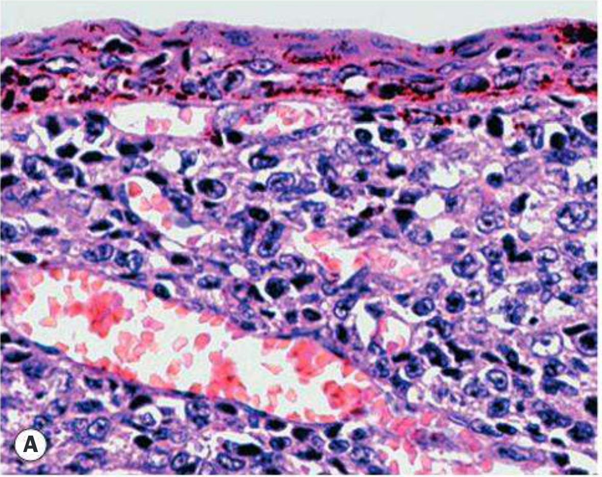
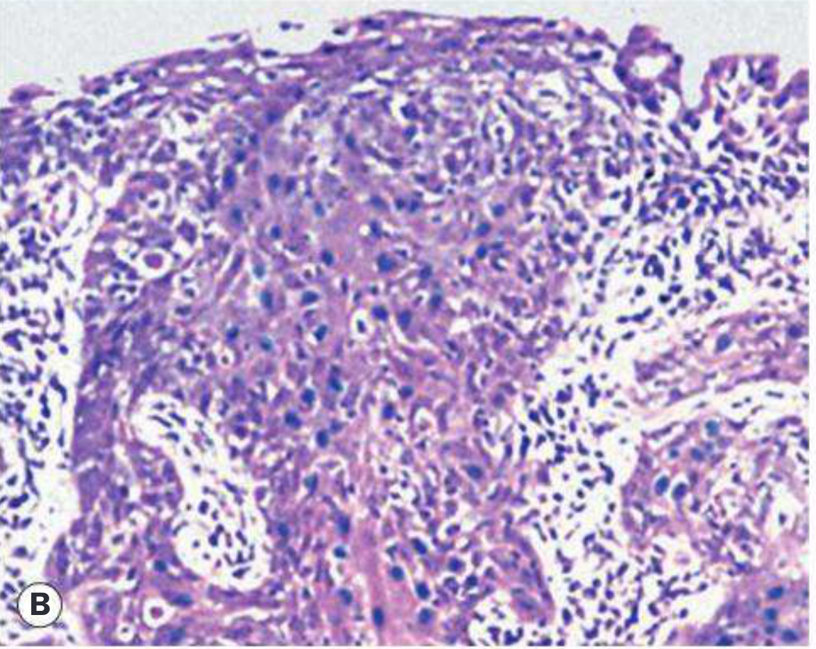
Does a 2 cm Conjunctival SCC Need Orbital Exenteration?
The T-Staging Framework (AJCC) and Exenteration Threshold
| T Stage | Definition | Exenteration? |
|---|---|---|
| Tis | Carcinoma in situ | No |
| T1 | Tumor ≤5 mm, limited to conjunctiva/cornea | No |
| T2 | Tumor >5 mm, limited to conjunctiva/cornea, no orbital invasion | No - wide excision |
| T3 | Tumor invades adjacent structures (eyelid, cornea, orbit, sinuses) WITHOUT intraocular invasion | Possible - depends on extent |
| T4 | Tumor invades orbit with bone invasion, or invades globe, CNS, or paranasal sinuses | Yes - exenteration often required |
- Exenteration was performed in 10/44 cases (23%)
- It was strongly associated with T3 or more advanced disease at presentation (p <0.001)
- T2 disease: 8 patients - none required exenteration in that series
- Exenteration and nodal metastasis were both independently associated with disease-related death
Standard Treatment for a 2 cm (T2) Conjunctival SCC
Primary: Wide Local Excision with Frozen Section Control
- Excision with 2-4 mm clear margins (no-touch technique)
- Intraoperative frozen section of all margins
- Critical: do not cut below Bowman's membrane - invasive recurrence can penetrate the corneal stroma
- Margins confirmed clear → conjunctival reconstruction same session
- Margins positive → re-excise until clear
Adjuvant Therapy Options
| Agent | Regimen | Effect | Notes |
|---|---|---|---|
| Topical interferon alfa-2b | Drops 1 MIU/mL QID; or subconj. injection 3 MIU/0.5 mL | ~80% complete resolution | Drug of choice; expensive; requires refrigeration |
| 5-Fluorouracil (5-FU) | 1% drops QID for 1 week, 3-week rest; repeat cycles until clear | <10% recurrence rate | Eyelid edema, keratopathy as side effects |
| Mitomycin C (MMC) | 0.02-0.04% drops | Effective but more ocular side effects | Second-line after 5-FU/IFN |
| Cryotherapy | Double freeze-thaw at excision margins | Reduces local recurrence | Applied to conjunctival edges at time of surgery |
| Brachytherapy | Radiation plaque | Adjunct in selected cases | Used for recurrences |
- Kanski's Clinical Ophthalmology, p. 868
- Goodman & Gilman's Pharmacological Basis of Therapeutics (IFN-alfa2b and 5-FU for OSSN)
When Does This 2 cm Mass Require Exenteration?
- Orbital invasion - tumor penetrates the periorbita into orbital fat, extraocular muscles, or optic nerve (T4)
- Intraocular extension (though uncommon in conjunctival SCC)
- Failure of other modalities - persistent/recurrent disease after multiple attempts at local excision and topical chemotherapy, where no organ-preserving option remains
- Immunosuppressed patients (e.g., organ transplant recipients): these patients have significantly higher rates of local recurrence and progression to exenteration (p<0.01 in El-Hadad et al.)
- Heavy smokers with inferior fornix involvement: tend to have larger, more aggressive tumors
The Periorbita Frozen Section Decision
- Tumor has NOT penetrated periorbita → resect and repair periosteum; orbit preserved
- Tumor HAS penetrated periorbita into fat or EOM → exenteration required
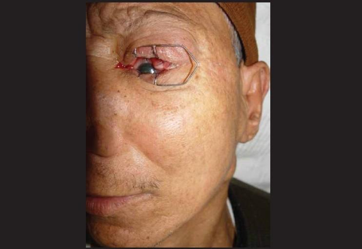
Clinical Image: Conjunctival SCC with Regional Metastasis
Staging Workup for a 2 cm Conjunctival SCC Mass
- Anterior segment OCT or UBM - estimate depth of stromal invasion
- MRI orbits (fat-suppressed T1+gadolinium) - periorbital or orbital fat infiltration
- CT orbits - bony erosion
- Sentinel lymph node biopsy / neck ultrasound - regional spread (preauricular, submandibular, cervical nodes); overall nodal metastasis rate ~9% in conjunctival SCC
- HIV/immunosuppression status - significantly alters prognosis and recurrence risk
- Exfoliative or impression cytology - if excision not yet performed
Summary: Decision Algorithm
2 cm Conjunctival SCC Mass
│
▼
Staging (OCT/MRI/CT)
│
┌─────┴──────┐
Confined to Orbital/intraocular
conjunctiva invasion on imaging
(T2/T3 limited) │
│ ▼
▼ EXENTERATION candidate
Wide excision (confirm with intraop
+ frozen section frozen section)
+ adjuvant
(IFN/5-FU/MMC)
+ cryotherapy
│
If positive margins / recurrence
│
▼
Topical chemo ± radiation
│
If fails / orbital invasion develops
│
▼
EXENTERATION
Key Points
- A 2 cm conjunctival SCC (T2) does not routinely require exenteration - wide excision with 2-4 mm margins and adjuvant topical therapy is the standard approach
- Exenteration is reserved for T3-T4 disease with orbital invasion, failed prior treatment, or uncontrollable recurrence
- Immunocompromised patients (especially organ transplant recipients) have significantly higher risk of progression requiring exenteration
- Intraoperative frozen section at the periorbita is the critical decision point when orbital invasion is suspected
- Exenteration and nodal metastasis are both independently associated with disease-related death in conjunctival SCC - El-Hadad et al., MD Anderson 2021, PMID 33009325