
Uric acid and gout
gout monosodium urate crystals joint inflammation podagra
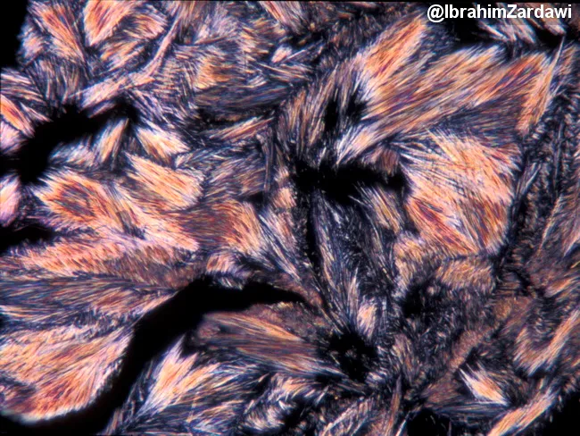
Polarized light microscopy image of monosodium urate crystals in synovial fluid from a joint, illustrating classic gout crystallography. Under cross-polarized illumination, slender, needle-shaped crystals appear colorless individually but generate bright interference colors when overlapped. These crystals are elongated with pointed termini and align to yield yellow-blue birefringence, creating a spectrum. The image showcases monosodium urate deposition, a hallmark of gout, with characteristic needle morphology and negative birefringence. The sample is not stained; polarization highlights crystalline structure and inter-crystal interference. Diagnostic significance includes definitive confirmation of gout and differentiation from calcium pyrophosphate crystals (pseudogout), which are rhomboid and weakly birefringent. Clinical relevance encompasses acute gout assessment, urate-lowering therapy decisions, and teaching crystal pathology in rheumatology education. Techniques: high magnification polarized light imaging with standard filters; interference colors reflect crystal overlap rather than stain. Credit: image courtesy of Dr. Ibrahim Zardawi. In PLM, monosodium urate crystals exhibit strong negative birefringence; their yellow-to-blue color shift depends on crystal orientation relative to the slow axis. By contrast, calcium pyrophosphate crystals in pseudogout are rhomboid and show weak positive birefringence, aiding differential diagnosis. This image is valuable for education, research on crystal pathophysiology, and confirming joint effusion analysis in rheumatologic clinics. Cited for teaching laboratories.
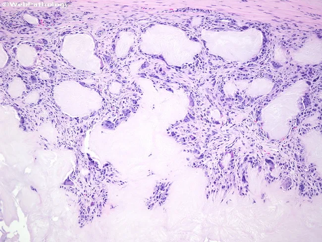
Light microscopy of a soft-tissue biopsy surrounding a gouty tophus shows extensive chalky, eosinophilic to basophilic amorphous deposits with adjacent granulomatous inflammation. Monosodium urate crystals are deposited within the periarticular soft tissue and synovium, appearing as refractile needle-shaped material that often dissolves during routine processing, leaving voids. The tophus is surrounded by a dense inflammatory capsule composed of macrophages, histiocytes, and numerous foreign body giant cells actively attempting to phagocytose crystalline material. Fibroblasts and scant lymphocytes form a fibrous stroma, with variable vascular proliferation. The gross histology may mimic a tumor or infection, but localization near joints and the characteristic granulomatous reaction supports chronic tophaceous gout. Polarized light examination would demonstrate negative birefringent crystals consistent with monosodium urate. Clinically, this pattern confirms chronic hyperuricemia with urate overproduction or reduced excretion and correlates with long-standing gout. The diagnostic significance lies in distinguishing inflammatory tophaceous gout from rheumatoid nodules, infectious arthritis, or soft-tissue neoplasms. Potential clinical use cases include confirming gout in patients with hyperuricemia, evaluating painful soft-tissue swellings around joints, and guiding urate-lowering therapy or surgical planning. This image emphasizes the pathognomonic granulomatous response to urate crystals and its impact on joint function. Correlation with serum uric acid improves diagnostic accuracy.
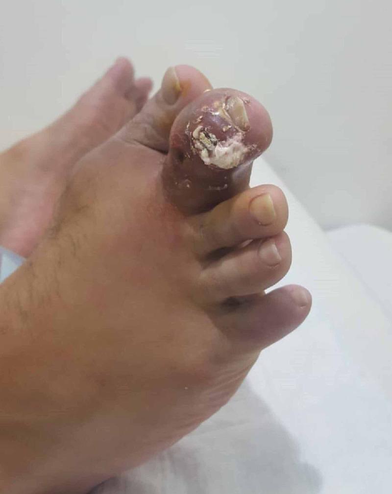
Clinical photograph of a human foot, focusing on the second toe, which exhibits severe gouty tophus formation. The distal portion of the toe is significantly enlarged with tense, erythematous skin, indicating acute inflammation. At the distal interphalangeal joint level, there is a visible rupture of the skin with the extrusion of chalky, white, toothpaste-like material characteristic of monosodium urate crystals. The surrounding soft tissue is edematous, and the nail appears dystrophic and partially elevated due to the underlying pressure of the tophaceous deposit. This image serves as a classic clinical representation of chronic tophaceous gout with an acute flare and ulceration. The condition demonstrates the typical presentation of urate crystal deposition in peripheral small joints, resulting in significant joint deformity and soft tissue involvement.
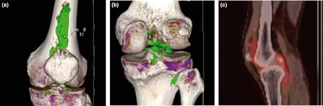
This composite diagnostic image features three panels illustrating the left knee of a patient with chronic gouty arthritis. Panels (a) and (b) present 3D-reconstructed Dual-Energy Computed Tomography (DECT) scans in anterior and posterior views, respectively. The DECT software uses color-coding to identify monosodium urate (MSU) crystals, shown in green. Panel (a) clearly demonstrates a large, compact, elongated green tophus located in the suprapatellar region, extending superiorly from the patella. Panel (b) shows scattered green patches representing MSU deposits within the joint space and posterior structures. Panel (c) is a sagittal fusion image of Positron Emission Tomography and Computed Tomography (PET/CT). The red/orange heat map indicates intense 18F-fluorodeoxyglucose (FDG) uptake, signifying high metabolic activity and active inflammation associated with the urate tophi. These modalities collectively provide evidence of the metabolic burden and structural localization of tophaceous gout that may be occult on conventional radiography.
"gout" AND "urate-lowering therapy"
Uric Acid and Gout
1. Biochemistry of Uric Acid
- De novo pathway: purine nucleotides built from non-purine precursors
- Salvage pathway: free purine bases (from dietary intake or DNA/RNA degradation) recycled into nucleotides via HGPRT (hypoxanthine-guanine phosphoribosyltransferase)
- Normal serum urate: < 6.8 mg/dL (the solubility threshold for monosodium urate in plasma)
- Hyperuricemia is defined as serum urate > 6.8 mg/dL
2. Hyperuricemia: Causes
| Mechanism | Primary | Secondary |
|---|---|---|
| Overproduction | Enzyme defects (HGPRT deficiency), increased PRPP amidotransferase activity | Tumor lysis syndrome, hemolytic anemia, myeloproliferative disorders, psoriasis |
| Underexcretion | Idiopathic (majority ~80%) | CKD, thiazide diuretics, cyclosporine, salicylates (low-dose), DKA, lactic acidosis |
Primary gout accounts for ~90% of cases and in most patients the basis for reduced renal excretion is unknown. Secondary gout accounts for ~10% - Robbins & Kumar Basic Pathology
3. Pathogenesis of Gouty Arthritis
- Crystal formation: When serum urate exceeds 6.8 mg/dL, monosodium urate (MSU) crystals precipitate in joints (favored by low temperature in peripheral joints, low pH, and mechanical trauma)
- Inflammasome activation: Resident synovial macrophages phagocytose MSU crystals → activate the NLRP3 inflammasome → caspase-1 activation → active IL-1β release
- Neutrophil recruitment: IL-1β drives massive neutrophil influx into the joint space; neutrophils also phagocytose crystals
- Phagolysosomal damage: Needle-shaped crystals rupture phagolysosomal membranes → leakage of lysosomal enzymes, free radicals, and other cytokines → acute inflammatory arthritis
- Self-limitation: Attacks typically resolve spontaneously in 7-14 days
- Chronic disease: Repeated attacks → tophi (aggregates of MSU crystals surrounded by macrophages, foreign body giant cells, and fibrous tissue) → cartilage destruction and joint deformity
4. Clinical Stages
Stage 1 - Asymptomatic Hyperuricemia
- Elevated urate without symptoms; can persist for decades
- Only 1 in 8 patients with urate 7-8 mg/dL develops clinical gout over 14 years
Stage 2 - Acute Intermittent Gout (Flares)
- Onset: sudden, often nocturnal, peaking in 12-24 hours
- Site: first metatarsophalangeal (MTP) joint = "podagra" in 50% of cases; also ankle, knee, midfoot, wrist
- Appearance: exquisite pain, warmth, erythema, swelling - can mimic cellulitis
- Up to 20% of patients have polyarticular involvement
- Self-limited: peaks in 1-2 days, resolves within a week without treatment
- Systemic features (fever) may occur and should raise concern for co-existing septic arthritis
Stage 3 - Intercritical Gout
- Asymptomatic intervals between attacks; attacks become more frequent and severe over time
Stage 4 - Chronic Tophaceous Gout
- No pain-free intervals; persistent joint damage
- Tophi: firm, chalky nodules in subcutaneous tissue, bursae (olecranon), joints, tendons, ear pinnae
- Cartilage erosion, joint deformity, and functional loss
5. Genetics
- Lesch-Nyhan Syndrome: Complete absence of HGPRT (X-linked recessive) → severe hyperuricemia + neurologic manifestations (self-mutilation, spasticity, intellectual disability). This is a form of secondary gout.
- Partial HGPRT deficiency (Kelley-Seegmiller syndrome): Gout only, no neurologic features
- Primary gout has a polygenic basis in most patients
6. Diagnosis
| Test | Findings |
|---|---|
| Synovial fluid analysis | Gold standard: MSU crystals - needle-shaped, strongly negative birefringence under polarized light (yellow when parallel, blue when perpendicular to slow axis); WBC >2,000/µL (neutrophilic) |
| Serum uric acid | Often normal during acute attack - NOT part of diagnostic criteria; may be elevated |
| X-ray | Late finding: "punched-out" periarticular erosions with overhanging edges, soft tissue tophi, normal joint space early |
| Ultrasound | Double-contour sign (urate on cartilage), hyperechoic tophi, erosions |
| Dual-energy CT (DECT) | Non-invasively identifies MSU crystal deposits (color-coded green) - excellent for tophus mapping |
Important: serum uric acid can be normal during an acute gout attack and does not exclude the diagnosis - Tietz Textbook of Laboratory Medicine
7. Associated Conditions
- Metabolic syndrome: obesity, hypertension, hyperlipidemia, diabetes
- Cardiovascular disease: independent risk factor for MI and stroke
- Chronic kidney disease: bidirectional - CKD raises urate; urate may accelerate CKD progression
- Nephrolithiasis: uric acid kidney stones (~10-20% of gout patients)
- Drug-induced hyperuricemia: thiazides, loop diuretics, low-dose aspirin, cyclosporine, pyrazinamide
8. Management
Acute Flare Treatment
| Agent | Mechanism | Notes |
|---|---|---|
| NSAIDs (e.g., indomethacin, naproxen) | COX inhibition | First-line; avoid in renal impairment, PUD, CV disease |
| Colchicine | Inhibits neutrophil migration, tubulin polymerization, NLRP3 inflammasome | Most effective if started within 36 hours; low-dose as effective as high-dose with fewer GI side effects |
| Corticosteroids (oral or intra-articular) | Broad anti-inflammatory | Useful when NSAIDs/colchicine contraindicated; oral prednisone or intra-articular injection |
| IL-1 inhibitors (anakinra, canakinumab) | Block IL-1β signaling | Reserved for refractory cases |
| Topical ice, joint rest | Supportive | Adjunctive |
Urate-Lowering Therapy (ULT) - Long-term Prevention
| Agent | Mechanism | Dose/Notes |
|---|---|---|
| Allopurinol | Xanthine oxidase inhibitor (XOI) | First-line; start low (100 mg/day), titrate; dose-adjust for renal impairment; risk of DRESS/SJS especially in HLA-B*58:01 carriers (screen in Han Chinese, Thai, Korean patients) |
| Febuxostat | Non-purine XOI | Alternative when allopurinol intolerant; 40-80 mg/day; no renal dose adjustment needed; FDA boxed warning for increased CV mortality vs. allopurinol in high-CV-risk patients |
| Probenecid / Sulfinpyrazone | Uricosuric agents | Block tubular urate reabsorption; avoid in CKD, nephrolithiasis |
| Pegloticase | Recombinant pegylated uricase (converts urate → allantoin) | Reserved for refractory tophaceous gout; IV every 2 weeks; anti-drug antibodies can cause loss of efficacy and infusion reactions |
| Lesinurad (+ XOI) | URAT1 inhibitor (uricosuric) | Used in combination with XOI |
Lifestyle Modifications
- Reduce purine-rich foods: organ meats, red meat, shellfish, anchovies, sardines
- Avoid fructose-sweetened drinks and beer (wine appears less problematic)
- Adequate hydration
- Weight loss, control hypertension and dyslipidemia
- Avoid/switch diuretics and low-dose aspirin when possible
9. Special Populations
- Renal transplant patients: CSA (cyclosporine) is a major cause of post-transplant hyperuricemia; allopurinol must be used cautiously if patient is on azathioprine (XOI inhibits azathioprine metabolism - reduce aza dose by 75%, or switch to mycophenolate)
- CKD: Allopurinol dose must be adjusted; NSAIDs contraindicated
- Pregnancy: All standard ULT agents are relatively contraindicated; management is largely supportive
- Postmenopausal women: Loss of estrogen (which is uricosuric) increases gout risk substantially
Summary Table
| Feature | Detail |
|---|---|
| Cause | Monosodium urate crystal deposition from hyperuricemia |
| Threshold | Serum urate > 6.8 mg/dL |
| Most common site | 1st MTP joint (podagra) |
| Crystal morphology | Needle-shaped, negatively birefringent |
| Key mediator | IL-1β (via NLRP3 inflammasome) |
| Acute Rx | NSAIDs, colchicine, corticosteroids |
| ULT target | Serum urate < 6 mg/dL |
| First-line ULT | Allopurinol |