
Embryology of Fertilization anatomy
Reading File
Now let me search the medical library for fertilization embryology content.
Finding Sources
Finding Sources
Excellent results. Now let me read the key embryology sections and fetch images in parallel.
Reading File
Reading File
Reading File
Now let me get more content and fetch the key images.
Reading File
Reading File
Loading Image
Now let me fetch the acrosome reaction diagram image.
Loading Image
Loading Image
I now have comprehensive content from multiple authoritative embryology textbooks with detailed diagrams. Let me compile the complete answer.
Embryology of Fertilization
Fertilization is the complex sequence of coordinated molecular and physical events that begins with contact between a sperm and a secondary oocyte, and ends with the intermingling of maternal and paternal chromosomes at metaphase of the first mitotic division of the zygote.
Site of Fertilization
Fertilization normally occurs in the ampulla of the uterine (fallopian) tube - the widest part, usually reached by only a few hundred of the millions of ejaculated sperm. Of 200-300 million spermatozoa deposited in the female genital tract, only 300-500 reach the site of fertilization, and only one fertilizes the egg. Chemical signals (attractants) secreted by the oocyte and surrounding follicular cells guide capacitated sperm via sperm chemotaxis to the oocyte.
- The Developing Human: Clinically Oriented Embryology, p. 117
- Langman's Medical Embryology, p. 58
The Oocyte at the Time of Fertilization
The secondary oocyte is arrested in metaphase II of meiosis at the time of ovulation. It is surrounded by:
- Corona radiata - an outer layer of follicular (granulosa) cells
- Zona pellucida - a glycoprotein shell between the corona radiata and the oocyte plasma membrane
- A first polar body within the perivitelline space
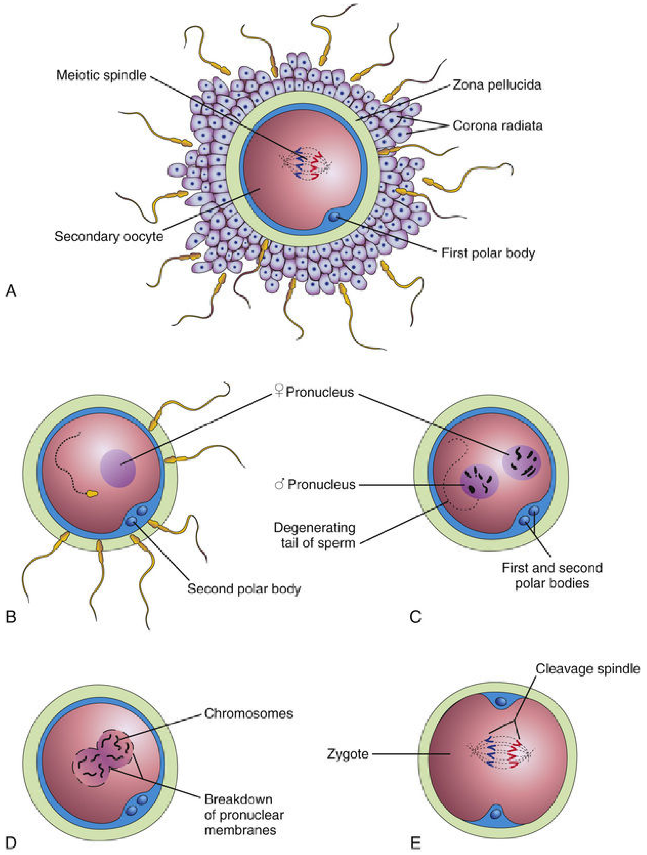
FIG. 2.15 (The Developing Human): A) Secondary oocyte with corona radiata, zona pellucida, first polar body; B) Sperm entry - second polar body forms, female pronucleus appears; C) Both pronuclei visible with degenerating sperm tail; D) Pronuclear membranes break down, chromosomes align; E) Zygote with cleavage spindle.
Step-by-Step Events of Fertilization
Phase 1: Sperm Capacitation
Before sperm can fertilize an oocyte, they must undergo capacitation - a period of conditioning lasting approximately 7 hours in the female reproductive tract (uterus or uterine tubes).
- During capacitation, a glycoprotein coat and seminal proteins are removed from the sperm acrosome surface
- Membrane components are extensively altered
- Capacitated sperm show no morphologic changes but are more active
- Molecules secreted by the oocyte (resact, speract) orient and stimulate sperm via guanylate cyclase
- Completion of capacitation is required for the acrosome reaction to occur
Phase 2: Penetration of the Corona Radiata
- Capacitated sperm pass relatively freely between the corona radiata cells
- Sperm release hyaluronidase by exocytosis, which dissolves hyaluronic acid between coronal cells, allowing easier passage to the zona pellucida
Phase 3: Penetration of the Zona Pellucida - The Acrosome Reaction
This is the critical barrier-crossing step:
- The sperm acrosome binds to ZP3 (a zona pellucida glycoprotein) on the zona pellucida surface via the SED1 protein
- Acrosomal reaction is triggered: multiple perforations form in the acrosomal wall, and the outer acrosomal membrane fuses with the overlying sperm plasma membrane
- This causes exocytosis of hydrolytic enzymes from the acrosome, including:
- Acrosin (main acrosomal protease)
- Hyaluronidase
- Other hydrolytic enzymes
- These enzymes locally degrade the zona pellucida, allowing the sperm to tunnel through
- As sperm penetrates, sperm pre-acrosin binds ZP2 - mediating forward movement
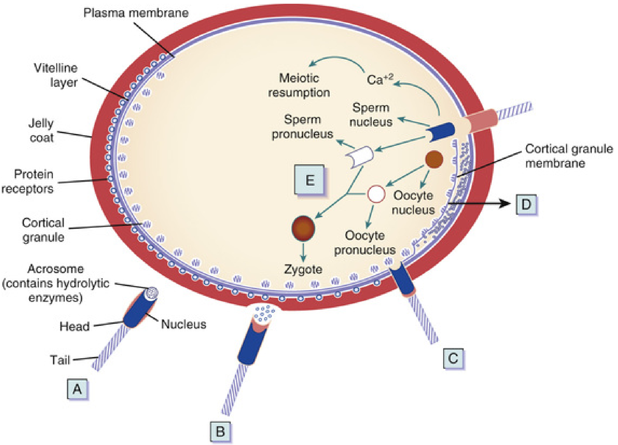
FIG. 2.13 (The Developing Human): Molecular events - A) Capacitation with oocyte-secreted attractants; B) Acrosome reaction; C) Sperm-oocyte fusion via IZUMO/Juno proteins; D) Cortical reaction with Ca²⁺ wave; E) Male pronucleus formation and second meiotic division completion.
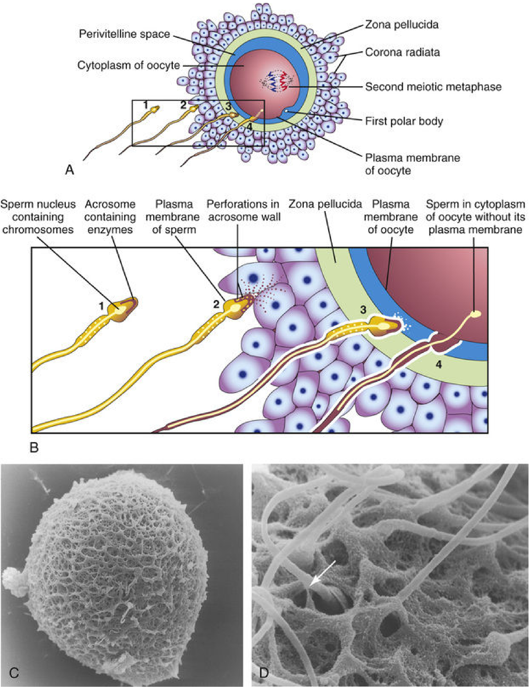
FIG. 2.14 (The Developing Human): Detailed anatomy of sperm approaching the secondary oocyte. 1) Capacitation; 2) Acrosome reaction with perforations; 3) Zona penetration; 4) Sperm in oocyte cytoplasm without its plasma membrane. C&D) Scanning electron micrographs of human oocyte with zona pellucida.
Phase 4: Sperm-Oocyte Membrane Fusion
- The first sperm to completely penetrate the zona pellucida fuses its cell membrane with the oocyte plasma membrane
- Actual fusion occurs between the oocyte membrane and the posterior membrane of the sperm head (the acrosomal cap membrane was shed during the acrosome reaction)
- Key molecular mediators of fusion:
- IZUMO protein on sperm binds Juno receptor on oocyte
- ADAMs 1, 2, 3 and CRISP1 on sperm bind integrins, CD9, CD81 on oocyte
- Disintegrin-integrin interactions mediate initial adhesion
- In humans, both the head and tail of the sperm enter the oocyte cytoplasm; the sperm's plasma membrane is left behind on the oocyte surface
Phase 5: Three Oocyte Responses to Sperm Entry
The oocyte immediately responds in three ways:
1. Cortical Reaction (Block to Polyspermy)
- Sperm entry triggers a wave of Ca²⁺ release from vesicles in the oocyte
- This causes exocytosis of proteases from cortical granules lining the oocyte membrane (cortical reaction)
- The wave spreads across the entire oocyte surface
- Released enzymes hydrolyze ZP3 and ZP2, altering the zona pellucida (zona reaction) - converting it into the impenetrable perivitelline barrier
- This permanent block prevents polyspermy (fertilization by more than one sperm)
- The fast block involves immediate membrane depolarization; the slow block is the cortical/zona reaction
2. Completion of Meiosis II
- The Ca²⁺ rise causes the oocyte to complete its second meiotic division (previously arrested at metaphase II)
- This produces:
- The second polar body (with little cytoplasm, expelled into perivitelline space)
- The haploid female pronucleus (23 chromosomes + X)
3. Metabolic Activation of the Egg
- The sperm-borne activating factor triggers initial cellular and molecular events of early embryogenesis
Pronuclear Stage and Zygote Formation
- The sperm head moves toward the female pronucleus, loses its protamines, and its chromatin decondenses - forming the male pronucleus (23 chromosomes)
- Both pronuclei are surrounded by their own membranes and are visible as distinct vesicular nuclei
- Maternal and paternal chromosomes do not fuse directly - instead, the pronuclear membranes break down (syngamy)
- The 46 chromosomes (23 from each pronucleus) align on the first cleavage spindle
- Fertilization is complete when chromosomes intermingle at metaphase of the first mitotic division - forming the zygote
Results of Fertilization
| Result | Significance |
|---|---|
| Restoration of diploid number (46 chromosomes) | Half from father, half from mother; new genetic combination |
| Determination of chromosomal sex | X-bearing sperm → female (XX); Y-bearing sperm → male (XY) |
| Initiation of cleavage | Without fertilization, oocyte degenerates within ~24 hours |
| Block to polyspermy | Cortical reaction prevents abnormal development |
Key Molecular Players Summary
| Molecule | Location | Function |
|---|---|---|
| ZP3 | Zona pellucida | Binds sperm; triggers acrosome reaction |
| ZP2 | Zona pellucida | Secondary sperm receptor post-acrosome reaction |
| Acrosin | Acrosome | Protease enabling zona penetration |
| Hyaluronidase | Acrosome | Disperses corona radiata cells |
| IZUMO | Sperm membrane | Gamete membrane fusion |
| Juno | Oocyte membrane | Receptor for IZUMO; essential for fusion |
| Ca²⁺ wave | Oocyte cytoplasm | Triggers cortical reaction + meiosis II completion |
| Cortical granule enzymes | Oocyte periphery | Zona reaction; block to polyspermy |
Clinical Correlates
- Ectopic (tubal) pregnancy: If the embryo cannot be transported to the uterus (e.g., due to chronic salpingitis/tubal adhesions), it implants in the tube, which ruptures causing potentially fatal hemorrhage
- Polyspermy: Penetration by more than one sperm results in triploidy (69 chromosomes) and abnormal, non-viable development
- Male infertility: Evaluated by semen analysis; >20 million sperm/mL needed for fertility; 50% should be motile at 2 hours
- In vitro fertilization (IVF): Capacitation is induced artificially in defined media for several hours; ICSI (intracytoplasmic sperm injection) bypasses zona penetration by directly injecting a single sperm
- Capacitation and src kinase: Both capacitation and the acrosome reaction appear regulated by tyrosine kinase (src kinase)
Sources:
- The Developing Human: Clinically Oriented Embryology (Moore & Persaud), pp. 113-120
- Langman's Medical Embryology, pp. 58-60
- Junqueira's Basic Histology - Text and Atlas, 17e, pp. 1127-1128
- Guyton and Hall Textbook of Medical Physiology, pp. 4061-4068
This is a shared conversation. Sign in to Orris to start your own chat.