
Diff bw facial paralysis and bells palsy
Bell's palsy facial paralysis comparison peripheral central
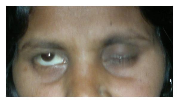
This clinical photograph displays a close-up view of the upper face and eyes of a patient demonstrating Bell's phenomenon, a hallmark sign of peripheral facial nerve paralysis (Bell's palsy). The patient is attempting to close both eyes. On the patient's left side, the eyelid closes normally. However, on the patient's right side, there is evidence of lagophthalmos (incomplete eyelid closure). Upon the attempt to close the right eye, the eyeball rotates upward and outward, a physiological movement that is unmasked by the failure of the orbicularis oculi muscle. This upward displacement exposes a significant portion of the inferior sclera. Additionally, the right side of the forehead appears smooth with a lack of expressive wrinkling compared to the contralateral side, further indicating lower motor neuron facial nerve involvement. This visual finding is critical for differentiating peripheral facial nerve lesions from central causes, where forehead movement is typically spared.
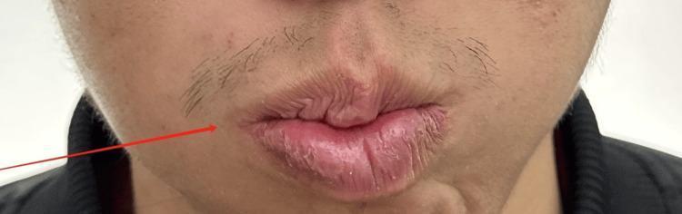
This clinical photograph shows a close-up view of the lower face of a patient with peripheral facial paralysis (Bell's palsy), specifically during a post-treatment evaluation. The image captures the patient attempting to whistle, a maneuver used to assess the function of the orbicularis oris muscle. A prominent red arrow points to the right side of the mouth (anatomical right), highlighting persistent asymmetry. The lips are pursed but deviate toward the left side of the image, indicating residual weakness or impaired motor control on the patient's right side. While the skin texture is similar bilaterally, there is a visible loss of muscle tone and fullness on the affected right side, resulting in an uneven labial seal and distorted philtrum during exertion. This image serves as a clinical demonstration of cranial nerve VII (facial nerve) dysfunction and is used to monitor recovery progress, often graded using the House-Brackmann scale.
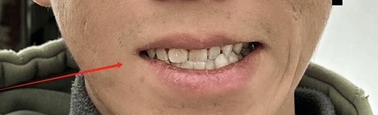
This clinical photograph displays a close-up view of the lower face and mouth of a patient with peripheral facial paralysis (Bell's palsy), likely of the right cranial nerve VII. The image demonstrates significant facial asymmetry during a mid-treatment evaluation. A red arrow points to the patient's right side (viewer's left), where there is evident muscle weakness. Key visual findings include a deviation of the mouth towards the unaffected left side (viewer's right) due to unopposed muscular contraction. The right side shows a flattening of the nasolabial fold and reduced muscle tone, resulting in less tooth exposure compared to the left. The patient is attempting a maximal effort smile or baring of the teeth, which accentuates the paralysis of the circumoral muscles. This image is an educational example of lower motor neuron facial nerve dysfunction and its clinical manifestation in the perioral region.
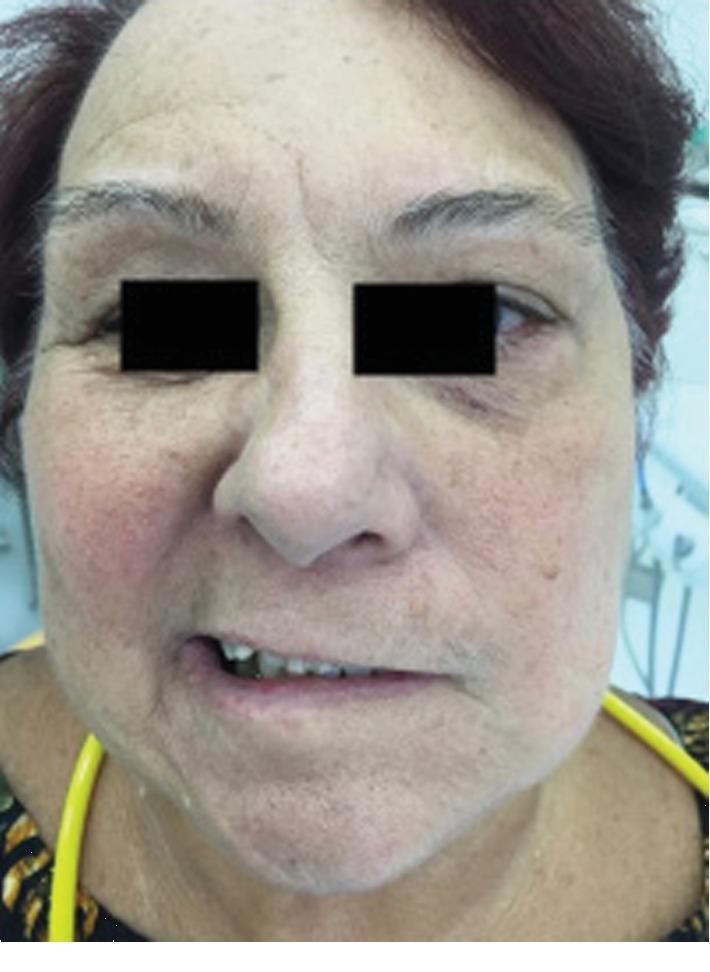
This clinical photograph displays a 71-year-old female patient with a primary presentation of Bell's palsy, demonstrating significant facial nerve (cranial nerve VII) paralysis. The image shows a frontal view of the face with the eyes obscured for privacy. Notable clinical findings include severe unilateral facial asymmetry at rest. The patient's left side exhibits a loss of muscle tone, characterized by a flattened nasolabial fold and downward drooping of the labial commissure (mouth corner). In contrast, the right side retains normal muscle contraction, causing the mouth to be pulled toward the unaffected side. This visual serves as an educational example of Grade V (severe) facial paralysis on the House-Brackmann Scale, highlighting the characteristic 'sagging' appearance and muscle weakness associated with idiopathic peripheral facial palsy before treatment. The image is a valuable tool for medical students and clinicians for recognizing diagnostic clinical signs of facial nerve dysfunction.
Facial Paralysis vs. Bell's Palsy
The Core Relationship
"The term Bell palsy should be reserved for cases of facial paralysis that have signs and symptoms consistent with the disease and in which a diligent search for another cause is negative." - Cummings Otolaryngology
Side-by-Side Comparison
| Feature | Facial Paralysis (general) | Bell's Palsy (specific) |
|---|---|---|
| Definition | Any weakness/paralysis of facial muscles | Idiopathic, acute, unilateral peripheral facial nerve paralysis |
| Scope | Umbrella term for many causes | One specific diagnosis within facial paralysis |
| Etiology | Multiple (see below) | Unknown/idiopathic; likely HSV-1 reactivation |
| Diagnosis | Requires workup to find cause | Diagnosis of exclusion after ruling out other causes |
| Forehead involvement | Depends on cause (spared in central lesions) | ALWAYS involves forehead (peripheral LMN lesion) |
| Side affected | Unilateral or bilateral, depends on cause | Almost always unilateral |
| Onset | Varies - sudden, gradual, or progressive | Sudden to rapid; complete within 1-7 days |
| CNS signs | May be present (e.g., stroke) | Absent |
| Ear exam | Varies | Normal (no vesicles, effusion, or cholesteatoma) |
| Prognosis | Depends entirely on cause | Generally good; 85% recover fully |
| Treatment | Cause-specific | Prednisolone (60 mg x 5 days then taper); eye care |
Causes of Facial Paralysis (Not Bell's Palsy)
- Herpes zoster oticus (Ramsay Hunt syndrome) - vesicles in ear canal
- Guillain-Barre syndrome
- Lyme disease
- HIV infection
- Otitis media (acute bacterial, chronic with cholesteatoma)
- Trauma (temporal bone fracture, birth trauma, barotrauma)
- Sarcoidosis
- Melkersson-Rosenthal syndrome
- Cerebrovascular disease (stroke)
- Parotid malignancy or metastatic tumor
- Facial nerve schwannoma
- Glomus tumor
- Cholesteatoma
Key Diagnostic Distinction: Central vs. Peripheral
| Central (UMN) Palsy - e.g., stroke | Peripheral (LMN) Palsy - e.g., Bell's palsy | |
|---|---|---|
| Forehead | Spared (bilateral cortical representation) | Affected (entire ipsilateral face involved) |
| Eye closure | Intact or mildly weak | Weak - Bell's phenomenon (eye rolls up on attempted closure) |
| Site of lesion | Motor cortex / corticobulbar tract | Facial nerve nucleus or anywhere along CN VII |
| Associated features | Hemiplegia, dysphasia, other CNS signs | Hyperacusis, dysgeusia, decreased tearing, post-auricular pain |
Diagnostic Criteria for Bell's Palsy (Taverner's Criteria)
- Paralysis or paresis of all muscle groups of one side of the face
- Sudden onset
- Absence of CNS disease signs
- Absence of ear or cerebellopontine angle disease signs
Features That Rule Against Bell's Palsy
- Slowly progressive paralysis over weeks/months (suggests tumor)
- Facial twitching
- Sensorineural hearing loss
- Vestibular dysfunction
- Otorrhea
- Severe, unrelenting otalgia
- Vesicles in ear canal (points to Ramsay Hunt syndrome instead)
- Bilateral simultaneous facial paralysis
- Recurrent episodes (raises suspicion for tumor or Melkersson-Rosenthal syndrome)
- Intact forehead movement (points to a central/UMN cause)