
Explain about action potential in nerve muscle
Reading File
Now let me search the medical library for action potential content, in parallel with a web search.
Finding Sources
Searching the Web
action potential nerve muscle physiology mechanism
Finding Sources
Excellent - found great sources. Let me read the key sections from Guyton & Hall (the gold standard physiology text) and Neuroscience: Exploring the Brain in parallel.
Reading File
Reading File
Now let me get more detail on voltage-gated channels, propagation, and the neuromuscular junction, and also fetch the action potential diagram image.
Reading File
Loading Image
Let me get more content on propagation, refractory periods, and the voltage-gated channel diagram.
Reading File
Loading Image
I now have rich content from Guyton & Hall covering all phases of the action potential, voltage-gated channels, refractory periods, propagation, and muscle-specific features. Let me compile the full answer.
Action Potential in Nerve and Muscle
Source: Guyton and Hall Textbook of Medical Physiology
What Is an Action Potential?
An action potential is a rapid, self-propagating change in membrane voltage that serves as the fundamental electrical signal in nerve and muscle cells. It begins as a sudden shift from the resting negative membrane potential to a positive value, then returns almost equally quickly to negative - the entire event lasting only 1-2 milliseconds in nerve fibers.
1. Resting Membrane Potential
Before an action potential occurs, the nerve fiber maintains a resting membrane potential of approximately -70 mV (inside negative relative to outside). This is established by:
- High intracellular K+ (maintained by the Na+-K+ ATPase pump)
- K+ leak channels allowing K+ to diffuse out
- Large impermeant anions (proteins, phosphates, sulfates) trapped inside the cell
The membrane is said to be polarized at rest.
2. Phases of the Action Potential
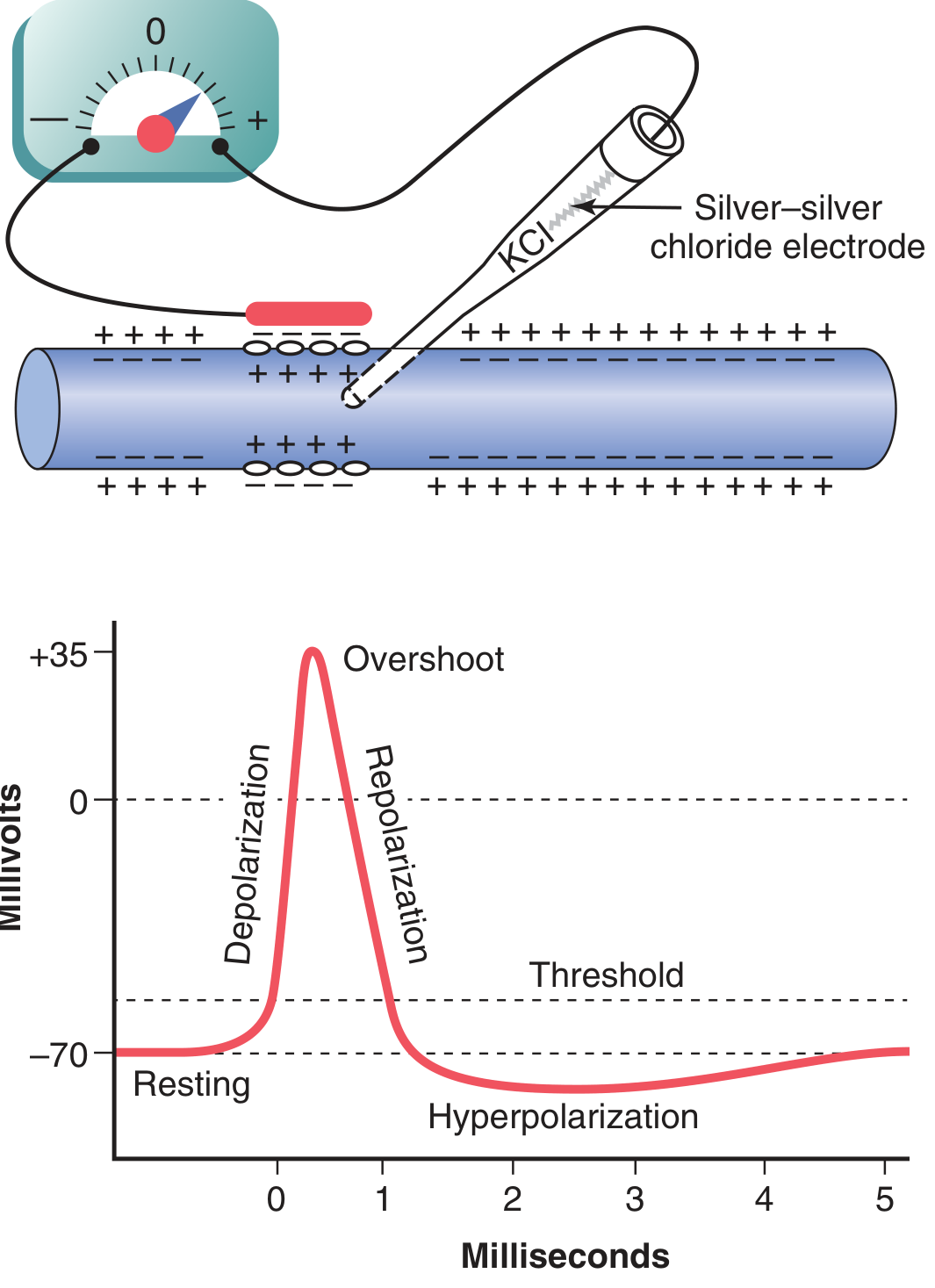
Phase 1 - Depolarization
- A stimulus raises the membrane potential from -70 mV toward the threshold (~-55 mV, about 15 mV above resting)
- Once threshold is reached, voltage-gated Na+ channels open explosively
- Na+ rushes INTO the cell (down both its concentration and electrical gradients)
- Membrane potential rapidly rises to approximately +35 mV (overshoot)
- This is an all-or-nothing event - once threshold is hit, the full action potential fires regardless of stimulus strength
Phase 2 - Repolarization
- Within a fraction of a millisecond after opening, Na+ channels inactivate (the inactivation gate closes)
- Simultaneously, voltage-gated K+ channels open (with a slight delay)
- K+ rushes OUT of the cell, driving the membrane potential back toward negative
- Repolarization restores the membrane to ~-70 mV
Phase 3 - Hyperpolarization (Afterhyperpolarization / Undershoot)
- K+ channels may remain open slightly longer than needed
- This drives the membrane potential briefly below -70 mV (to about -80 mV)
- Once K+ channels close, the potential returns to the true resting level
3. Voltage-Gated Channels - The Molecular Basis
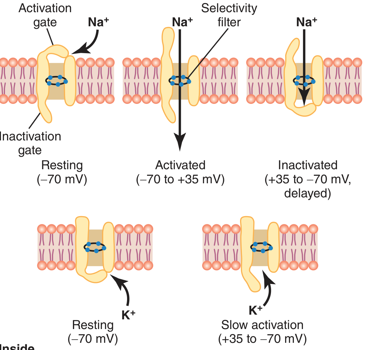
Voltage-Gated Na+ Channel (3 states)
| State | Gate Status | When |
|---|---|---|
| Resting | Activation gate CLOSED, Inactivation gate open | At -70 mV |
| Activated | Activation gate OPEN, Inactivation gate open | During depolarization |
| Inactivated | Activation gate open, Inactivation gate CLOSED | Shortly after activation |
The activation gate opens fast when threshold is reached. The inactivation gate closes a few ten-thousandths of a second later, self-terminating Na+ influx. The Na+ channel cannot reopen until the membrane repolarizes - this is the molecular basis of the refractory period.
Voltage-Gated K+ Channel (2 states)
- Has only one gate; opens slowly during depolarization
- Provides the main outward current driving repolarization
- Sodium permeability increases 500-5000 fold during depolarization; K+ permeability increases only ~30-fold, but its delayed opening is what drives the membrane back negative
4. Refractory Periods
| Period | Mechanism | Effect |
|---|---|---|
| Absolute Refractory Period | Na+ channels inactivated (cannot reopen) | No stimulus of any strength can fire another AP |
| Relative Refractory Period | Channels recovering; K+ channels still open | A stronger-than-normal stimulus can fire an AP |
The refractory period ensures APs travel in one direction only and limits firing frequency.
5. Propagation Along the Nerve Fiber
When an action potential fires at one point on the membrane, local positive charges flow laterally to adjacent resting membrane areas, raising their potential to threshold and triggering the AP there. This self-regenerating process propagates the signal from one end of the fiber to the other.
Saltatory conduction (in myelinated fibers): The action potential "jumps" between nodes of Ranvier, making propagation much faster (~70-120 m/s in large myelinated fibers vs. ~0.5 m/s in unmyelinated fibers) and more energy-efficient.
6. Action Potential in Skeletal Muscle - Key Differences
Muscle APs follow the same basic mechanism as nerve APs but with important quantitative differences (Guyton & Hall):
| Feature | Nerve Fiber | Skeletal Muscle Fiber |
|---|---|---|
| Resting membrane potential | ~-70 mV | -80 to -90 mV |
| Duration of AP | ~1 ms | 1-5 ms |
| Conduction velocity | ~70-120 m/s | 3-5 m/s |
7. Excitation-Contraction Coupling (Muscle-Specific)
After an AP is generated at the muscle fiber surface, it must reach the deep contractile machinery:
- Surface AP travels along the sarcolemma
- T-tubules (transverse tubules) - open extensions of the cell membrane that penetrate deep into the fiber - carry the electrical signal inward to every myofibril
- T-tubule depolarization triggers voltage-sensing dihydropyridine receptors (DHPRs) on the T-tubule wall
- DHPRs mechanically activate ryanodine receptors (RyRs) on the sarcoplasmic reticulum (SR)
- Ca2+ is released from SR terminal cisternae into the cytoplasm
- Ca2+ binds troponin C, allowing actin-myosin cross-bridge cycling → muscle contraction
This entire sequence is called excitation-contraction coupling.
Summary Flow
Stimulus → Membrane reaches threshold (-55 mV)
       ↓
Voltage-gated Na+ channels open → Na+ influx → Depolarization (+35 mV)
       ↓
Na+ channels inactivate + K+ channels open → K+ efflux → Repolarization
       ↓
Hyperpolarization → Return to resting (-70 mV)
       ↓
[In muscle]: AP travels via T-tubules → SR releases Ca2+ → Contraction
Key Clinical Points:
- Local anesthetics (lidocaine, bupivacaine) block voltage-gated Na+ channels, preventing AP generation and propagation
- Tetrodotoxin (TTX) from puffer fish specifically blocks Na+ channels - a classic tool in neurophysiology research
- Hyperkalemia raises resting membrane potential, initially depolarizing then inactivating Na+ channels, causing muscle weakness or paralysis
- Guyton and Hall Textbook of Medical Physiology
This is a shared conversation. Sign in to Orris to start your own chat.