I now have comprehensive content from multiple authoritative textbooks. Let me compile the full detailed notes.
Hepatic Encephalopathy (HE) - Comprehensive Clinical Notes
Definition
Hepatic encephalopathy is a wide array of transient and reversible neuropsychiatric and neuromuscular manifestations resulting from the diseased liver's failure to perform its normal metabolic functions. It encompasses a spectrum from mild neurocognitive disturbances to overt coma, occurring in the setting of acute liver failure (ALF) or chronic liver disease with portal hypertension.
- Develops in 50-70% of patients with cirrhosis
- Projected 1-year survival without liver transplant: 42%, 3-year survival: 23%
- About 20,000 patients/year hospitalized for HE (USA, 2005-2009)
- Minimal HE affects approximately 50% of all cirrhotics (commonly underdiagnosed)
Types (Classification - West Haven / World Gastroenterological Association)
The WGA uses a multiaxial classification based on the underlying liver disease and portosystemic shunting:
Type A - Acute Liver Failure
- Occurs in patients with previously normal livers
- Neurological signs appear within 8 weeks of developing liver disease
- Examples: viral hepatitis, acetaminophen toxicity, Amanita poisoning
Type B - Bypass (Porto-systemic shunting without hepatocellular disease)
- Small group of patients free of hepatocellular disease
- Have significant portacaval shunting of blood
- Rare variant
Type C - Cirrhosis (Most common)
- Patients with both hepatocellular disease and portosystemic shunts
- Further subclassified by temporal pattern:
- Episodic HE - discrete attacks
- Persistent HE - continuous behavioral abnormalities
- Minimal HE (MHE) - subtle impairment detectable only on neuropsychological testing
Grading - West Haven Criteria
| Grade | Features |
|---|
| 0 (MHE) | No clinical signs; detectable only on neuropsychological testing / EEG |
| 1 | Trivial lack of awareness, euphoria or anxiety, shortened attention span, impaired addition/subtraction |
| 2 | Lethargy, disorientation to time, obvious personality change, inappropriate behavior, asterixis |
| 3 | Somnolence to semi-stupor, responsive to stimuli, gross disorientation, bizarre behavior, hyperreflexia, extensor plantar response |
| 4a | Coma - response to pain present |
| 4b | Coma - no response to pain; may have decerebrate/decorticate posturing |
Grade 4 is invariably associated with intracranial hypertension and cerebral edema, which can lead to brainstem herniation (the most common cause of death).
Pathophysiology / Causes
HE is multifactorial. Key mechanisms include:
1. Ammonia Toxicity (Central mechanism)
- Ammonia is produced primarily in the colon where bacteria metabolize proteins and nitrogen-based products
- Enterocytes also synthesize ammonia from glutamine
- Normally cleared by hepatocytes via the urea cycle
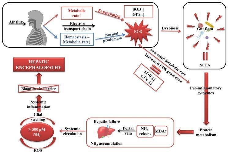
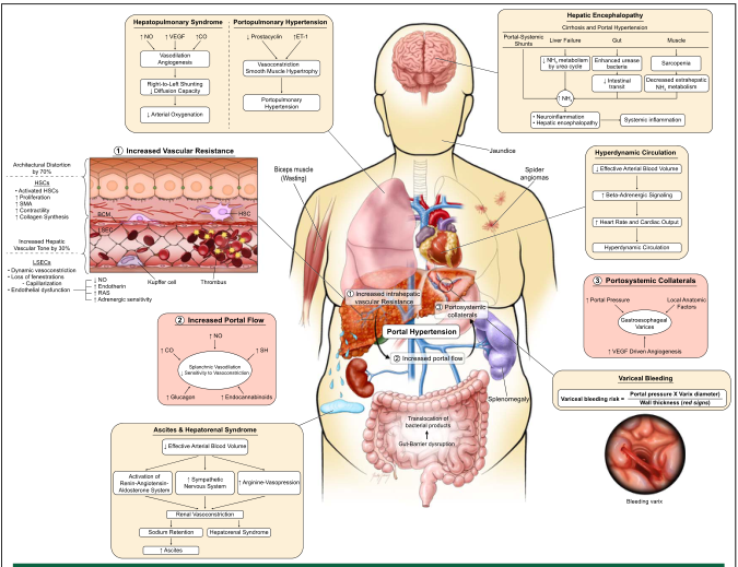
- In cirrhosis: reduced hepatocyte function + portosystemic shunting → elevated circulating ammonia
- Arterial hyperammonemia in up to 90% of HE patients
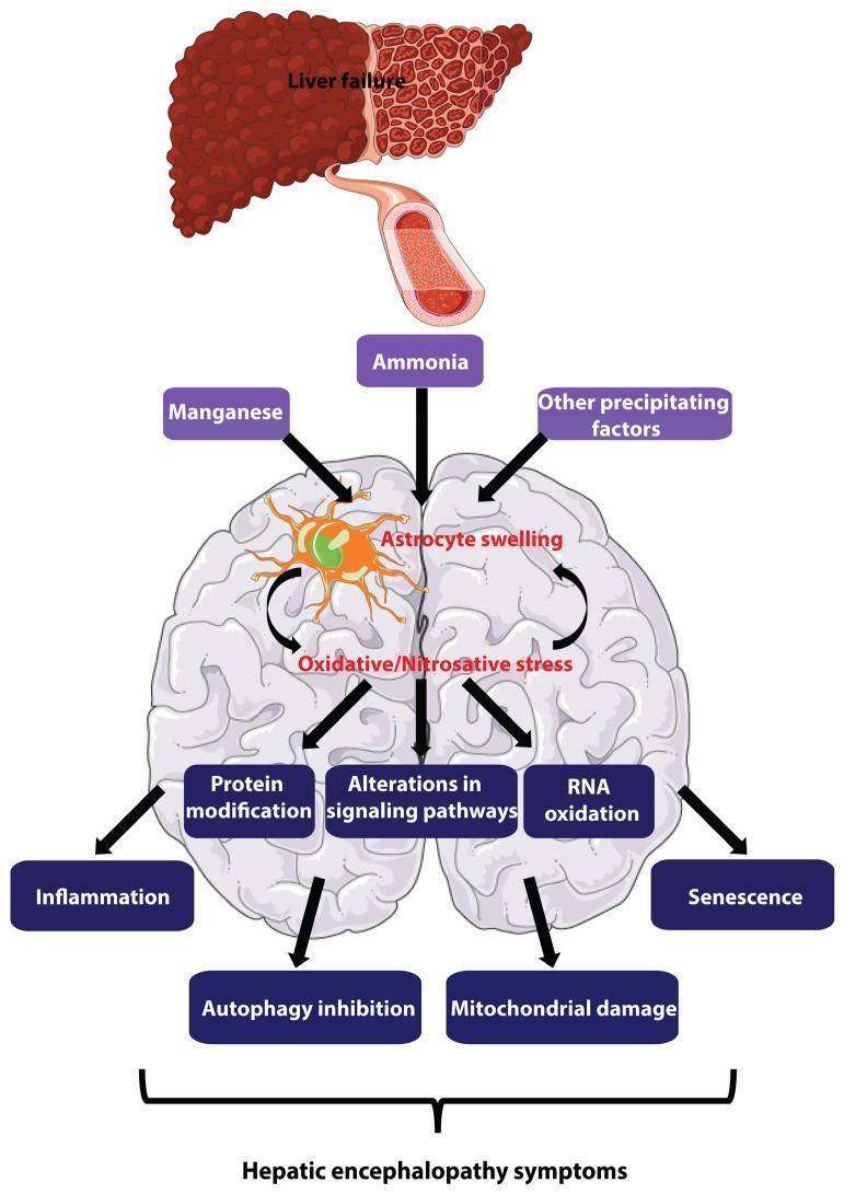
- Ammonia crosses the blood-brain barrier → enters astrocytes → combines with glutamate → forms glutamine (via glutamine synthetase) → astrocyte swelling and cytotoxic brain edema
- Direct neuronal membrane dysfunction
- Note: Serum ammonia level does not directly correlate with severity of encephalopathy (normal in ~10% of HE patients)
2. GABA-Benzodiazepine System
- Increased sensitivity of the peripheral-type (astrocyte) benzodiazepine receptor
- Enhanced activation of GABA-A/benzodiazepine receptor complex
- Endogenous neurosteroids (allopregnanolone, tetrahydrodeoxycorticosterone) further activate this system
- Results in inhibitory neurotransmission → altered consciousness
3. Other Neurotoxins
- Mercaptans (products of methionine metabolism) - responsible for fetor hepaticus
- Manganese - causes dopaminergic dysfunction, deposits in globus pallidus
- False neurotransmitters: β-phenylethylamine, tyramine, octopamine
- Serotonin (5-HT), nitric oxide, circulating opioid peptides
4. Blood-Brain Barrier (BBB) Changes
- Increased BBB permeability → increased uptake of ammonia and other toxins by cerebellum and basal ganglia
5. Genetic Factors
- Allelic mutations in the glutaminase gene increase risk of overt HE independent of hepatic synthetic function
6. Gut Microbiome
- Differences in colonic mucosal microbiota in cirrhotic patients with vs. without HE influence production of neurotoxic substances
Precipitating Causes (Triggers for HE in Known Liver Disease)
These are especially important in Type C / episodic HE:
| Category | Examples |
|---|
| GI bleeding | Variceal hemorrhage, peptic ulcer - increases nitrogen load |
| Electrolyte disturbances | Hypokalemia, metabolic alkalosis |
| Infection | SBP, UTI, pneumonia, sepsis |
| Dehydration / Hypovolemia | Over-diuresis, vomiting, diarrhea |
| Renal impairment | Acute/chronic kidney injury, hepatorenal syndrome |
| Constipation / Ileus | Increases colonic ammonia production |
| Drugs | Sedatives, opioids, benzodiazepines |
| Dietary excess | High protein intake |
| Venous thrombosis | Portal vein thrombosis |
| Portosystemic shunts | TIPS, spontaneous splenorenal shunts |
| Hypovolemia | Dehydration |
Symptoms
Symptoms range along a spectrum. The onset may be insidious or subacute (in chronic liver disease):
Early (Grade 1-2):
- Mild confusion, irritability
- Sleep-wake cycle reversal (daytime somnolence, nighttime insomnia)
- Shortened attention span, difficulty concentrating
- Impaired calculation (addition/subtraction)
- Euphoria or anxiety
- Personality changes, inappropriate behavior
- Slurred speech
Later (Grade 2-3):
- Increasing lethargy and drowsiness
- Disorientation (first to time, then place)
- Obvious personality and behavioral changes
- Impaired motor coordination
- Nausea, vomiting (more common in ALF)
Severe (Grade 3-4):
- Stupor, unresponsiveness to commands
- Gross disorientation
- Profound confusion, delirium
- Coma
Signs
Neurological Signs:
| Sign | Description |
|---|
| Asterixis | "Flapping tremor" - low-amplitude alternating flexion/extension of wrist when held in extension; also elicited at dorsiflexed foot or extended neck. Characteristic of mild-to-moderate HE. Can occur in other metabolic encephalopathies. |
| Altered consciousness | From mild confusion → stupor → coma |
| Hyperreflexia | Grade 3+ HE |
| Extensor plantar response (Babinski) | Grade 3+ HE |
| Decerebrate/decorticate posturing | Grade 4 HE |
| Seizures | Occur in 10-30% of cases, especially in children |
| Pyramidal signs | May transition to hypotonia; focal findings, tremor, dysarthria |
| Ataxia / Parkinsonism | Early parkinsonian features in chronic HE |
Hepatic/Systemic Signs:
| Sign | Description |
|---|
| Fetor hepaticus | Musty/sweet breath odor from mercaptans (methionine metabolites cleared by the liver) - indicates severe disease |
| Jaundice | Yellow discoloration of skin/sclera |
| Spider angiomata | Signs of chronic liver disease |
| Gynecomastia | Chronic liver disease |
| Testicular atrophy | Chronic liver disease |
| Muscle wasting | Chronic liver disease/sarcopenia |
| Ascites | Portal hypertension |
| Superficial bruising | Coagulopathy |
Investigations
Bloods (Laboratory Tests)
| Test | Finding / Purpose |
|---|
| Serum ammonia | Usually elevated; does not reliably correlate with severity |
| LFTs (AST, ALT, ALP, GGT) | Assess degree of liver disease |
| Serum albumin | Low - marker of hepatic synthetic function |
| PT/INR | Prolonged - marker of hepatic synthetic function |
| Electrolytes | Identify hypokalemia, alkalosis (precipitants) |
| Urea / Creatinine | Assess renal function; detect hepatorenal syndrome |
| Blood glucose | Hypoglycemia possible in ALF |
| CBC | Thrombocytopenia, anaemia |
| Toxicology screen | Exclude drug/alcohol causes |
| Blood cultures | Identify infection precipitant |
| Arterial blood gas | Acid-base status |
Neuroimaging
| Modality | Finding |
|---|
| CT brain | Rule out structural causes (bleed, SOL); cerebral edema in severe HE |
| MRI brain | T1 hyperintensity of globus pallidus (manganese deposition) - suggestive of chronic HE; cerebral edema; brain atrophy |
| MR Spectroscopy | Elevation in glutamine/glutamate peak; decreased myoinositol and choline |
| Diffusion Tensor Imaging (DTI) | Increased mean diffusivity - useful for detecting minimal HE |
Electrophysiology
| Test | Finding |
|---|
| EEG | Early: slowing of alpha rhythm → theta frequencies → delta waves; Triphasic waves - late sign, poor prognosis |
Neuropsychological Tests (for Minimal HE)
- Number Connection Test (Trail-Making Test)
- Digit Symbol Test
- Critical Flicker Frequency (CFF)
- Psychometric Hepatic Encephalopathy Score (PHES)
CSF Analysis (if needed to exclude other causes)
- Elevated glutamine concentration
Management
1. General Supportive Measures
- ABC - Ensure airway protection (intubation if Grade 3-4)
- IV access, monitoring, positioning (30-degree head elevation for cerebral edema)
- Identify and remove precipitating factors - this is the single most important step
- Correct underlying electrolyte abnormalities (especially hypokalemia)
- Avoid sedatives, benzodiazepines, opioids
- Treat infection aggressively (sepsis workup)
- Manage GI bleeding (proton pump inhibitors, endoscopy, vasoactive drugs)
- Adequate hydration
2. Dietary Management
- Restrict dietary protein cautiously in acute severe HE (short-term only; avoid prolonged restriction as it worsens sarcopenia)
- Preferred protein sources: vegetable protein and dairy protein over animal protein (less ammoniogenic)
- Branched-chain amino acids (BCAA) supplementation: shown to benefit without increased mortality; replaces aromatic amino acids competing for BBB entry
- Small, frequent meals; avoid prolonged fasting
3. Ammonia-Lowering Therapies (Pharmacological)
Lactulose (First-line)
- Dose: 30-60 g/day orally (titrate to 2-3 soft stools/day)
- Mechanism: Non-absorbable disaccharide → fermented in colon to organic acids → lowers colonic pH → converts NH₃ to NH₄⁺ (trapped in colon, not absorbed) → cathartic effect reduces nitrogen load
- Also given rectally as enema in patients unable to take orally
- Avoid overuse (diarrhoea → dehydration → worsens HE)
Rifaximin (First-line, especially for secondary prophylaxis)
- Dose: 400 mg PO every 8 hours (1200 mg/day); alternatively 550 mg BD
- Mechanism: Non-absorbable antibiotic → reduces ammonia-producing gut bacteria
- Approved for prevention of recurrent HE - significantly reduces hospitalizations
- A 2024 network meta-analysis (PMID 38439005) confirms rifaximin is effective for both prevention and treatment across different dosing strategies
- Superior to lactulose alone for secondary prophylaxis
Other Antibiotics (alternative/adjunctive)
- Neomycin, metronidazole - historical use; limited by side effects (nephrotoxicity, neurotoxicity)
4. Additional Pharmacological Options
| Drug | Mechanism/Use |
|---|
| L-Ornithine L-Aspartate (LOLA) | Stimulates urea cycle and glutamine synthesis → reduces ammonia |
| Zinc | Cofactor for urea cycle enzymes; often deficient in cirrhosis |
| Sodium benzoate / phenylacetate | Alternative nitrogen scavengers - increase urinary nitrogen excretion |
| Flumazenil | Benzodiazepine receptor antagonist - short-term improvement in HE (not routinely used) |
5. Management of Cerebral Edema (Grade 3-4 / ALF)
- Mannitol 20% IV (0.5-1 g/kg) - osmotic diuretic
- Hypertonic saline
- Elevate head of bed to 30°
- Avoid hypoxia, hypotension, hyperthermia
- ICP monitoring in selected ALF patients
- Hyperventilation (short-term measure only)
6. Interventional Procedures
- TIPS (Transjugular Intrahepatic Portosystemic Shunt): can precipitate or worsen HE
- Portosystemic shunt embolization: Spontaneous portosystemic shunts (SPSS) can be embolized - a 2026 meta-analysis (PMID 41162300) confirms efficacy of SPSS embolization for refractory HE
7. Liver Transplantation (Definitive Treatment)
- Liver transplantation (LT) generally reverses HE - the only definitive cure
- Indicated in patients with recurrent/refractory HE unresponsive to medical therapy
- Outcomes are worse if persistent neurological injury has occurred before transplant
- Model for End-Stage Liver Disease (MELD) score guides transplant allocation
Summary Algorithm
HE Suspected
     ↓
Assess grade (West Haven Criteria)
     ↓
Grade 1-2 → Oral lactulose + identify/treat precipitant
     ↓
Grade 3-4 → Secure airway + ICU monitoring + IV lactulose/enema + rifaximin + treat precipitant + assess for cerebral edema
     ↓
Refractory/Recurrent → Rifaximin (secondary prophylaxis) + consider TIPS evaluation / shunt embolization
     ↓
End-stage → Liver transplant assessment
Pathophysiology Diagram
Key Points to Remember
- Ammonia is central but not the whole story - serum level doesn't correlate with severity
- Always look for a precipitant - treating it is the most important management step
- Lactulose + rifaximin are the pharmacological cornerstones
- Asterixis is characteristic but not specific to HE
- Fetor hepaticus (musty breath) = mercaptans = severe disease marker
- Globus pallidus T1 hyperintensity on MRI = manganese deposition = chronic HE
- Liver transplant is the only definitive treatment
- Minimal HE is far more common than overt HE and is often missed
Sources: Sleisenger and Fordtran's Gastrointestinal and Liver Disease, 11th ed.; Bradley and Daroff's Neurology in Clinical Practice, 8th ed.; ROSEN's Emergency Medicine, 9th ed.; recent meta-analyses PMID 38439005 (rifaximin) and PMID 41162300 (shunt embolization)