
Assignment on moment analysis in biomechanics ...write task...phases...joints involved....muscles involved....plane and axis....kinetics....kinematics....functional significance
Asking for Preferences
Which movement or task should the moment analysis focus on? · What is your academic level? This helps set the depth of the assignment.
I'll use gait (walking) as it's the most clinically relevant, well-studied movement for BPT-level biomechanics. Let me pull information from the medical library and current sources simultaneously.
Reading File
Finding Sources
Finding Sources
Searching the Web
moment analysis gait biomechanics joint moments phases muscles kinetics kinematics
Finding Sources
Finding Sources
Reading Web Page
https://now.aapmr.org/biomechanics-normal-gait
Reading File
Reading File
Reading File
Reading File
I now have rich textbook content. Let me fetch the gait cycle diagram image for inclusion in the assignment.
Loading Image
Reading File
I now have comprehensive data from textbooks and clinical sources. Let me compile the full assignment.
MOMENT ANALYSIS IN BIOMECHANICS
Task: Normal Human Gait (Walking)
BPT Undergraduate Assignment
1. DEFINITION OF MOMENT IN BIOMECHANICS
A moment (also called torque) is the rotational effect produced by a force acting at a perpendicular distance from an axis of rotation. In biomechanics:
Moment (M) = Force (F) × Moment Arm (d)
where the moment arm is the perpendicular distance from the line of action of the force to the joint axis.
At every joint during gait, the total moment is the sum of:
- Ground Reaction Moment - from the ground reaction force (GRF) acting at a distance from the joint
- Joint Reaction Moment - from the joint contact forces
- Internal (Muscle) Moment - from active muscle contraction
Ground reaction moment + Joint reaction moment + Internal joint moment = Total joint moment
(Winter, 1987; OUHSC Gait Analysis)
Moments are expressed in Newton-meters (Nm) or Nm/kg (normalized to body weight for comparison between individuals).
2. THE TASK: NORMAL GAIT (WALKING)
Normal human walking is the most studied task in biomechanics because it is the foundation of functional independence. The gait cycle is defined as the sequence of events from the initial contact (IC) of one foot to the subsequent IC of the same foot.
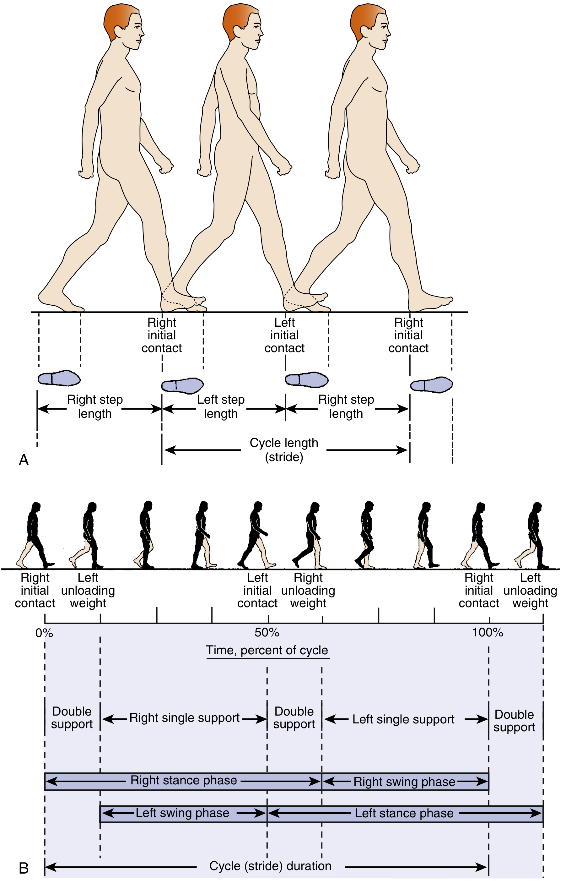
Key Spatiotemporal Terms:
| Parameter | Definition |
|---|---|
| Step length | Distance from IC of one foot to IC of the opposite foot |
| Stride length | Distance from IC of one foot to the next IC of the same foot (= 1 full gait cycle) |
| Cadence | Steps per unit time |
| Velocity | Distance / Time |
3. PHASES OF THE GAIT CYCLE
The gait cycle is divided into two major phases:
A. STANCE PHASE (60% of gait cycle)
Weight-bearing period - foot is in contact with the ground.
| Sub-phase | % Gait Cycle | Key Event |
|---|---|---|
| Initial Contact (IC) / Heel Strike | 0-2% | Heel touches ground; weight acceptance begins |
| Loading Response (LR) | 2-12% | Body weight transfers onto limb; "first rocker" (heel rocker); knee flexes ~15° |
| Mid-Stance (MSt) | 12-31% | Body passes over the supporting foot; "second rocker" (ankle rocker) |
| Terminal Stance (TSt) | 31-50% | Heel rises; body weight moves forward; "third rocker" (forefoot rocker) |
| Pre-Swing (PSw) | 50-60% | Push-off; limb unloads; preparation for swing |
B. SWING PHASE (40% of gait cycle)
Non-weight-bearing period - foot is off the ground.
| Sub-phase | % Gait Cycle | Key Event |
|---|---|---|
| Initial Swing (ISw) | 60-73% | Foot leaves ground; limb accelerates forward |
| Mid-Swing (MSw) | 73-87% | Limb advances forward; foot clears the ground |
| Terminal Swing (TSw) | 87-100% | Tibia perpendicular to ground; limb decelerates; foot prepares for IC |
Double limb support occurs during IC+LR and PSw (20-30% of cycle). Running eliminates double limb support and adds a "float phase."
(Source: Miller's Review of Orthopaedics, 9th ed., p. 865; General Anatomy THIEME Atlas)
4. JOINTS INVOLVED IN GAIT
The primary joints participating in gait moment generation are:
| Joint | Role |
|---|---|
| Hip joint | Ball-and-socket; controls limb advancement, trunk stability, sagittal & frontal plane moments |
| Knee joint | Modified hinge; shock absorption in LR, propulsion assistance, swing clearance |
| Ankle joint | Hinge (talocrural); three rockers (heel, ankle, forefoot); major propulsive moment at push-off |
| Subtalar joint | Triplanar motion; pronation in LR dampens impact; supination stabilizes push-off |
| Metatarsophalangeal (MTP) joints | Extension during terminal stance/push-off (third rocker) |
| Lumbopelvic region | Pelvic rotation (transverse), pelvic list (frontal), trunk counterrotation |
5. PLANE AND AXIS OF MOTION
Each joint action during gait occurs in a specific anatomical plane around a specific axis:
| Plane | Axis | Motion at Joint |
|---|---|---|
| Sagittal | Mediolateral (coronal) axis | Flexion/Extension at hip, knee, ankle - the dominant plane in gait |
| Frontal (Coronal) | Anteroposterior axis | Hip abduction/adduction; pelvic list |
| Transverse (Horizontal) | Vertical (longitudinal) axis | Hip/pelvis rotation; subtalar pronation/supination |
Clinical note: While knee motion primarily occurs in the sagittal plane, the hip, pelvis, and subtalar joint exhibit motion in all three planes. The knee adduction moment (KAM) - a frontal plane moment - is of major clinical interest as a surrogate for medial compartment loading in knee osteoarthritis.
(Source: Firestein & Kelley's Textbook of Rheumatology, p. 692-710)
6. MUSCLES INVOLVED AND THEIR MOMENT-GENERATING ROLE
Most muscle activity in gait is eccentric (muscle active while lengthening to control motion and absorb energy). Concentric activity occurs mainly during push-off and initial swing.
HIP MUSCLES
| Phase | Muscle Group | Type of Contraction | Moment Produced |
|---|---|---|---|
| IC - LR | Gluteus maximus, Hamstrings | Eccentric | Hip extension moment; decelerates hip flexion |
| LR - MSt | Hip abductors (Gluteus medius, minimus, TFL) | Isometric/Eccentric | Frontal plane: stabilizes pelvis (prevents contralateral drop = Trendelenburg) |
| TSt - PSw | Hip flexors (Iliopsoas, rectus femoris) | Concentric | Hip flexion moment; initiates limb advancement |
| MSw - TSw | Hamstrings | Eccentric | Decelerates hip flexion; prepares for IC |
KNEE MUSCLES
| Phase | Muscle Group | Type of Contraction | Moment Produced |
|---|---|---|---|
| IC - LR | Quadriceps femoris | Eccentric | Knee extension moment; controls 15° flexion for shock absorption |
| MSt - TSt | Quadriceps | Near isometric | Maintains knee extension during single limb support |
| PSw - ISw | Hamstrings | Concentric | Knee flexion moment for foot clearance |
| TSw | Quadriceps | Eccentric | Decelerates knee; prepares for IC |
ANKLE MUSCLES
| Phase | Muscle Group | Type of Contraction | Moment Produced |
|---|---|---|---|
| IC | Tibialis anterior | Eccentric | Dorsiflexion moment; controls foot lowering (heel rocker) |
| LR - MSt | Tibialis anterior | Eccentric | Controls plantar flexion; "second rocker" |
| MSt - TSt | Gastrocnemius-Soleus | Eccentric then Concentric | Plantarflexion moment; controls forward tibial progression then propels push-off |
| PSw | Gastrocnemius-Soleus | Concentric (push-off) | Maximum plantarflexion moment; major propulsive force |
| Swing | Tibialis anterior | Concentric | Dorsiflexion moment; foot clearance |
(Source: Miller's Review of Orthopaedics 9th ed.; Firestein & Kelley's Textbook of Rheumatology)
7. KINETICS OF GAIT (Force and Moment Analysis)
Kinetics refers to the forces and moments that cause motion.
A. Ground Reaction Force (GRF)
The GRF is the force exerted by the ground on the body (Newton's 3rd Law equal and opposite to body weight). It changes in magnitude and direction throughout the gait cycle.
- Normal walking GRF shows a characteristic double-hump pattern in the vertical direction:
- First peak (~120% body weight) at LR - weight acceptance
- Valley (~80% BW) at mid-stance
- Second peak (~110% BW) at TSt/push-off
The moment arm of the GRF vector relative to each joint centre determines the external moment at that joint.
B. Joint Moments During Gait
Hip Moments:
- Sagittal plane: Hip extension moment dominates during LR and stance (counteracts hip flexion tendency from GRF passing posterior to hip). Hip flexion moment during PSw and swing.
- Frontal plane: Hip abductor moment throughout stance - prevents pelvic drop (Trendelenburg). Magnitude ~0.6-0.8 Nm/kg.
Knee Moments:
- LR: Knee flexion moment (GRF passes posterior to knee) - controlled by eccentric quadriceps.
- MSt - TSt: Knee extension moment (GRF passes anterior to knee) - passive stability.
- KAM (Knee Adduction Moment): Frontal plane; the GRF passes medial to the knee - resultant medial compartment loading. Normal: ~0.4-0.5 Nm/kg.
Ankle Moments:
- Plantarflexion moment dominates throughout stance from LR to push-off.
- Peak plantarflexion moment at PSw: ~1.5-1.8 Nm/kg - the largest joint moment in gait. This drives the propulsive push-off.
Support Moment: The sum of extensor moments at hip + knee + ankle. This remains relatively constant during stance, demonstrating that if one joint provides less extension moment, others compensate (inter-joint coordination).
(Source: Winter DA 1987; OUHSC Gait Analysis; Miller's Review of Orthopaedics)
C. Moment Arm and Mechanical Advantage
As muscles typically insert close to the joint, their internal moment arm (d) is small. External forces (GRF, body weight) act at much larger moment arms. This means:
- Muscles must generate large forces (poor mechanical advantage)
- Result: Joint contact forces often 2-5x body weight during normal gait
Example - Ankle at push-off:
- Gastrocnemius-soleus pulls on the calcaneus ~5 cm posterior to the ankle axis
- GRF acts at the forefoot ~15 cm anterior to the axis
- To balance these moments: Achilles tendon force ≈ 3 × Body weight
(Source: Firestein & Kelley's Textbook of Rheumatology, p. 944-950; Eric Kandel's Principles of Neural Science)
8. KINEMATICS OF GAIT (Motion Analysis)
Kinematics describes the geometry of motion (angles, velocities, accelerations) without reference to the forces causing it. Kinematics provides the data needed to calculate moments via inverse dynamics.
Sagittal Plane Kinematics (Primary Plane of Gait)
Hip:
| Phase | Motion | Range |
|---|---|---|
| IC | Flexed ~30° | - |
| MSt | Moving toward neutral | 0-5° |
| TSt | Hyperextension | ~10-15° |
| Swing | Flexion | 0° → ~30° |
Knee:
| Phase | Motion | Range |
|---|---|---|
| IC | Near full extension | 0-5° |
| LR | Flexion (shock absorption) | peaks at ~15-20° |
| MSt - TSt | Extension | returns to near 0° |
| PSw - ISw | Rapid flexion | peaks ~60-65° (foot clearance) |
| TSw | Extension | returns to ~0° before IC |
The knee exhibits two flexion-extension cycles per gait cycle - one for shock absorption in stance, one for clearance in swing. (Firestein & Kelley's Textbook of Rheumatology, p. 667-685)
Ankle:
| Phase | Motion | Range |
|---|---|---|
| IC | Neutral (0°) | - |
| LR | Plantarflexion | ~10-15° (heel rocker) |
| MSt | Dorsiflexion | peaks ~10° (ankle rocker) |
| TSt | Increasing dorsiflexion | 10-15° |
| PSw | Plantarflexion | ~20° (push-off) |
| Swing | Dorsiflexion | returns to neutral for IC |
Center of Mass (COM) Kinematics
- Vertical displacement of COM: sinusoidal, amplitude ~5 cm (highest at MSt, lowest at double support)
- Lateral displacement: sinusoidal, amplitude ~6 cm
- Minimizing these displacements = energy efficiency (six determinants of gait)
Center of Pressure (COP)
Tracks from heel posteriorly at IC, progresses forward to the hallux at push-off. Provides the point of application of the GRF - essential for moment arm calculations.
9. FUNCTIONAL SIGNIFICANCE OF MOMENT ANALYSIS IN GAIT
Understanding joint moments in gait has direct clinical and rehabilitation relevance:
1. Energy Efficiency
The body minimizes COM displacement through coordinated joint moments (six determinants of gait). The support moment remains nearly constant - if one joint fails, others compensate.
2. Shock Absorption
The eccentric knee extensor moment during LR (quadriceps controlling ~15° flexion) dissipates impact energy equivalent to a significant percentage of body weight. Loss of this eccentric control (quadriceps weakness) leads to stiff-knee or crouched gait.
3. Propulsion
The ankle plantarflexion moment at push-off (peak ~1.5-1.8 Nm/kg) is the primary propulsive mechanism in gait. Calf muscle weakness (e.g., in stroke, peripheral neuropathy) dramatically reduces walking speed and cadence.
4. Frontal Plane Stability
The hip abductor moment (gluteus medius) counteracts the tendency for the pelvis to drop on the swing side. Weakness produces Trendelenburg gait and abnormally high KAM (increasing medial knee compartment stress).
5. Clinical Applications
| Condition | Altered Moment | Clinical Effect |
|---|---|---|
| Knee OA | Elevated KAM | Increased medial compartment loading; disease progression |
| Stroke / Hemiplegia | Reduced ankle push-off moment | Reduced propulsion; asymmetric gait |
| ACL injury | Reduced knee extension moment | Altered quadriceps function; joint instability |
| Hip abductor weakness | Reduced hip abductor moment | Trendelenburg gait; increased contralateral pelvic drop |
| Cerebral palsy | Reduced/altered ankle moment | Equinus gait; crouch gait |
| Prosthetic gait | Absent push-off at ankle | Energy inefficiency; compensatory strategies |
6. Clinical Gait Analysis Tools
- Force plates - measure GRF (needed for external moment calculation)
- Motion capture systems - measure kinematics
- EMG - records muscle activity timing and intensity
- Inverse dynamics - combines kinematics + GRF + anthropometric data to calculate joint moments
(Sources: Springer Nature - Interpreting Joint Moments in Gait; PM&R KnowledgeNow - Biomechanics of Normal Gait; Firestein & Kelley's Textbook of Rheumatology)
10. SUMMARY TABLE: MOMENT ANALYSIS AT MAJOR JOINTS DURING GAIT
| Phase | Hip Moment | Knee Moment | Ankle Moment | Key Muscles |
|---|---|---|---|---|
| IC (0-2%) | Ext moment | Small flex moment | Dorsiflexion moment | Glut. max, Hamstrings, Tibialis anterior |
| LR (2-12%) | Ext moment | Flex moment (eccentric) | Plantar flex (eccentric) | Quadriceps (eccentric), Tibialis anterior |
| MSt (12-31%) | Abductor moment (frontal) | Ext moment | Increasing dorsiflexion | Glut. medius, Gastrocnemius-soleus (eccentric) |
| TSt (31-50%) | Ext → flex transition | Ext moment | Peak plantar flex moment | Gastrocnemius-soleus (concentric) |
| PSw (50-60%) | Flex moment | Flex moment | Plantar flex (push-off) | Hip flexors, Gastroc-soleus |
| ISw (60-73%) | Flex moment | Flex moment | Dorsiflexion | Iliopsoas, Tibialis anterior |
| MSw (73-87%) | Flex moment | Extension moment | Dorsiflexion | Hamstrings (decel), Tibialis anterior |
| TSw (87-100%) | Ext moment | Ext moment (decel) | Neutral | Hamstrings (eccentric), Quadriceps (eccentric) |
REFERENCES
- Miller MD et al. Miller's Review of Orthopaedics, 9th ed. Elsevier; 2024. (Gait cycle and dynamics, pp. 860-870)
- Firestein GS, Kelley WN. Firestein & Kelley's Textbook of Rheumatology, 2-Volume Set. Elsevier; 2022. (Kinematics and kinetics, Chapter 6, pp. 660-960)
- Kandel ER et al. Principles of Neural Science, 6th ed. McGraw-Hill; 2021. (Muscle torque and moment arm, pp. 921-937)
- Winter DA. Biomechanics and Motor Control of Human Movement. 1987. (Foundational joint moment data)
- Thompson D. Joint Moments in Gait. University of Oklahoma HSC.
- PM&R KnowledgeNow. Biomechanics of Normal Gait. AAPM&R; 2024.
- Jafarnezhadgero AA et al. Step-by-step insight into gait analysis: knee biomechanics. Knee Surgery, Sports Traumatology, Arthroscopy. 2024.
Assignment tip: When drawing moment diagrams, always identify: (1) the joint axis, (2) the direction of the external force (GRF or gravity), (3) the perpendicular moment arm, and (4) whether the resulting external moment tends to flex or extend the joint - then the internal (muscle) moment must oppose it for controlled motion.
This is a shared conversation. Sign in to Orris to start your own chat.