
give me clinical features , causes , diagnosis and mangement of drug induced lupus erythamatosis
"drug-induced lupus erythematosus"[MeSH Terms]
drug induced lupus erythematosus skin rash butterfly malar
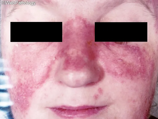
Comprehensive Description: This clinical dermatology photograph depicts the classic malar, or butterfly, rash of systemic lupus erythematosus (SLE). Modality: Clinical photography of facial skin in a frontal/anterior view. The malar distribution spans the nasal bridge and both malar prominences with symmetric erythematous, violaceous, and edematous plaques. The skin surface appears mildly scaly in places, and photoexposed regions may show accentuated erythema consistent with photosensitivity. The nasolabial folds are relatively spared in this pattern, and the overall appearance supports cutaneous lupus erythematosus when correlated with systemic signs. While not pathognomonic, the image demonstrates a prototypical manifestation that can mimic rosacea, dermatitis, or dermatomyositis, emphasizing the importance of clinical context and serologic testing for diagnosis. Alopecia or scarring alopecia may accompany cutaneous disease but is not evident in this frame. This photo highlights the diagnostic significance of a photosensitive, malar eruption and its role in assessing disease activity and treatment response. Potential clinical uses include dermatology and rheumatology education, reference for malar rash recognition, differential diagnosis training, and documentation for patient counseling and longitudinal follow-up. The image also serves as a teaching aid for recognizing sun-induced exacerbations, differentiating inflammatory rashes, and discussing psychosocial impact and cosmetic considerations in lived care for SLE patients.
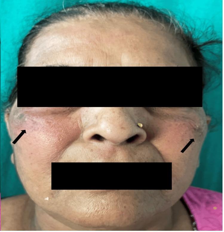
Clinical photograph of a 59-year-old female patient showing a characteristic malar rash. The image displays confluent, erythematous patches distributed over the bilateral cheeks and extending towards the temporal regions, as indicated by black arrows. The rash exhibits a symmetric, butterfly-like distribution, though the nasolabial folds appear relatively spared. The skin within the affected areas shows noticeable texture changes and erythema. Additional visible features include forehead rhytids, perioral lines consistent with age-related skin changes, and uneven pigmentation. The patient has a nose ring in the left nostril. The clinical presentation is consistent with cutaneous lupus erythematosus, specifically a drug-induced manifestation. The educational focus is on identifying classic dermatological signs of systemic inflammatory or drug-induced conditions through facial skin morphology and distribution patterns.
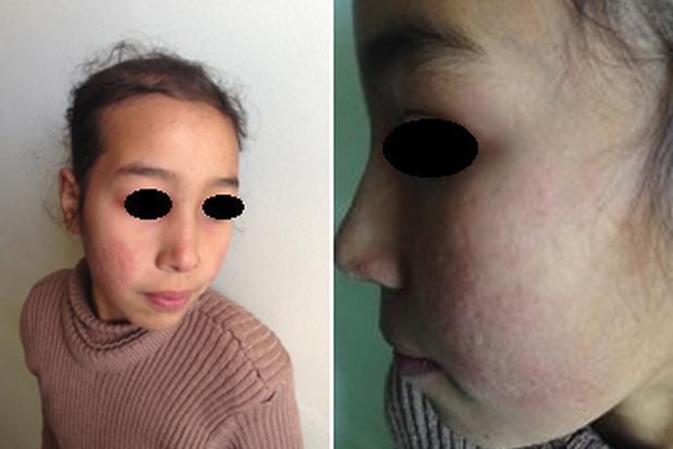
This clinical photograph displays a pediatric patient with dermatological manifestations consistent with Systemic Lupus Erythematosus (SLE). The image consists of two panels: a frontal view and a lateral close-up of the face. A distinctive malar rash, often referred to as a 'butterfly rash,' is visible across the nasal bridge and bilateral cheeks, notably sparing the nasolabial folds. The affected skin exhibits erythematous discoloration and an irregular texture. Higher magnification reveals atrophic scarring within the malar region, characterized by thin, slightly depressed skin with altered pigmentation. The forehead and peri-orbital regions appear relatively unaffected. These visual features are classic diagnostic markers for lupus-related skin involvement, illustrating the chronic inflammatory and sequela phases of the disease. The content is relevant for medical students and clinicians studying rheumatology, dermatology, and autoimmune connective tissue disorders.
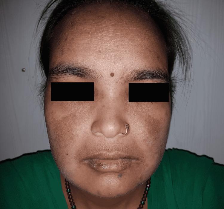
This clinical photograph of a 38-year-old female patient displays a classic malar rash (butterfly rash), a hallmark sign of Systemic Lupus Erythematosus (SLE). The image illustrates a symmetric, erythematous maculopapular eruption extending across the nasal bridge and both malar eminences (cheeks). A key diagnostic feature visible is the distinct sparing of the nasolabial folds. The affected skin shows reddish discoloration with both flat (macular) and slightly raised (papular) textures. Additional observations include generalized facial hyperpigmentation, uneven skin tone on the forehead, and mild dryness or discoloration at the vermillion border of the lips. The photograph serves as a primary educational resource for medical students and clinicians to recognize LE-specific cutaneous manifestations. The target audience is intermediate-level medical learners focusing on rheumatology and dermatology. Educational concepts highlighted include the distribution patterns of photosensitive rashes and the clinical distinction between lupus-related malar eruptions and other facial dermatoses like rosacea, which typically involves the nasolabial folds.
Drug-Induced Lupus Erythematosus (DILE)
Causes / Implicated Drugs
High-Risk Drugs (most commonly implicated)
| Drug | Class | Risk |
|---|---|---|
| Procainamide | Antiarrhythmic | Up to 30% risk; nearly 80% develop ANA after prolonged use |
| Hydralazine | Antihypertensive | Well-established; higher risk in slow acetylators |
| Isoniazid (INH) | Antitubercular | Classic causative agent |
| Minocycline | Antibiotic | Notable - often anti-histone NEGATIVE |
| Quinidine | Antiarrhythmic | Classic association |
Other Implicated Drugs
- Antihypertensives: methyldopa, ACE inhibitors, beta-blockers, hydrochlorothiazide
- Antiarrhythmics: other class I agents
- Antibiotics: rifampin, nitrofurantoin
- Anticonvulsants: phenytoin, carbamazepine
- Antipsychotics: chlorpromazine, lithium
- Anti-rheumatic: sulfasalazine, D-penicillamine
- Antithyroid: propylthiouracil
- Biologics: Anti-TNF-alpha agents (infliximab, etanercept), IFN-alpha
- Others: NSAIDs, oral contraceptives, proton pump inhibitors
Pathogenesis
-
Acetylator status: Slow acetylators have lower levels of acetyltransferase, the enzyme that detoxifies drugs like hydralazine and procainamide. Slow acetylators develop ANA and symptoms more rapidly. Rapid acetylators are not protected - they just require a higher cumulative dose. - Henry's Clinical Diagnosis and Management by Laboratory Methods
-
DNA hypomethylation: Procainamide directly inhibits DNMT1 (the maintenance DNA methyltransferase), and hydralazine inhibits it indirectly - leading to demethylation of T cells and resulting T cell autoreactivity. - Firestein & Kelley's Rheumatology
-
Hapten mechanism: Drugs may act as haptens, prompting an autoimmune response.
-
Direct cytotoxicity: Some drugs cause direct cellular damage triggering immune activation.
-
HLA association: HLA-DR4 is associated with susceptibility.
Clinical Features
Typical Symptoms
- Fever
- Arthralgia / arthritis (most common)
- Myalgia
- Rash (including malar rash, photosensitivity)
- Serositis: pleuritis, pericarditis - pleural effusions occur in ~40% of lupus (including DILE)
Typically ABSENT or Rare in DILE
- Renal involvement (lupus nephritis is uncommon)
- CNS involvement (neuropsychiatric lupus is uncommon)
- Severe cytopenias (mild cytopenias may be present)
Hepatic Involvement
- Hepatic involvement is notably common in DILE compared to idiopathic SLE. - Firestein & Kelley's Rheumatology
Onset
- Symptoms may begin weeks to several months after drug initiation - making recognition difficult.
Diagnosis
Serologic Findings
| Test | Finding in DILE |
|---|---|
| ANA | Positive; most commonly homogeneous pattern; may appear before symptoms |
| Anti-histone antibodies | Hallmark - positive in 75-95% of cases; directed against H2A-H2B dimer complex |
| Anti-dsDNA | Rare (distinguishes DILE from SLE, where it is common) |
| Anti-Smith (Sm) | Rare (also helps distinguish from SLE) |
| Anti-ssDNA / chromatin | Can be present |
| Complement (C3/C4) | Hypocomplementemia less common than in SLE |
| Cytopenias | Mild if present |
Important note: Minocycline-induced DILE is a notable exception - it is often anti-histone NEGATIVE. - Firestein & Kelley's Rheumatology
Diagnostic Approach
- Identify the offending drug and timeline of exposure
- Check ANA (homogeneous pattern typical), anti-histone, anti-dsDNA
- Skin biopsy may be indistinguishable from idiopathic SLE
- Confirmation: Resolution of symptoms after drug withdrawal strongly supports the diagnosis
- Absence of anti-dsDNA, anti-Sm, and hypocomplementemia help distinguish from SLE
Management
Step 1: Withdraw the Offending Drug
- This is the primary and most important treatment
- Symptoms typically resolve within weeks after discontinuation
- However, complete resolution of clinical findings may take months to years - Firestein & Kelley's Rheumatology
- ANA titers may persist for months even after clinical resolution
Step 2: Symptomatic / Supportive Treatment
- NSAIDs - for arthralgia, fever, serositis
- Hydroxychloroquine (HCQ) - for skin and joint manifestations
Step 3: Immunosuppressive Therapy (if severe)
- Corticosteroids - for significant serositis, cytopenias, or persistent symptoms after drug withdrawal
- Further immunosuppression is rarely needed given the generally mild nature of DILE
Step 4: Long-term Monitoring
- Monitor for persistence or recurrence if re-exposure occurs
- Anti-histone antibodies and ANA may persist long after clinical resolution - do not use serology alone to gauge treatment success
Key Differences: DILE vs. Idiopathic SLE
| Feature | DILE | SLE |
|---|---|---|
| Sex predominance | Equal (M = F) | Female predominance (9:1) |
| Renal involvement | Rare | Common |
| CNS involvement | Rare | Common |
| Hepatic involvement | Common | Less common |
| Anti-histone Ab | 75-95% positive | ~50% (less specific) |
| Anti-dsDNA | Rare | Common (marker of activity) |
| Anti-Sm | Rare | Characteristic |
| Hypocomplementemia | Uncommon | Common |
| Course | Reversible on drug withdrawal | Chronic, relapsing |