
IHD patient posted for TKR 20 mark answer
Reading File
Quick answer: An IHD (Ischemic Heart Disease) patient posted for TKR (Total Knee Replacement) requires thorough preoperative cardiac risk stratification (using tools like the Revised Cardiac Risk Index), optimization of medications, careful anesthetic planning (regional vs. general), and vigilant intraoperative/postoperative hemodynamic monitoring to prevent MACE (Major Adverse Cardiac Events).
Searching sources...
Finding Sources
Finding Sources
Finding Sources
Reading File
Finding Sources
Finding Sources
Reading File
Reading File
Reading File
Reading File
Reading File
Finding Sources
Reading File
Finding Sources
Reading File
Finding Sources
Reading File
Now I have comprehensive data from authoritative sources. Let me compile the full 20-mark answer.
Loading Image
Here is the complete 20-mark structured answer:
Anesthetic Management of an IHD Patient Posted for Total Knee Replacement (TKR)
Introduction
A patient with Ischemic Heart Disease (IHD) undergoing Total Knee Replacement (TKR) - an intermediate-to-high risk orthopedic procedure - presents significant perioperative challenges. The primary goal is to prevent Major Adverse Cardiovascular Events (MACE) including myocardial infarction, arrhythmia, acute heart failure, and cardiac arrest. Management is best organized into preoperative, intraoperative, and postoperative phases.
I. PREOPERATIVE ASSESSMENT AND OPTIMIZATION
A. Cardiac Risk Stratification
TKR is classified as an intermediate-risk surgical procedure (perioperative MACE risk >1%). The stepwise ACC/AHA algorithm (Fig 492-1) guides evaluation:
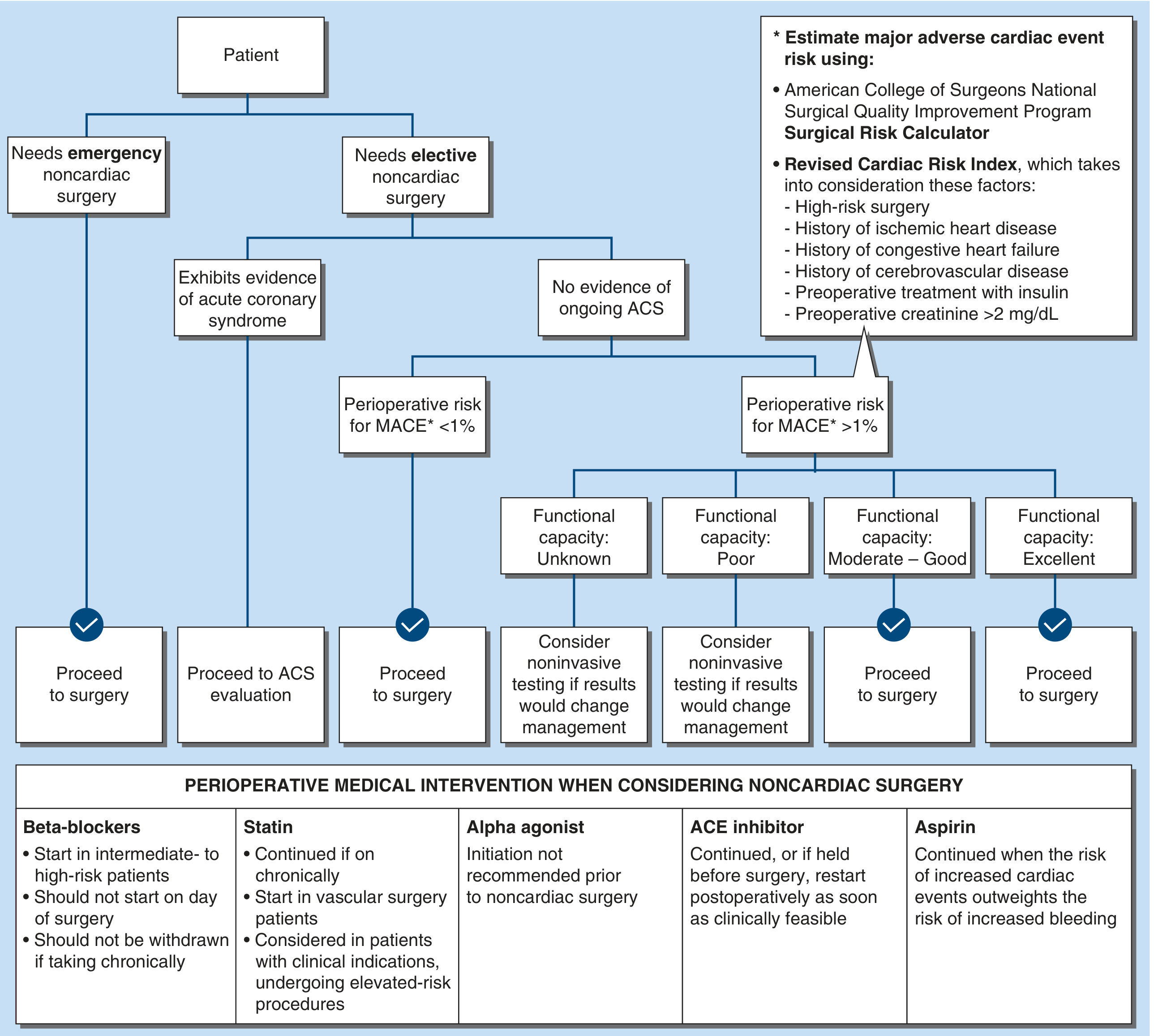
Step 1 - Urgency: TKR is elective - full evaluation is warranted. Emergency surgery proceeds without full evaluation.
Step 2 - Acute Coronary Syndrome (ACS): Exclude active/unstable ACS. If present, delay surgery and treat per ACS guidelines.
Step 3 - Revised Cardiac Risk Index (RCRI) (Lee Index) - the preferred validated tool:
Each factor scores 1 point:
- High-risk surgery (intrathoracic, intraabdominal, suprainguinal vascular)
- Ischemic heart disease (history of MI, angina, positive stress test)
- Congestive heart failure
- Cerebrovascular disease (TIA/stroke)
- Insulin-dependent diabetes mellitus
- Serum creatinine >2.0 mg/dL
Predicted MACE rates: 0 factors = 0.4%, 1 factor = 0.9%, 2 factors = 7%, ≥3 factors = 11%.
An IHD patient with TKR (high-risk surgery) already scores ≥2 points (MACE risk ~7%), and additional factors push this higher - Harrison's Principles of Internal Medicine 22E, p. 3949-3950.
Step 4 - Functional Capacity: Expressed as Metabolic Equivalents (METs).
- Poor/unknown (<4 METs): Cannot walk 4 blocks or climb 2 flights of stairs - proceed to noninvasive testing if it would change management.
- Moderate-Good (≥4 METs): Proceed to surgery.
- This is particularly problematic in TKR patients due to limited mobility from knee disease - the pre-existing orthopedic condition often masks poor cardiorespiratory reserve.
B. Investigations
| Investigation | Indication |
|---|---|
| 12-lead ECG | Mandatory for all IHD patients |
| Echocardiography (TTE) | Assess LV function (EF), wall motion abnormalities, valvular disease |
| Cardiac stress test (pharmacologic - dobutamine stress echo / nuclear scan) | If functional capacity unknown/poor and result will change management |
| Cardiac biomarkers (hsTroponin, BNP) | Baseline for perioperative comparison; elevated baseline = high risk |
| CBC, RFT, LFT, blood glucose, HbA1c | Comorbidity screen |
| CXR | Pulmonary congestion, cardiomegaly |
| Coagulation profile | Baseline, especially if on anticoagulants |
Coronary angiography is NOT routinely recommended for the sole purpose of reducing perioperative risk. Revascularization (PCI/CABG) before noncardiac surgery provides no short-term benefit in stable CAD without left main disease or 3-vessel CAD with poor LV function - Harrison's, p. 3950.
C. Medication Management
Beta-blockers:
- Continue if the patient is already on chronic beta-blocker therapy (abrupt withdrawal is dangerous - causes rebound tachycardia and hypertension).
- Initiate in high-risk patients (≥3 RCRI factors or intermediate/high-risk ischemia on stress testing), ideally days-to-weeks before surgery to assess tolerability.
- Never initiate on the day of surgery (POISE trial showed high-dose acute initiation increased stroke and mortality despite reducing MI) - Harrison's, p. 3951.
Statins:
- Continue all statin therapy perioperatively - reduces perioperative cardiac events.
- Initiation is reasonable in statin-naïve patients undergoing elevated-risk procedures.
Antiplatelet agents (Aspirin/DAPT):
- Aspirin: Continue unless bleeding risk outweighs cardiac benefit (risk-benefit decision with surgeon).
- DAPT (if patient has a coronary stent):
- Bare Metal Stent (BMS): Delay elective surgery ≥1 month; ideally 6 months.
- Drug-Eluting Stent (DES): Delay elective surgery ≥6 months; ideally 12 months after ACS.
- If surgery cannot be delayed and DAPT must be stopped: maintain aspirin, restart P2Y12 inhibitor ASAP postoperatively.
- Premature discontinuation risks stent thrombosis - Harrison's, p. 3950.
ACE Inhibitors/ARBs:
- Generally continue; if held preoperatively (due to hypotension risk), restart as soon as clinically feasible postoperatively.
SGLT-2 Inhibitors:
- Discontinue 3-4 days before surgery to prevent euglycemic diabetic ketoacidosis from perioperative fasting - Harrison's, p. 3950.
Nitrates: Continue if on long-acting nitrates for angina prophylaxis.
II. INTRAOPERATIVE MANAGEMENT
A. Choice of Anesthesia
Regional anesthesia (Spinal/Subarachnoid Block - SAB) is the preferred technique for TKR in IHD patients for the following reasons:
- Avoids the hemodynamic stress of laryngoscopy and intubation
- Provides excellent surgical anesthesia and postoperative analgesia
- Reduces systemic opioid requirements
- Meta-analyses show lower rates of pneumonia and respiratory failure with neuroaxial vs. general anesthesia
- However: no significant difference in cardiac events between neuroaxial and general anesthesia has been demonstrated - Harrison's, p. 3951
Spinal anesthesia considerations:
- Causes sympathetic block → hypotension → compensate with cautious IV fluids and vasopressors (phenylephrine, ephedrine)
- Bradycardia may occur - have atropine ready
- Level of block: T12-L1 adequate for TKR
General anesthesia (if regional contraindicated):
- All inhaled agents cause dose-dependent vasodilation, myocardial depression, and decreased cardiac output - use cautiously
- Avoid tachycardia (HR <80 bpm target to reduce myocardial oxygen demand)
- TIVA (propofol-based) may be preferable in LV dysfunction
- Careful titration of induction agents (reduced doses in compromised patients)
Combined spinal-epidural (CSE):
- Allows intraoperative spinal anesthesia plus postoperative epidural analgesia
- Caution: epidural hematoma risk if systemic anticoagulation used for VTE prophylaxis
Peripheral nerve blocks:
- Femoral nerve block / Adductor canal block as part of multimodal analgesia - reduces opioid use
B. Intraoperative Monitoring
Standard monitoring:
- Continuous ECG (5-lead preferred, with ST-segment analysis in leads II and V5)
- Pulse oximetry
- NIBP (5-minute intervals minimum)
- Capnography (if GA)
- Temperature monitoring
- Urine output
Additional monitoring (if high-risk):
- Invasive arterial line (IBP) - continuous BP monitoring, arterial blood sampling
- Central venous catheter - if poor IV access, major fluid shifts expected
- Pulmonary artery catheter / TEE - only in severely compromised LV function
C. Hemodynamic Goals
| Parameter | Target |
|---|---|
| Heart rate | 60-80 bpm (avoid tachycardia) |
| Blood pressure | Maintain within 20% of baseline MAP |
| SpO2 | >95% |
| Hematocrit | >25-30% (avoid anemia-driven demand ischemia) |
- Treat hypotension promptly: IV fluids, vasopressors (phenylephrine if sinus rhythm, ephedrine if bradycardic)
- Treat hypertension: deepen anesthesia, opioids, nitroglycerin infusion
- Avoid anemia (TKR involves significant blood loss - use tourniquet judiciously, cell saver, tranexamic acid)
- Tourniquet inflation causes sudden afterload increase and BP spike - monitor closely; deflation causes sudden BP drop and reperfusion arrhythmias
D. Special Considerations in TKR
- Cement implantation (bone cement implantation syndrome - BCIS): Polymethylmethacrylate cement can cause hypotension, hypoxia, arrhythmias, and even cardiac arrest due to fat/marrow/cement embolism to pulmonary circulation. More dangerous in IHD patients with poor cardiac reserve. Have resuscitation drugs ready.
- Tourniquet pain/hypertension: Intraoperative tourniquet use causes progressive hypertension - ensure adequate analgesia/anesthesia depth.
- VTE: TKR carries very high DVT/PE risk - mechanical and pharmacological prophylaxis essential.
III. POSTOPERATIVE MANAGEMENT
A. Recovery and Monitoring
- ICU/HDU admission for high-risk IHD patients (RCRI ≥3, EF <40%, recent MI)
- Continue continuous ECG monitoring for 24-48 hours
- Serial high-sensitivity troponins for 48-72 hours postoperatively in high-risk patients - to detect Perioperative Myocardial Injury (PMI)
PMI: Defined as absolute increase in hsTroponin ≥14 ng/L from preoperative to postoperative values. In TKR patients (intermediate-risk orthopedic surgery), PMI incidence is 20% and carries 30-day mortality of up to 9% and 1-year mortality of up to 22% - Miller's Anesthesia 10e, p. 9148-9149.
Canadian Cardiovascular Society guidelines recommend daily troponin measurements for 48-72 hours after noncardiac surgery in high-risk patients.
B. Analgesia
- Multimodal analgesia: paracetamol + NSAIDs (caution with NSAIDs in IHD - COX-2 inhibitors increase cardiovascular events) + opioids (titrated) + regional blocks (adductor canal block)
- Epidural analgesia (if CSE placed): Provides superior pain control; maintain for >24 hours
- Avoid pain-induced tachycardia and hypertension - both increase myocardial oxygen demand
C. VTE Prophylaxis
- Mechanical: Graduated compression stockings + pneumatic compression devices - start immediately
- Pharmacological: LMWH (enoxaparin 40 mg OD or 30 mg BD) or fondaparinux, or direct oral anticoagulants (rivaroxaban 10 mg OD preferred in some guidelines)
- Balance VTE prophylaxis with risk of epidural hematoma if epidural catheter is in place (follow ASRA neuraxial-anticoagulation guidelines for timing)
- Aspirin is NOT recommended as the sole agent for VTE prophylaxis - Harrison's, p. 3953
D. Resumption of Cardiac Medications
- Restart all cardiac medications as soon as oral intake resumes
- ACE inhibitors/ARBs: restart when hemodynamically stable
- Antiplatelet agents: restart as per the bleeding/stent risk balance
- Beta-blockers: do NOT miss doses - rebound can precipitate MI
E. Watch for Postoperative Complications
- Myocardial Infarction (highest risk in first 6 weeks post TKR based on MI incidence data - Miller's Anesthesia, p. 9148)
- Acute Heart Failure
- Arrhythmias (AF incidence ~3% after noncardiac surgery)
- Pulmonary Embolism
- Acute kidney injury
IV. SUMMARY TABLE
| Phase | Key Actions |
|---|---|
| Preoperative | RCRI scoring, ECG, echo, functional capacity, optimize medications, DAPT/stent timing |
| Intraoperative | Prefer regional/spinal anesthesia, 5-lead ECG, arterial line, avoid tachycardia, treat BCIS |
| Postoperative | Serial troponins, ICU monitoring, multimodal analgesia, VTE prophylaxis, restart medications |
Key References
- Harrison's Principles of Internal Medicine 22E (2025), Chapter 492: Perioperative Medical Care - Cardiac Risk Assessment
- Miller's Anesthesia 10E, Chapter 60: Orthopedic Anesthesia - Cardiac Risk in TKR
- Sabiston Textbook of Surgery: Cardiac-Specific Preoperative Evaluation
- ACC/AHA 2014 Guidelines on Perioperative Cardiovascular Evaluation for Noncardiac Surgery
- 2022 ESC Guidelines on Cardiovascular Assessment for Noncardiac Surgery
For THR
The framework for an IHD patient posted for Total Hip Replacement (THR) is largely the same as TKR - same cardiac risk stratification, same medication management principles. The key differences lie in patient positioning, surgical approach, specific complications (fat embolism, BCIS pattern), and some anesthetic nuances. Let me pull the specific THR data from the textbooks.
Searching sources...
Finding Sources
Finding Sources
Reading File
Reading File
Reading File
Reading File
Reading File
Reading File
Reading File
Finding Sources
Reading File
I now have all the data needed. Here is the complete 20-mark answer for IHD patient posted for THR, with emphasis on the specific differences from TKR:
Anesthetic Management of an IHD Patient Posted for Total Hip Replacement (THR)
Introduction
Total Hip Replacement (THR / Total Hip Arthroplasty - THA) is an intermediate-to-high risk elective orthopedic procedure. In a patient with Ischemic Heart Disease (IHD), the perioperative risk is significantly elevated due to the combination of patient-related cardiac risk and surgery-specific factors including lateral decubitus positioning, substantial blood loss, bone cement implantation, fat embolism risk, and high VTE incidence. The perioperative goal is to prevent MACE (Major Adverse Cardiovascular Events) while ensuring safe surgical and anesthetic conditions.
I. PREOPERATIVE ASSESSMENT AND OPTIMIZATION
A. Surgical Risk Classification
THR is classified as an intermediate-to-elevated risk procedure (MACE risk >1%). It carries specific risks compared to TKR:
- Greater blood loss (500-1500 mL for primary THR vs. 500-1000 mL for TKR)
- Lateral decubitus positioning with significant hemodynamic implications
- Higher incidence of fat embolism syndrome (FES) due to femoral canal reaming
- Bone cement implantation syndrome (BCIS) in cemented prostheses
- No intraoperative tourniquet (unlike TKR) - therefore more continuous blood loss
B. Cardiac Risk Stratification (RCRI)
The Revised Cardiac Risk Index is applied identically to THR as for TKR. An IHD patient with THR scores at minimum 2 points (IHD + high-risk surgery), putting MACE risk at ~7%:
| RCRI Factor | Score |
|---|---|
| High-risk surgery (THR qualifies) | 1 |
| History of IHD (angina, prior MI, +ve stress test) | 1 |
| History of CCF | +1 |
| Cerebrovascular disease | +1 |
| Insulin-dependent DM | +1 |
| Creatinine >2.0 mg/dL | +1 |
MACE risk: 0 = 0.4%, 1 = 0.9%, 2 = 7%, ≥3 = 11% - Harrison's Principles of Internal Medicine 22E, p. 3949-3950
C. Functional Capacity Assessment
- Expressed in METs (Metabolic Equivalents)
- THR patients often have limited mobility from hip disease/pain, making functional capacity assessment difficult - similar to TKR
- Pharmacologic stress testing (dobutamine stress echocardiography or nuclear perfusion imaging) is indicated in patients with poor or unknown functional capacity where results will change management
- Myocardial infarction peaks in the first 6 weeks post-THR based on nationwide cohort data - Miller's Anesthesia 10E, p. 9148
D. Investigations
| Investigation | Rationale |
|---|---|
| 12-lead ECG | Mandatory; assess for old MI, ST changes, LVH, arrhythmia |
| 2D Echocardiography | LV EF, wall motion abnormality, diastolic dysfunction, pulmonary artery pressure |
| Pharmacologic stress test | If functional capacity <4 METs and result changes management |
| hsTroponin baseline | Elevated baseline predicts postoperative PMI |
| BNP/NT-proBNP | Elevated = higher perioperative cardiac risk |
| CBC | Baseline Hb (THR involves significant blood loss; preoptimize anemia) |
| Coagulation profile, LFT, RFT | Anticoagulant/statin safety; renal function impacts LMWH dosing |
| Blood group & cross-match | Type and screen; autologous blood donation considered |
| CXR | Cardiomegaly, pulmonary edema |
Preoperative anemia optimization is especially important in THR (autologous blood donation, IV iron, erythropoietin if time permits) to reduce allogenic transfusion risk in an IHD patient.
E. Medication Management
Identical principles to TKR with these key points:
| Drug | Action |
|---|---|
| Beta-blockers | Continue if on chronic therapy; do not withdraw; initiate in very high-risk patients weeks before surgery |
| Statins | Continue perioperatively - reduces perioperative cardiac events |
| Aspirin | Continue if cardiac benefit outweighs bleeding risk |
| DAPT / Coronary stent | Delay elective THR ≥6 months post-DES, ≥1 month post-BMS; never stop DAPT prematurely (stent thrombosis risk) |
| ACE inhibitors/ARBs | Hold on the day of surgery if hypotension anticipated; restart postoperatively ASAP |
| SGLT-2 inhibitors | Stop 3-4 days before surgery (euglycemic DKA risk) |
| Nitrates | Continue if on antianginal therapy |
II. INTRAOPERATIVE MANAGEMENT
A. Patient Positioning - Key Difference from TKR
THR uses either:
-
Lateral decubitus position (most common - posterior approach): The patient lies on the non-operative side with the operative hip uppermost.
- Anesthetic implications: ventilation-perfusion mismatch (dependent lung receives more perfusion, non-dependent lung is better ventilated)
- Axillary roll must be placed to protect the brachial plexus and axillary artery
- Head and cervical spine kept strictly neutral
- If spinal/epidural: can be placed in lateral or sitting position before positioning
- If GA: airway must be secured before final lateral positioning
-
Supine/Anterior approach (increasingly popular - tissue-sparing, faster recovery, no axillary roll needed): Patient lies supine on a specialized hip table (e.g., Mizuho OSI Hana table) - Barash Clinical Anesthesia 9E, p. 4378
No tourniquet is used in THR (unlike TKR), so there are no tourniquet-related pressure spikes or reperfusion events. However, blood loss is more continuous and can be substantial.
B. Choice of Anesthesia
Neuraxial anesthesia (spinal/epidural) is preferred for THR in IHD patients based on evidence:
- Large database studies show neuraxial anesthesia for THA is associated with:
- Lower 30-day mortality
- Decreased thromboembolic events
- Less blood loss and lower transfusion requirements
- Shorter length of stay
- Lower in-hospital complications
- Barash Clinical Anesthesia 9E, p. 4377
However, a systematic review and a randomized controlled trial found no significant difference in morbidity/mortality between neuraxial and general anesthesia - results are still debated. In practice, neuraxial is preferred for IHD patients due to hemodynamic benefits and avoidance of intubation stress.
Options:
| Technique | Details |
|---|---|
| Spinal (SAB) | Drug of choice: heavy bupivacaine 0.5% 2.5-3.5 mL; onset in 5 min; level T10 required; add intrathecal fentanyl/morphine for postoperative analgesia |
| Combined Spinal-Epidural (CSE) | Intraoperative spinal + epidural catheter for postoperative analgesia; ideal for IHD patients needing good pain control |
| General Anesthesia | If neuraxial contraindicated (severe spinal stenosis, coagulopathy, anticoagulation, patient refusal); use TIVA (propofol-based) preferred in LV dysfunction; careful induction to avoid hemodynamic instability |
| Peripheral Nerve Blocks | Lumbar plexus block (LPB/psoas compartment block) provides powerful analgesia for hip; femoral nerve block (FNB) as an alternative; avoid LPB in anticoagulated patients (risk of deep hematoma); fascia iliaca block, PENG block as motor-sparing alternatives |
Caution with neuraxial sympathectomy in IHD: Spinal anesthesia causes sudden sympatholysis - hypotension is more pronounced in:
- Hypovolemic patients (preoperative IV fluid preloading advised)
- Patients on chronic ACE inhibitors/ARBs
- Patients with IHD (hypotension = reduced coronary perfusion pressure = ischemia)
- A preload fluid bolus before spinal block is recommended; have vasopressors (phenylephrine, ephedrine) ready - Barash Clinical Anesthesia 9E, p. 4379
C. Intraoperative Monitoring
Standard:
- Continuous 5-lead ECG with ST-segment monitoring (leads II + V5)
- SpO2
- NIBP every 3 minutes (more frequent during cement insertion)
- Capnography (if GA or sedation)
- Temperature
Advanced (for high-risk IHD - EF <40%, RCRI ≥3, recent MI):
- Invasive arterial line (IBP): Mandatory for continuous BP monitoring especially during:
- Induction of anesthesia
- Cement insertion (BCIS risk - sudden catastrophic hypotension)
- Joint reduction
- Central venous catheter: For CVP monitoring and vasoactive drug infusion
- TEE or PAC: Reserved for severely compromised LV function (EF <30%) or pulmonary hypertension
D. Hemodynamic Goals
| Parameter | Target |
|---|---|
| Heart rate | 60-80 bpm (avoid tachycardia - increases O2 demand) |
| Mean Arterial Pressure | Within 20% of baseline (avoid hypotension - reduces coronary perfusion) |
| SpO2 | >95% |
| Hematocrit | >25-30% (avoid anemia; use TXA, cell saver) |
| Temperature | Normothermia (hypothermia worsens coagulopathy and cardiac arrhythmias) |
E. Blood Conservation - Specific to THR
THR involves more blood loss than TKR (no tourniquet), making blood conservation critical for IHD patients (who tolerate anemia poorly):
- Tranexamic Acid (TXA): IV 10-15 mg/kg before incision, then 1 mg/kg/hr during surgery; or topical 1-3 g; significantly reduces blood loss and transfusion requirements in THA/TKA. Safe even in IHD patients - a retrospective study of 765,011 patients showed TXA in those with MI, stroke, AF, and renal disease was not associated with increased complications - Miller's Anesthesia 10E, p. 9198
- Cell salvage (autologous transfusion)
- Deliberate hypotension (MAP 55-65 mmHg) during neuraxial anesthesia reduces blood loss - but use with extreme caution in IHD patients (may precipitate ischemia)
- Maintain normothermia to preserve coagulation
F. Surgery-Specific Intraoperative Hazards in IHD Patients
1. Bone Cement Implantation Syndrome (BCIS)
The most dangerous intraoperative event. Occurs during cemented femoral prosthesis insertion:
- Mechanism: Pressurization of cement into the femoral medullary canal forces bone marrow debris, fat globules, air, and methyl methacrylate monomer into the venous circulation → pulmonary embolization → acute right heart strain → cardiovascular collapse
- Clinical features: Sudden profound hypotension, hypoxia, bronchoconstriction, arrhythmias, increased pulmonary vascular resistance (PVR), right ventricular failure, and potentially cardiac arrest
- Risk factors specific to IHD patients: Pre-existing poor LV function, pulmonary hypertension, right ventricular dysfunction, long-stem prosthesis, first-time femoral canal instrumentation, pathological fractures, large cement volume - Miller's Anesthesia 10E, p. 9200
- Prevention: Pulsatile lavage of medullary canal; distal venting holes in the femur before cement; consider uncemented/cementless prosthesis in very high-risk IHD patients
- Management of BCIS:
- Immediately increase FiO2 to 1.0
- Vigorous IV fluid resuscitation
- Vasopressors: Epinephrine is the drug of choice (inotropic + vasopressor effect, offloads the right ventricle)
- Norepinephrine / vasopressin for refractory hypotension
- If cardiac arrest: CPR per ACLS protocol
- Inform surgeon immediately
The anesthesiologist must anticipate BCIS - have epinephrine drawn and ready before cement insertion in every IHD patient.
2. Fat Embolism Syndrome (FES)
- Occurs during femoral canal reaming - intramedullary pressure forces fat and marrow debris into circulation
- Subclinical form in nearly all THR patients; clinical FES in up to 30%
- In IHD patients with patent foramen ovale (PFO): paradoxical fat embolism to coronary and cerebral circulation can cause acute MI or stroke
- Clinical triad: Hypoxemia + neurological changes + petechial rash (conjunctival/axillary/neck)
- Intraoperatively: presents as cardiovascular collapse during or after reaming
- Management: Supportive (O2, ventilation, fluids); no proven role for steroids, heparin, or dextran; mortality up to 20% - Miller's Anesthesia 10E, p. 9199
3. Joint Reduction
- Reduction of the new prosthetic hip joint causes a sudden vasovagal reflex → severe bradycardia and hypotension
- In IHD patients: can precipitate acute myocardial ischemia
- Have atropine (0.6 mg IV) drawn and ready
III. POSTOPERATIVE MANAGEMENT
A. Recovery and Monitoring
- ICU/HDU admission for high-risk IHD patients (RCRI ≥3, EF <40%, recent MI, perioperative instability)
- Continuous ECG monitoring for minimum 24-48 hours
- Serial high-sensitivity troponins (hsTnI/hsTnT) at 24 and 48 hours post-surgery - to detect Perioperative Myocardial Injury (PMI)
PMI in THR: Defined as an absolute rise in hsTroponin ≥14 ng/L from baseline. Incidence in hip/knee replacement patients (intermediate-risk orthopedic surgery) = 20%. Carries:
- 30-day mortality: up to 9%
- 1-year mortality: up to 22%
- Miller's Anesthesia 10E, p. 9148-9149
Canadian Cardiovascular Society guidelines recommend daily troponin for 48-72 hours post-noncardiac surgery in high-risk patients.
B. Postoperative Analgesia
Optimal pain control is critical - uncontrolled pain causes tachycardia, hypertension, and sympathetic activation, all of which increase myocardial oxygen demand.
Multimodal analgesia strategy:
- Epidural analgesia (if CSE placed): 0.1-0.125% bupivacaine ± fentanyl; continue >24 hours; most effective pain control; but risk of epidural hematoma with anticoagulation - follow ASRA timing guidelines
- Peripheral nerve blocks:
- Lumbar plexus (psoas compartment) block - most effective for hip surgery; covers femoral, obturator, and lateral femoral cutaneous nerves; avoid in anticoagulated patients
- Fascia iliaca block - easier, safer alternative; widely used
- PENG block (Pericapsular Nerve Group block) - motor-sparing, targets articular branches of the hip capsule
- FNB (femoral nerve block) - useful but causes quadriceps weakness, increasing fall risk
- Periarticular injection by surgeon: local anesthetic ± epinephrine ± ketorolac ± steroid cocktail; does not outperform peripheral nerve blocks but is a useful adjunct
- Systemic: Paracetamol + celecoxib (NSAIDs with caution in IHD - COX-2 inhibitors increase CV risk) + low-dose opioids (titrated)
Avoid COX-2 inhibitors/non-selective NSAIDs in IHD patients if possible (increased risk of MI and platelet inhibition); use paracetamol + gabapentinoids as safer alternatives.
C. VTE Prophylaxis
THR carries very high DVT and PE risk. Prophylaxis is mandatory:
Mechanical:
- Graduated compression stockings (thigh-high)
- Intermittent pneumatic compression (IPC) devices - start immediately in recovery
Pharmacological:
- LMWH (enoxaparin 40 mg OD or 30 mg BD SC) - start 12 hours postoperatively
- Fondaparinux 2.5 mg OD SC
- Direct oral anticoagulants (DOACs): Rivaroxaban 10 mg OD - favored in many guidelines for THA/TKR (shown superior to enoxaparin with similar bleeding risk)
- Duration: Continue for 35 days post-THR (longer than TKR's 14 days)
ASRA Guidelines for Neuraxial Anesthesia + Anticoagulation timing:
| Drug | Time before neuraxial block | Time after block/catheter removal |
|---|---|---|
| LMWH (prophylactic) | ≥12 hours | ≥4 hours |
| LMWH (therapeutic) | ≥24 hours | ≥4 hours |
| Warfarin | ≥5 days; INR <1.5 | INR <1.5 before catheter removal |
| Rivaroxaban/Edoxaban | ≥72 hours | ≥5-6 hours after block |
| Apixaban | ≥72-120 hours | ≥6 hours after block |
| Aspirin | No restriction | No restriction |
Barash Clinical Anesthesia 9E, p. 4380-4382
Aspirin is NOT sufficient as sole VTE prophylaxis - must combine with pharmacological anticoagulant. Balance between VTE prophylaxis and epidural hematoma risk is the key challenge.
D. Resumption of Cardiac Medications
- Restart all IHD medications (beta-blockers, statins, aspirin, ACE inhibitors) as soon as oral intake is established
- Beta-blocker: Do not miss a single dose - rebound tachycardia can precipitate MI
- Statin: Restart immediately - perioperative statin continuation reduces MACE
- Antiplatelet / DAPT: Restart per the balance of bleeding risk (wound) vs. stent thrombosis risk - discuss with cardiologist
E. Watch for Postoperative Complications
| Complication | Time frame | Features |
|---|---|---|
| Myocardial Infarction / PMI | First 48-72 hours (peak first 6 weeks post-THR) | Chest pain, ECG changes, troponin rise, hemodynamic instability |
| Acute Heart Failure | First 24-48 hours | Fluid shifts, volume overload, pulmonary edema |
| Arrhythmias (AF) | 10-20% after noncardiac surgery | New-onset irregularity on ECG |
| DVT / Pulmonary Embolism | Days to weeks | Tachycardia, hypoxia, leg swelling |
| Fat Embolism Syndrome (delayed) | 12-72 hours post-op | Hypoxia, confusion, petechial rash |
| Dislocation / neurovascular compromise | Early | Severe hip pain, neurovascular check mandatory |
| Delirium | Especially in elderly | Related to opioids, hypotension, poor sleep |
IV. KEY DIFFERENCES: THR vs. TKR in IHD Patient
| Feature | THR | TKR |
|---|---|---|
| Position | Lateral decubitus / supine (anterior) | Supine |
| Tourniquet | No - continuous blood loss | Yes - BP spike on inflation, hypotension on release |
| Blood loss | More (500-1500 mL) | Less (500-1000 mL) |
| BCIS risk | Higher (femoral canal, long stem) | Present but lower |
| FES risk | Higher (femoral canal reaming) | Lower |
| Nerve blocks | LPB, PENG, fascia iliaca | Adductor canal, IPACK |
| VTE prophylaxis duration | 35 days | 14 days |
| Joint reduction reflex | Vasovagal bradycardia/hypotension | Less pronounced |
| Position-related airway/brachial plexus risk | Yes (lateral position) | No |
V. SUMMARY
Management of an IHD patient for THR requires:
- Preoperative: RCRI scoring, cardiac workup, medication optimization (especially DAPT/stent timing), anemia correction, informed consent for transfusion
- Intraoperative: Neuraxial anesthesia preferred, invasive arterial monitoring, anticipate and prepare for BCIS and FES, strict hemodynamic targets, TXA for blood conservation, consider cementless prosthesis in very high-risk patients
- Postoperative: ICU/HDU monitoring, serial troponins, multimodal analgesia (avoid COX-2 inhibitors), ASRA-compliant VTE prophylaxis for 35 days, early restart of cardiac medications
Sources:
- Barash, Cullen & Stoelting's Clinical Anesthesia 9E, Chapter 51: Orthopedic Anesthesia
- Miller's Anesthesia 10E, Chapter 60: Orthopedic Anesthesia - BCIS, FES, TXA
- Harrison's Principles of Internal Medicine 22E, Chapter 492: Perioperative Cardiac Risk Assessment
This is a shared conversation. Sign in to Orris to start your own chat.