
Right side upper ocular mass felt swelling in the occular region can be seen
upper eyelid ocular mass swelling clinical photo
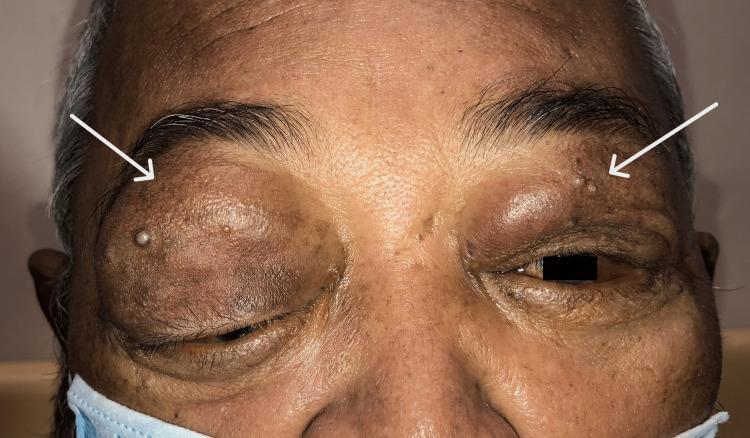
A clinical photograph showing the periorbital region of a patient with bilateral upper eyelid swelling and mechanical ptosis. The swelling is asymmetrical, being significantly more pronounced on the right side, where the mass effect has caused inferior dystopia (downward displacement) of the globe. The skin of the upper eyelids is hyperpigmented and appears firm and lobulated, consistent with underlying orbital masses. White arrows point to small, discrete, flesh-colored papular skin lesions located on the upper eyelids and supraorbital brow area. The clinical presentation is indicative of bilateral orbital involvement, which in this context represents diffuse large B-cell lymphoma (DLBCL) affecting the lacrimal glands. The image illustrates the external ocular manifestations of orbital lymphoma, including proptosis, lid edema, and mechanical secondary effects on eye position and eyelid function.
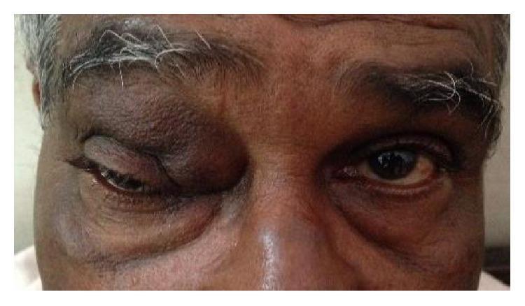
This clinical photograph shows a close-up, frontal view of an adult's face, highlighting significant facial asymmetry due to a mass in the left periocular region. The left upper eyelid displays a pronounced, firm-appearing swelling located superior and medial to the globe, consistent with an angular dermoid cyst. This mass causes mechanical ptosis, significantly narrowing the palpebral fissure compared to the unaffected right eye. The skin overlying the swelling appears intact without acute inflammatory signs like erythema or ulceration, though normal eyelid creases are obliterated. In contrast, the right eye serves as a baseline, showing normal eyelid position and periocular anatomy. This image is used to demonstrate the clinical presentation of orbital or adnexal dermoid cysts and their effect on eyelid mechanics and ocular symmetry within ophthalmology and dermatology training.
Clinical photography of the left periorbital skin demonstrating an erythematous, scaly plaque along the upper eyelid margin. The lesion is well demarcated, with red to pink erythema and fine superficial desquamation; mild crusting is present at the center and along the lid edge. Surrounding skin shows mild irritation without confluent swelling. The ocular surface appears not to be visibly involved; no purulent discharge is evident. The pattern is unilateral, localized to the periorbital region, compatible with common inflammatory dermatoses affecting the eyelid, rather than a primary ocular pathology. Differential diagnoses include seborrheic dermatitis of the eyelids, atopic or contact dermatitis, blepharitis with dermatitis, and less likely psoriasis or fungal infection. The image emphasizes surface features such as hyperemia, scaling, fissuring, and crusts, with the lid margin appearing slightly thickened. This clinical photo supports assessment of periorbital dermatitis, eyelid dermatitis, and blepharitis variants, and can guide topical therapy decisions. While histology is not available from a photograph, correlating with history and, if needed, skin scraping or biopsy could clarify diagnosis. Potential clinical uses include education, documentation of eyelid dermatitis severity, and monitoring response to topical steroids, calcineurin inhibitors, or antiseborrheic shampoos. This image supports teaching, case discussions, and diagnostic reasoning sessions.
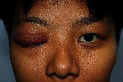
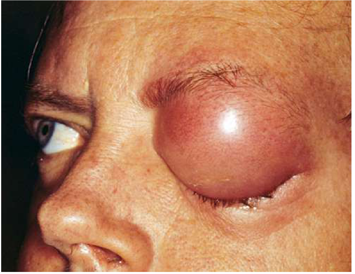
Clinical photograph of a patient's face showing a comparison between the right and left ocular regions. The right upper eyelid exhibits significant pathology characterized by marked edema (swelling), diffuse erythema, and complete mechanical ptosis, where the eyelid droops to entirely obscure the globe. The swelling appears localized to the right upper eyelid and superolateral orbital area, suggestive of a firm, underlying infiltrative mass. In contrast, the left eye appears normal with clear anatomical landmarks, healthy skin texture, and proper eyelid positioning. The image illustrates a unilateral orbital or eyelid inflammatory process, often associated with infectious or granulomatous conditions such as orbital cellulitis or zygomycosis (e.g., Saksenaea vasiformis). This visual is intended for ophthalmology and dermatology education to demonstrate clinical manifestations of severe eyelid lesions and differential diagnosis of mechanical ptosis.
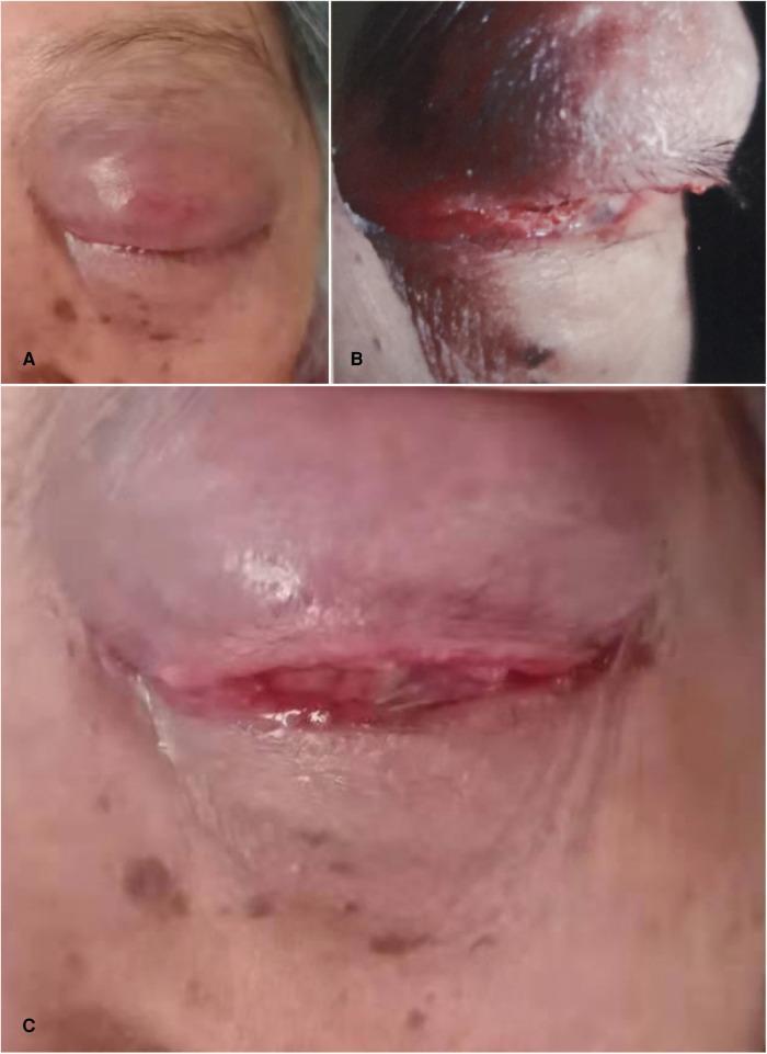
This set of clinical photographs illustrates severe ocular pathology in the left eye, later confirmed as orbital squamous cell carcinoma. Panel A shows significant erythematous swelling of the left upper eyelid with the loss of normal skin folds (dermatoglyphs), leading to mechanical ptosis. Panel B provides a profile view of a large, infiltrating mass measuring approximately 30 mm x 20 mm within the upper eyelid; the lesion exhibits ill-defined boundaries, firm texture, and overlying skin hyperpigmentation. Panel C demonstrates secondary ocular involvement including severe bulbar conjunctival chemosis (edema), conjunctival hyperemia, and corneal conjunctivalization. These findings represent advanced orbital malignancy with massive space-occupying effects, resulting in globe compression and severe secondary glaucoma (46.5 mmHg). The images provide high-level educational value for ophthalmology and oncology specialties regarding the clinical presentation of aggressive orbital adnexal tumors.
Right-Sided Upper Ocular (Superolateral) Mass - Clinical Overview
Clinical Features
- Persistent or progressive swelling of the outer one-third of the upper eyelid
- Pain or diplopia may or may not be present
- Dull aching pain over the forehead or temple = ominous sign suggesting malignancy
- Palpable mass in outer one-third of upper eyelid
- Globe displacement (inferiorly and medially) ± proptosis
- Restricted extraocular motility
- Conjunctival injection
Differential Diagnosis
Inflammatory / Infectious
| Condition | Features |
|---|---|
| Sarcoidosis | Bilateral, painless; may have lung/skin/renal disease; more common in African descent |
| Idiopathic Orbital Inflammatory Syndrome (IOIS) | Typically painful; acute onset |
| IgG4-related dacryoadenitis | Slowly progressive; check IgG4/IgG levels |
| Infectious dacryoadenitis | Purulent discharge (bacterial); bilateral viral (e.g., EBV, mumps) |
| GPA (Wegener's) | c-ANCA positive; may have systemic vasculitis |
Benign Neoplasms
| Condition | Features |
|---|---|
| Pleomorphic adenoma (benign mixed tumor) | Slowly progressive, painless proptosis; smooth margins on CT; pressure erosion of lacrimal fossa WITHOUT destruction |
| Dermoid cyst | Usually in young patients; arises from frontoethmoidal suture |
| Lacrimal gland cyst (dacryops) | Young/middle-aged adult; fluctuating size |
Malignant Neoplasms
| Condition | Features |
|---|---|
| Adenoid cystic carcinoma (ACC) | Subacute pain over 1-3 months; perineural invasion causing forehead/temple pain; bony erosion on CT |
| Pleomorphic adenocarcinoma | Elderly; rapid painful progression; may arise from pre-existing benign tumor |
| Lymphoproliferative tumor / Lymphoma | Slowly progressive; pink "salmon-patch" subconjunctival extension; "molds" to globe on CT; DLBCL may show bony erosion |
| Metastasis | Especially prostate, breast; history of primary malignancy |
NOTE from Wills Eye Manual: Primary epithelial neoplasms are almost always unilateral; inflammatory disease may be bilateral. A lesion in the outer one-third of the upper eyelid in an adult should prompt CT imaging to exclude malignancy.
Imaging - CT and MRI of Lacrimal Gland Lesions
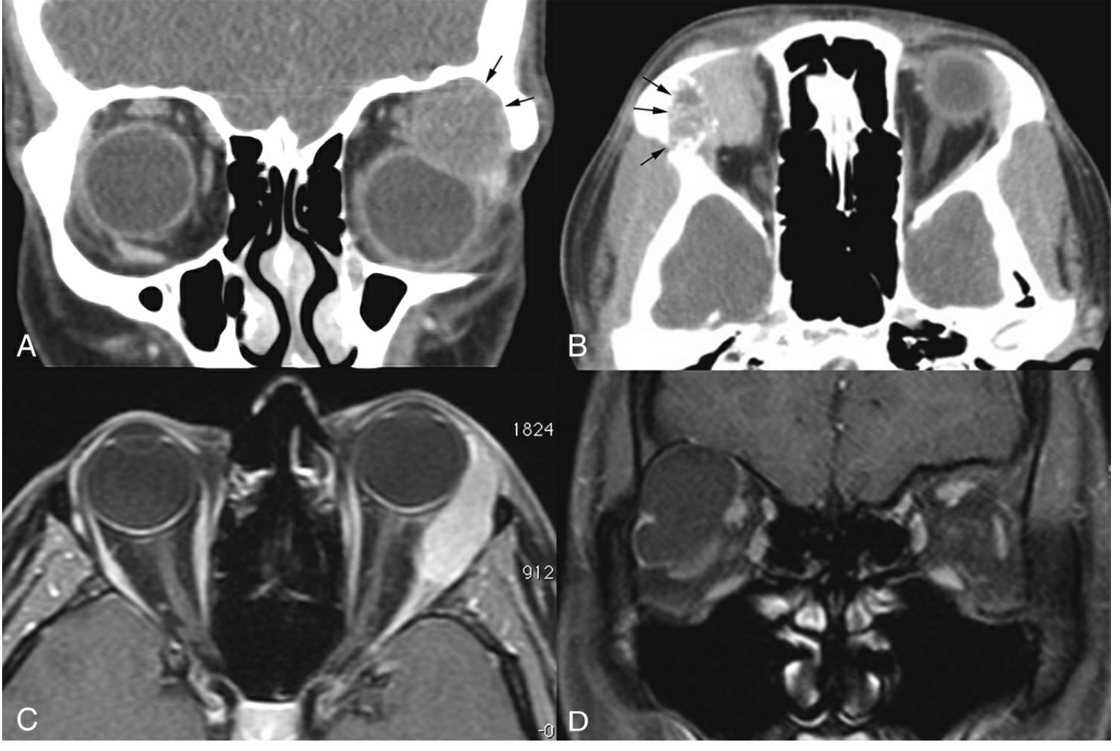
- A - Pleomorphic adenoma: smooth, pressure-induced lacrimal fossa changes (arrows) - no bone destruction
- B - Adenoid cystic carcinoma: bone destruction with intralesional calcifications (arrows) - aggressive
- C - Lymphoma: "molds" to the globe; well-circumscribed
- D - Dermoid cyst: arising from the frontoethmoidal suture
Clinical Examples
Workup (Recommended Approach)
- History - Duration, rate of progression, pain, diplopia, systemic symptoms (weight loss, fever, respiratory problems, skin rash, uveitis history)
- Complete ocular examination - Look for proptosis, globe displacement, motility restriction, conjunctival salmon patch
- Orbital CT (axial + coronal + parasagittal) - First-line imaging; defines bony anatomy. Use MRI if intracranial extension is suspected
- Chest CT - May diagnose sarcoidosis, lymphoma, primary malignancy, or metastases
- Blood tests to consider:
- CBC with differential
- ACE (sarcoidosis)
- c-ANCA/p-ANCA (GPA)
- SPEP, LDH, IgG4/IgG levels (lymphoma, IgG4 disease)
- PPD or QuantiFERON-TB Gold (TB)
- Biopsy - Required for definitive diagnosis in most cases
Red Flags Requiring Urgent Evaluation
- Pain over the forehead or temple (suggests ACC with perineural invasion)
- Rapid progression over weeks
- Bony destruction on CT
- Associated weight loss, fever, night sweats (lymphoma / systemic malignancy)
- Globe displacement or proptosis
Summary
- Painless + slow = benign (pleomorphic adenoma, sarcoidosis, IgG4) or lymphoma
- Painful + subacute = malignant epithelial tumor (ACC most feared)
- Inflammatory signs = IOIS, infectious, sarcoidosis
Swelling on the temporal region mass felt on temporal region and swelling lower eyelid
temporal region mass swelling lower eyelid facial tumor
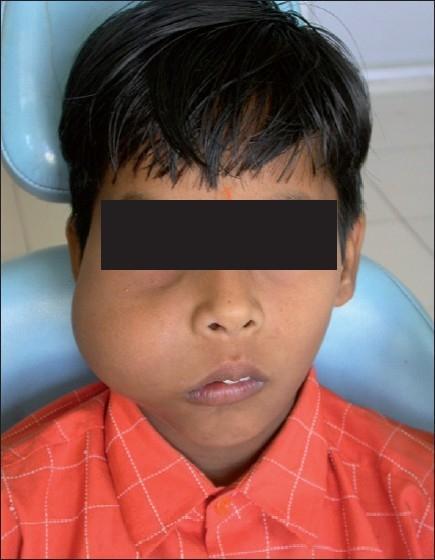
A clinical photograph of a pediatric male patient demonstrating significant facial asymmetry due to a large, diffuse swelling on the right midface. The swelling originates from the right maxilla and extends from the infraorbital region down to the mandible, and laterally from the nasolabial fold toward the preauricular area. Visually, the mass causes a superior displacement of the right lower eyelid and eyeball (proptosis/exophthalmos) and an upward elevation of the right corner of the mouth. The overlying skin appears intact with normal color and texture, suggesting a deep-seated, slow-growing pathology. The case is consistent with an odontogenic tumor or fibro-osseous lesion of the maxilla, such as an odontogenic myxoma, causing extensive bony expansion and secondary displacement of orbital and perioral soft tissue structures. This image serves as a clinical example of maxillo-facial pathology and its impact on cranial symmetry and orbital alignment.
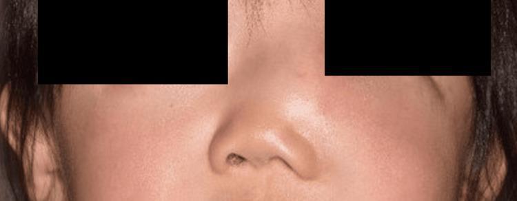
This clinical photograph shows a close-up frontal view of a patient's face, specifically focusing on the nasal and midface region. A prominent, localized swelling is visible on the left side of the nasal dorsum, extending toward the medial infraorbital area. The mass causes a significant loss of facial symmetry, with a visible bulge that displaces the normal contours of the nose and the lower medial eyelid. The overlying skin appears slightly erythematous with subtle fullness, though it remains intact without ulceration. This presentation is characteristic of a soft tissue tumor or cystic lesion of the sinonasal region, such as a solitary myofibroma (SNM), as suggested by the clinical history of recurrence and rapid growth. The image effectively demonstrates the clinical manifestation of a midfacial mass and its impact on regional anatomical landmarks, including the nasal bone and lacrimal drainage pathway.
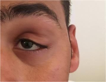
This clinical photograph shows the left periocular region of a patient, demonstrating significant soft tissue swelling at the superolateral angle of the left orbit. The mass is located in the anatomic region of the lacrimal gland, presenting as a visible fullness that distends the overlying skin. Associated clinical findings include a mechanical ptosis of the left upper eyelid, resulting in a narrowed palpebral fissure. The skin overlying the swelling appears slightly erythematous or discolored compared to the surrounding facial tissue. There is no evidence of significant proptosis or globe displacement visible from this frontal-oblique view. This clinical presentation is typical for orbital adnexal lesions, such as a lacrimal gland tumor or inflammatory myofibroblastic tumor (IMT). The image serves as an educational example of orbital mass localization and its secondary effects on eyelid positioning.
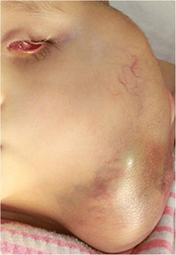
This clinical photograph shows a pediatric patient with a massive, firm, and expanding swelling on the left side of the face and neck. The mass involves the left orbital, parotid, and submandibular regions, causing significant facial asymmetry. Visible cutaneous findings over the tumor include prominent superficial telangiectasia (vascular markings) and ecchymotic discoloration (bruising) at the inferior aspect of the lesion. The left eye exhibits severe proptosis with conjunctival chemosis, erythema, and eyelid edema, consistent with orbital involvement. From an educational perspective, the image illustrates advanced-stage extraocular retinoblastoma following local recurrence. This represents a critical case study in pediatric oncology and ophthalmology, emphasizing the progression of intraocular malignancy into regional soft tissues when primary management is delayed or inadequate. The clinical presentation is highly suggestive of extensive lymph node and soft tissue metastasis in the cervical region.
dermoid cyst temporal region periorbital swelling
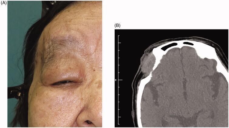
This composite image consists of a clinical photograph and a diagnostic radiological scan illustrating a chronic orbital dermoid cyst. Figure A is a clinical photograph of an elderly patient's right periorbital region. It demonstrates significant mechanical ptosis of the right upper eyelid and a visible mass in the lateral orbital area, causing focal swelling and redundant skin. Figure B is an axial computed tomography (CT) scan of the head at the level of the orbits. The scan reveals a well-circumscribed, hypo-dense mass in the right superolateral orbit. There is clear evidence of chronic pressure-related bone remodeling and erosion, specifically involving the frontal bone and frontosphenoidal suture area, resulting in an indented orbital margin. The clinical and radiological findings are consistent with a long-standing, slow-growing orbital cyst, such as a dermoid cyst, which has caused secondary structural deformation of the surrounding bony architecture.
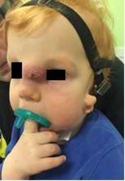
Clinical photograph of a pediatric patient presenting with an infected nasal dermoid cyst. The primary pathology is a prominent midline mass located on the nasal dorsum/glabella region. Visually, the mass exhibits an erythematous, reddish-purple hue with an irregular, textured surface suggestive of acute inflammation or secondary infection. Associated periorbital edema and soft tissue swelling are evident around the lesion. The image also demonstrates medical support and devices, including a tracheostomy tube positioned beneath the chin, a pacifier in the mouth, and a headgear strap, indicating possible complex airway management or comorbid conditions. This visual serves to educate on the typical midline presentation of congenital nasal dermoid cysts and the clinical signs of superimposed infection in a surgical candidate.
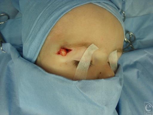
This intraoperative clinical photograph captures the surgical excision of a dermoid cyst in a pediatric patient. The image shows the periorbital region, specifically the lateral aspect of the left eyebrow, where a horizontal incision has been made. Protruding through the surgical opening is a well-circumscribed, spherical, yellowish-white cystic mass, typical of a dermoid cyst containing keratin and sebaceous material. The surrounding skin is clean and shows minimal erythema at the incision margins. The patient is prepared in a sterile field with blue surgical drapes, and the eyes are secured with protective adhesive tape. Surgical instruments are visible in the periphery, indicating an active operative procedure. This visual serves as an educational example of the anatomical presentation and surgical management of common benign head and neck neoplasms in children, highlighting the superficial location and distinct morphology of the cyst relative to local tissues.
temporal region mass lower eyelid swelling differential diagnosis orbital tumor
Temporal Region Mass + Lower Eyelid Swelling - Clinical Analysis
Anatomical Basis
- Temporalis muscle and fascia
- Zygomatic arch (inferiorly)
- Frontosphenoidal suture (connects to lateral orbital wall)
- Greater wing of sphenoid (forms part of the lateral orbital wall)
- Direct extension through the lateral orbital wall into the orbit/infraorbital rim
- Lymphatic spread (temporal → preauricular → periorbital nodes)
- Venous obstruction causing edema
- Deep temporal space infection tracking anteriorly
Differential Diagnosis
1. Orbital Lymphoma with Temporal Extension (Most Important to Exclude)
- Features: Slowly progressive, painless, rubbery/firm mass; may have lower eyelid fullness from orbital component
- CT/MRI: Homogeneous, isodense to muscle; molds to adjacent structures without bone destruction (in low-grade histology)
- Salmon-patch subconjunctival lesion may be visible
2. Dermoid Cyst (Lateral/Deep Variant)
- Features: Present in young adults; slow-growing; may enlarge suddenly if ruptures; smooth, non-tender, skin-colored
- CT: Well-circumscribed hypodense cyst; may show chronic pressure remodeling of adjacent bone (NOT destruction)
- (Wills Eye Manual, Fig. 7.6.1D)
3. Sebaceous Carcinoma of the Eyelid with Regional Spread
"Sebaceous carcinoma tends to spread first to the parotid and submandibular nodes."
- Robbins Pathologic Basis of Disease, p. 1206
- Features: Elderly patient; lower eyelid nodule initially mimicking chalazion; eyelid thickening; misdiagnosed as blepharitis
- Red flag: Recurrent "chalazion" that fails standard treatment = biopsy mandatory
- Mortality up to 22%; aggressive perineural and lymphatic spread
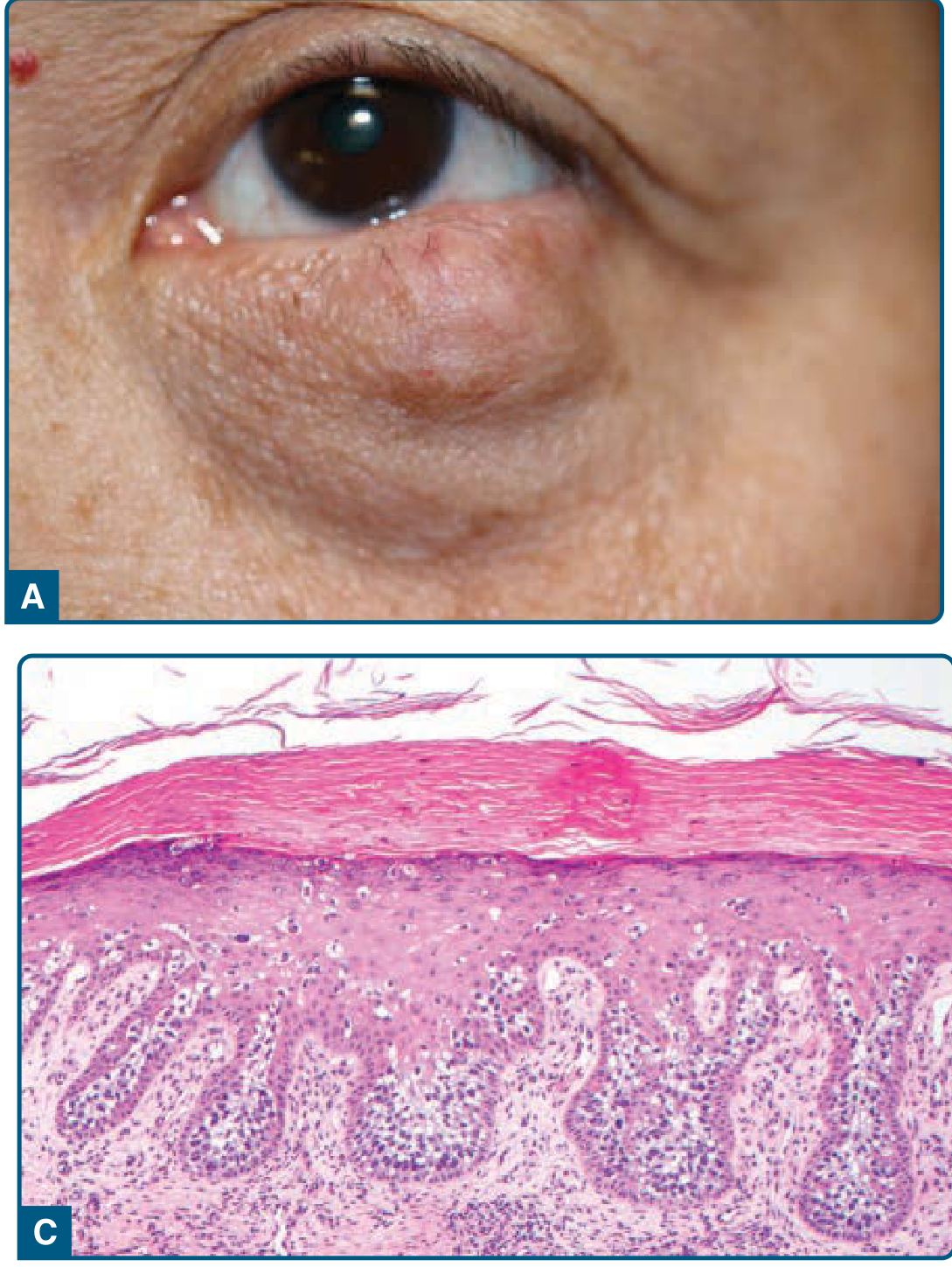
4. Temporal Space Infection / Deep Space Abscess
-
Fill the temporal fossa causing visible/palpable temporal swelling
-
Track anteriorly beneath the zygomatic arch to cause lower eyelid/infraorbital edema
-
Features: Trismus (jaw stiffness), pain, fever, overlying erythema, recent dental work or trauma
5. Parotid Gland Tumor with Superior Extension
-
Temporal/preauricular swelling
-
Lower eyelid edema via lymphatic obstruction or direct extension
-
Features: Firm, slow-growing preauricular/temporal mass; facial nerve involvement (VII palsy) suggests malignancy
6. Zygomatic/Maxillary Mass (Odontogenic or Fibro-Osseous)
- Facial asymmetry and temporal fullness
- Lower eyelid displacement superiorly from the underlying bony expansion
7. Basal Cell / Squamous Cell Carcinoma
- BCC has a distinct predilection for the lower eyelid and medial canthus - can invade deeply and spread to temporal lymphatics
- SCC is more aggressive; perineural spread along trigeminal branches can cause temporal region swelling
- (Robbins, p. 1334)
8. Other Causes to Consider
| Condition | Clue |
|---|---|
| Rhabdomyosarcoma | Children; rapid growth; proptosis |
| Metastasis (breast, lung, prostate) | Known primary malignancy |
| Lipoma / Epidermal cyst | Soft, fluctuant, very slow growing |
| Hemangioma | Compressible; may enlarge with Valsalva |
| Temporal arteritis | Tender, nodular, pulseless temporal artery; elderly; raised ESR/CRP |
Workup - Step by Step
-
History
- Duration and rate of growth (slow = benign/lymphoma; rapid = malignant/infectious)
- Pain (infection, ACC, perineural invasion), fever (infection)
- Dental history / recent procedures (deep space infection)
- Lower eyelid lesion history - any prior "chalazion" (sebaceous carcinoma)
- Age: children → dermoid/rhabdomyosarcoma; adults >60 → lymphoma/carcinoma
-
Examination
- Consistency: rubbery (lymphoma), firm (carcinoma), soft/fluctuant (dermoid/abscess)
- Skin changes: erythema (infection/BCC), ulceration (SCC/BCC), salmon patch conjunctiva (lymphoma)
- Globe position: proptosis, dystopia
- Facial nerve assessment (VII palsy = parotid malignancy)
- Eyelid: look for primary eyelid lesion (BCC, SCC, sebaceous carcinoma)
-
Imaging
- Orbital + facial CT with contrast (axial + coronal): first line - defines bony anatomy, soft tissue extent
- MRI if soft tissue characterization needed, intracranial extension suspected, or nerve involvement
- Ultrasound: useful for superficial cysts or parotid masses
-
Blood tests
- CBC, LDH (lymphoma screen)
- ESR, CRP, temporal artery biopsy if temporal arteritis considered
- ACE (sarcoidosis)
-
Biopsy
- Required for all lesions without a clear benign diagnosis
- Incisional biopsy (large lesions) or excisional biopsy (small, accessible)
- Avoid incisional biopsy of suspected benign mixed tumor (pleomorphic adenoma) - risk of seeding
Red Flags Requiring Urgent Referral
- Rapid growth
- Pain in the temporal/forehead region
- Facial nerve palsy
- Proptosis or vision change
- Constitutional symptoms (weight loss, night sweats, fever)
- Eyelid ulceration or recurrent "chalazion" not responding to treatment
History 1 week ago of congejectivitis and pain only when palpated
preseptal orbital cellulitis eyelid swelling conjunctivitis
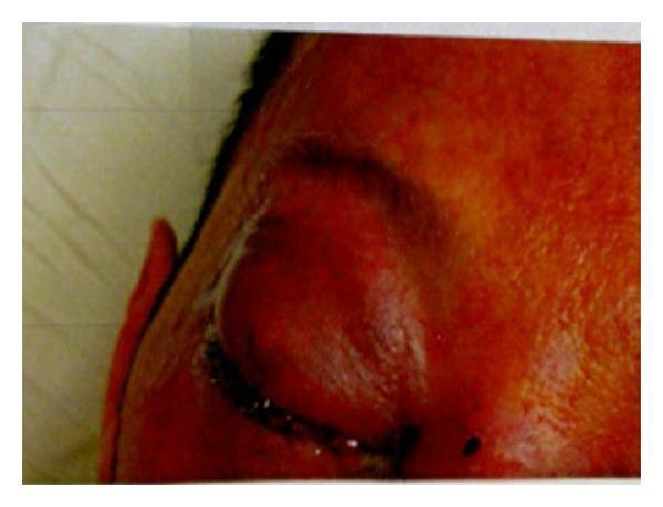
Clinical photograph of the right periorbital and midface region demonstrating features of preseptal cellulitis. The image shows significant erythema and marked edema (swelling) extending across the forehead, nose, and periorbital areas. The right upper and lower eyelids are severely edematous, characterized by chemosis (conjunctival swelling) that restricts the visualization of the globe. A prominent yellowish crust is visible along the lid margins, suggesting the presence of dried purulent exudate or discharge. The skin appears tense and inflamed, consistent with an acute infectious or inflammatory process. This visual serves as an educational example of the clinical presentation of orbital or preseptal infections and the associated soft tissue manifestations such as eyelid crusting and severe chemosis.

A comparative series of four axial Computed Tomography (CT) scans of the orbits, illustrating the radiological progression of orbital complications typically following sinusitis, categorized by severity. Panel A (Eyelid Cellulitis) shows preseptal soft tissue swelling and eyelid edema with preserved clarity of the post-septal orbital fat. Panel B (Orbital Cellulitis) demonstrates increased density and 'dirty' stranding of the intraconal and extraconal orbital fat, indicating post-septal inflammation without a focal collection. Panel C (Subperiosteal Abscess) reveals a localized, rim-enhancing fluid collection adjacent to the medial orbital wall (lamina papyracea), displacing the medial rectus muscle laterally. Panel D (Orbital Abscess) displays a more advanced, poorly defined heterogeneous collection within the orbital soft tissues, causing significant proptosis and mass effect. These diagnostic images provide clinical examples of the Chandler classification for orbital infections, emphasizing the importance of identifying organized abscesses for surgical planning.
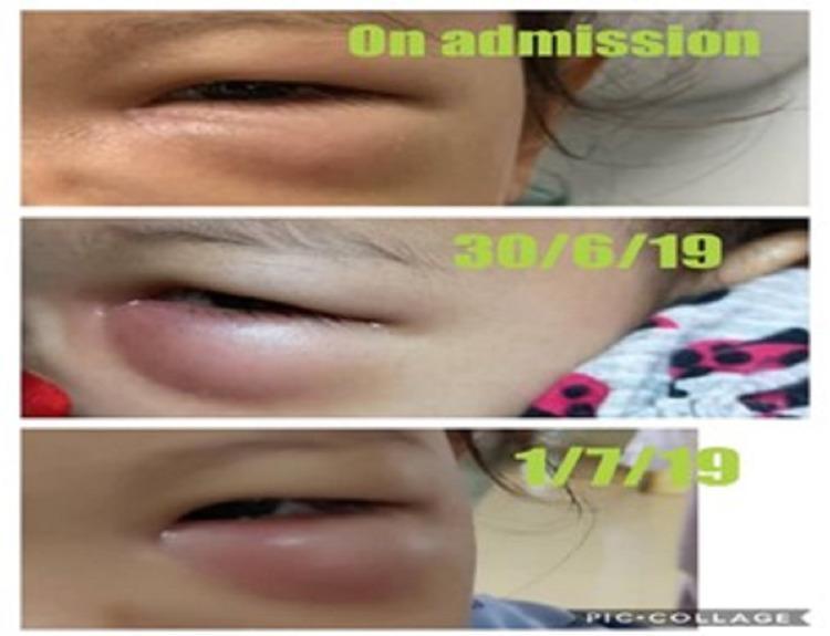
A series of three clinical photographs arranged as a vertical timeline demonstrating the progression of left-sided periorbital cellulitis in a pediatric patient. The first image ('On admission') shows moderate edema of the left upper and lower eyelids with mild erythema. The second image ('30/6/19') shows a significant increase in inflammatory swelling, characterized by tense, erythematous skin and worsening ptosis of the upper eyelid. The final image ('1/7/19') depicts generalized, severe periorbital edema that almost completely obscures the globe, with the skin appearing stretched and shiny due to underlying inflammation. This timeline illustrates the clinical worsening of preseptal cellulitis despite initial antibiotic treatment, which was later attributed to an odontogenic infection and orbital periosteal abscess. The images serve as an educational tool for identifying the signs of progressing orbital complications, including changes in eyelid morphology, distribution of swelling, and the urgent need for diagnostic imaging like CECT to differentiate between preseptal and orbital cellulitis.
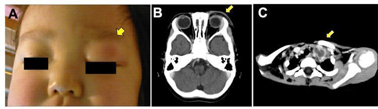
This composite medical image displays clinical and radiological findings in a pediatric patient. Figure A is a clinical photograph showing significant left supraorbital swelling and erythema (indicated by a yellow arrow) involving the left upper eyelid, initially suggestive of preseptal cellulitis. Figure B is an axial enhanced CT scan of the head at the orbital level, demonstrating thickening of the left supraorbital skin and irregular attenuation within the subcutaneous fatty tissue, though the orbital contents appear spared. Figure C is an axial enhanced CT scan of the lower neck/supraclavicular region, revealing a pathologically enlarged left supraclavicular lymph node with a characteristic low-density (hypodense) center, suggesting necrotic changes or metastatic involvement. These findings are pedagogically used to illustrate the clinical presentation of metastatic neuroblastoma, which can mimic inflammatory conditions like orbital cellulitis through orbital and lymph node spread.
Refined Clinical Assessment - Updated with New History
The Key Clinical Triad
| Feature | Significance |
|---|---|
| Conjunctivitis 1 week ago | Preceding infection - likely viral (adenoviral) or bacterial |
| Temporal region mass | Reactive lymphadenopathy OR spread from periorbital infection |
| Lower eyelid swelling | Preseptal cellulitis / reactive edema from adjacent infection |
| Pain only on palpation (no spontaneous pain) | Points AWAY from orbital cellulitis; suggests reactive/preseptal process |
Viral/Bacterial Conjunctivitis → Preseptal Cellulitis/Periorbital Inflammation → Reactive Preauricular/Temporal Lymphadenopathy
Most Likely Diagnoses (Ranked)
1. VIRAL CONJUNCTIVITIS WITH REACTIVE PREAURICULAR LYMPHADENOPATHY (Most Likely)
"Viral conjunctivitis: watery discharge, foreign body sensation, preauricular lymphadenopathy..."
- Goldman-Cecil Medicine, Table 916
- Acute conjunctivitis with watery discharge
- Palpable, tender preauricular / temporal lymph node - this is the "temporal mass"
- Lower eyelid chemosis and edema
2. BACTERIAL CONJUNCTIVITIS → PRESEPTAL CELLULITIS
- Lower eyelid swelling and erythema
- Tender periorbital/temporal soft tissue mass
- Follows an upper respiratory infection or conjunctivitis episode
3. OCULOGLANDULAR SYNDROME (Parinaud's) (Consider if atypical features)
| Pathogen | Clue |
|---|---|
| Cat-scratch disease (Bartonella henselae) | Cat exposure, scratch |
| Tularemia (Francisella tularensis) | Animal/tick exposure |
| Tuberculosis | Endemic area, systemic symptoms |
| Listeria | Immunocompromised |
| EBV / CMV | Systemic lymphadenopathy |
4. DACRYOADENITIS (Infectious) (If swelling is lateral upper not lower)
- Outer upper eyelid swelling + temporal tenderness
- May track to lower eyelid by gravity/edema
- Bacterial (S. aureus) or viral (EBV, mumps)
Distinguishing Preseptal from Orbital Cellulitis - CRITICAL
| Feature | Preseptal (Pre-septal) | Orbital (Post-septal) |
|---|---|---|
| Pain | On palpation only ✓ | Spontaneous, severe |
| Proptosis | Absent | Present |
| Eye movement | Normal | Restricted, painful |
| Vision | Normal | May be reduced |
| Fever | Mild/absent | High |
| Urgency | Outpatient IV/oral Abx | Hospitalize immediately |
Workup
- Full ocular exam - check for proptosis, restricted eye movement, and vision. If any present → CT orbit urgently + hospitalize
- Check temperature - fever suggests systemic infection
- Examine the "temporal mass" carefully:
- Soft, mobile, oval, preauricular → reactive lymph node (viral/bacterial)
- Fixed, hard → consider neoplasm (lower priority given acute history)
- Very large, fluctuant → lymph node abscess
- Conjunctival swab (if discharge still present) - bacterial culture + sensitivity
- Bloods if systemic features: CBC, CRP, Bartonella serology (if cat contact), Monospot
- CT orbit only if: no improvement in 24-48h on antibiotics, proptosis develops, or vision changes
Management Based on Most Likely Diagnosis
Viral Conjunctivitis + Reactive Lymphadenopathy
- Supportive: Cold compresses, lubricating eye drops (artificial tears)
- Lymph node usually resolves in 2-4 weeks as conjunctivitis clears
- No antibiotics needed (viral cause)
- Highly contagious (adenovirus) - hand hygiene, avoid sharing towels
Bacterial Conjunctivitis + Preseptal Cellulitis
- Topical antibiotics (moxifloxacin 0.5% drops or erythromycin ointment) for conjunctivitis
- Oral antibiotics for preseptal cellulitis: Amoxicillin-clavulanate 875/125 mg twice daily for 7-10 days (adults); cefalexin is an alternative
- Warm compresses to the eyelid
- Review in 24-48 hours - if worsening, CT orbit and IV antibiotics