
How to know hairfall is their and remedies for hair fall
"alopecia"[MeSH Terms] AND "therapy"
androgenetic alopecia male pattern hair loss Hamilton Norwood scale
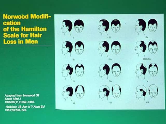
Imaging modality: Not applicable; this is a dermatology illustration. Primary subject: male pattern hair loss staged by the Norwood modification of the Hamilton scale. Specimen type: schematic illustration, not biological tissue. Imaging perspective: frontal and lateral head views shown in a grid to depict progressive hairline recession and vertex thinning. Anatomical context: scalp region, focusing on frontal hairline, temporal recession, crown/vertex, with multiple silhouettes representing different severity levels. Key visual features: a green background and a light-blue central panel displaying numerous head portraits labeled I through VII (and variants). Each portrait shows varying degrees of temple recession, bitemporal thinning, and/or vertex baldness. Text notes include the scale title: 'Norwood Modification of the Hamilton Scale for Hair Loss in Men' and the source citation: Adapted from Norwood OT, South Med J, 1951; 53:708-728. Clinical significance: the Norwood-Hamilton scale standardizes assessment of androgenetic alopecia in men, providing a repeatable, ordinal measure to stage disease, monitor progression, and guide therapy decisions. Differential considerations: other hair loss patterns (Alopecia areata patterns) are not depicted but may be considered when clinical history contradicts the pattern; use in research and education. Clinical correlation: used in dermatology practice and trichology studies to quantify severity, assess treatment outcomes, and facilitate patient counseling.
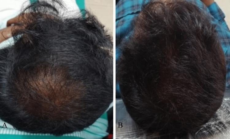
This comparison clinical photograph demonstrates treatment progression in a 30-year-old male with Grade III vertex androgenetic alopecia (AGA) according to the modified Norwood-Hamilton scale. Panel A (Baseline) provides a superior view of the scalp, showing localized thinning and significant reduction in hair density at the vertex. The underlying scalp is clearly visible through sparse, miniaturized hair follicles. Panel B (Post-treatment) shows the same anatomical region after four sessions of Platelet-Rich Plasma (PRP) therapy. This image illustrates a marked clinical improvement, characterized by increased hair density, thicker hair shaft diameter, and comprehensive coverage of the vertex. The comparison serves as an educational tool for assessing treatment efficacy in hair restoration, highlighting the visual transition from vertex baldness to fuller hair regrowth. The content is relevant for dermatology and trichology education, focusing on therapeutic outcomes in male pattern hair loss.
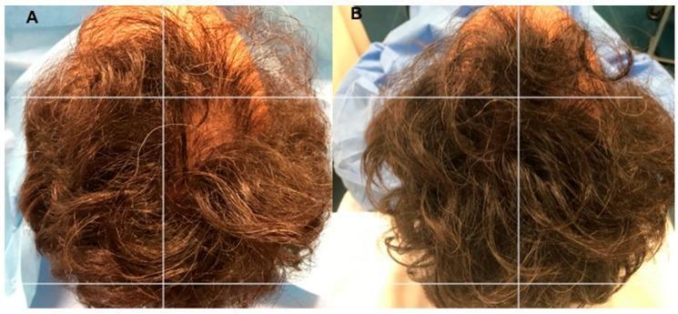
Clinical photograph showing a side-by-side comparison (A vs. B) of a 34-year-old male's scalp with Androgenetic Alopecia (AGA) stage 2 on the Norwood–Hamilton Scale. Image A (baseline, T0) illustrates significant thinning and hair loss localized to the vertex, parietal, temporal, and frontal regions, with high scalp visibility and a reddish-brown hair hue. Image B (T3, 12 weeks post-treatment) demonstrates the results after autologous platelet-rich plasma (A-PRP) injections. A white grid overlay is used for anatomical referencing. Visually, Image B shows a marked increase in hair density, particularly in the treated right parietal area, resulting in reduced scalp visibility and a darker, fuller hair appearance compared to baseline. The comparison serves to illustrate the clinical efficacy of A-PRP in stimulating hair regrowth and increasing follicular density in patients with male-pattern baldness.
Hair fall: how to tell, and what helps
1. Is it really hair fall?
- Many hairs on the pillow, in the shower drain, or in the brush each day.
- A thinner ponytail, or a wider parting.
- Visible scalp, or thinning at the crown or temples.
- Round bald patches, which suggest alopecia areata.
- Broken hairs, or scalp redness, scaling, itching or pain, which suggest a scalp disease or fungal infection.
- Pull test: Grasp about 40 hairs between thumb and forefinger and pull slowly. More than 4-6 hairs coming out is abnormal. Right after shampooing, 2-3 is already abnormal. - Andrews' Diseases of the Skin, p. 870
- Daily hair count: Collect all hairs from the pillow, comb and wash for a few days. Count the small ones too.
- Look at the root: In telogen shedding, the hair falls out with a pale, club-shaped bulb at the root.
- Look at the pattern:
- Men: receding temples and crown thinning (Norwood-Hamilton scale).
- Women: widening central parting with the frontal hairline kept.
- Diffuse shedding all over: telogen effluvium.
2. Common causes
- Androgenetic alopecia (pattern baldness): the most common cause, and it is genetic and hormonal. It progresses slowly over time.
- Telogen effluvium: shedding about 3-5 months after a trigger. Triggers include fever or illness, surgery, childbirth, crash dieting, major stress, and some drugs. - Andrews' Diseases of the Skin, p. 870
- Nutritional and medical causes: iron deficiency, thyroid disease, low vitamin D, low protein intake, PCOS.
- Drugs: retinoids, SSRIs, tamoxifen, anticoagulants, antithyroid drugs, chemotherapy, and others. - Goldman-Cecil Medicine, p. 4334
- Autoimmune or scalp disease: alopecia areata, seborrheic dermatitis, fungal infection (tinea capitis), lichen planus.
- Traction: tight hairstyles, harsh chemical treatments and heat.
3. Remedies
A. Fix the cause (especially for diffuse shedding)
- Telogen effluvium usually settles by itself once the trigger is gone. Regrowth takes months.
- Tests worth asking a doctor about: CBC, ferritin, TSH, vitamin D, vitamin B12, and hormones if you have irregular periods or acne/facial hair.
- Correct deficiencies only if a test shows one. Taking extra biotin or iron without a deficiency has little proven benefit, and too much of some nutrients can cause hair loss.
- Eat enough protein and calories. Avoid crash diets.
- Review your medicines with your doctor. Do not stop them yourself.
B. Treatment for pattern hair loss (androgenetic alopecia)
- Topical minoxidil: 2% for women, 5% for men. Apply it daily. It may cause a brief extra shed at the start.
- Oral finasteride 1 mg/day (men): Topical finasteride spray works similarly. A minority of men report persistent sexual dysfunction or depression (post-finasteride syndrome).
- Oral dutasteride 0.5 mg: more effective than finasteride in some countries where it is approved for this use.
- Low-dose oral minoxidil (0.25-2.5 mg/day) is effective and usually well tolerated. It can cause unwanted facial hair growth and, rarely, low blood pressure or fluid retention, so it needs medical supervision.
- Platelet-rich plasma (PRP) injections, low-level laser or light therapy, and hair transplant. Recent systematic reviews (injectable therapy review, management of androgenic alopecia) find some benefit, but studies are small and methods vary. They are adjuncts, not replacements for minoxidil or 5-alpha-reductase inhibitors.
C. Hair care
- Use a gentle shampoo, and treat dandruff if present.
- Avoid tight hairstyles, frequent bleaching or straightening, and very hot styling tools.
- Be gentle when the hair is wet, since wet hair breaks more easily.
- Do not smoke, and manage stress and sleep.
- Home remedies (onion juice, amla, coconut or rosemary oil, scalp massage) have little reliable evidence. They are harmless for most people, but they should not delay proper treatment.
4. When to see a doctor (dermatologist)
- Sudden or rapid loss, or round bald patches.
- Scalp pain, redness, scaling, pus or scarring.
- Loss together with fatigue, weight change, irregular periods, or hair loss elsewhere on the body.
- Shedding that lasts more than 3-6 months.
- Hair loss in a young person, which is easier to treat early. Pattern loss responds best when treatment starts early.