
Surfactant is essential for respiration why?
Reading File
Surfactant reduces surface tension in alveoli, preventing their collapse during expiration. Without it, the enormous surface tension at the air-water interface inside tiny alveoli would cause them to collapse after each breath, making re-inflation extremely difficult and requiring dangerously high respiratory effort.
Searching sources...
Finding Sources
Searching PubMed
pulmonary surfactant surface tension alveolar stability
Reading File
Reading File
Reading File
Now let me fetch the diagram image for visual clarity:
Loading Image
Here is a thorough explanation of why surfactant is essential for respiration, drawn from authoritative physiology and pulmonary medicine textbooks.
Why Surfactant is Essential for Respiration
1. The Problem: Surface Tension in Alveoli
The inner surface of every alveolus is coated with a thin layer of fluid. Liquid molecules attract each other, creating surface tension - a force that acts to minimize the surface area, which in a spherical alveolus means a force that tries to collapse it.
The Law of Laplace describes this relationship:
P = 2T / r
Where P = collapsing pressure, T = surface tension, r = radius of the alveolus.
This means:
- Large alveoli - bigger radius, lower collapsing pressure - relatively stable
- Small alveoli - smaller radius, higher collapsing pressure - prone to collapse (atelectasis)
Without something to counteract this, every small alveolus would collapse after each breath, making re-inflation require enormous force and making gas exchange impossible.
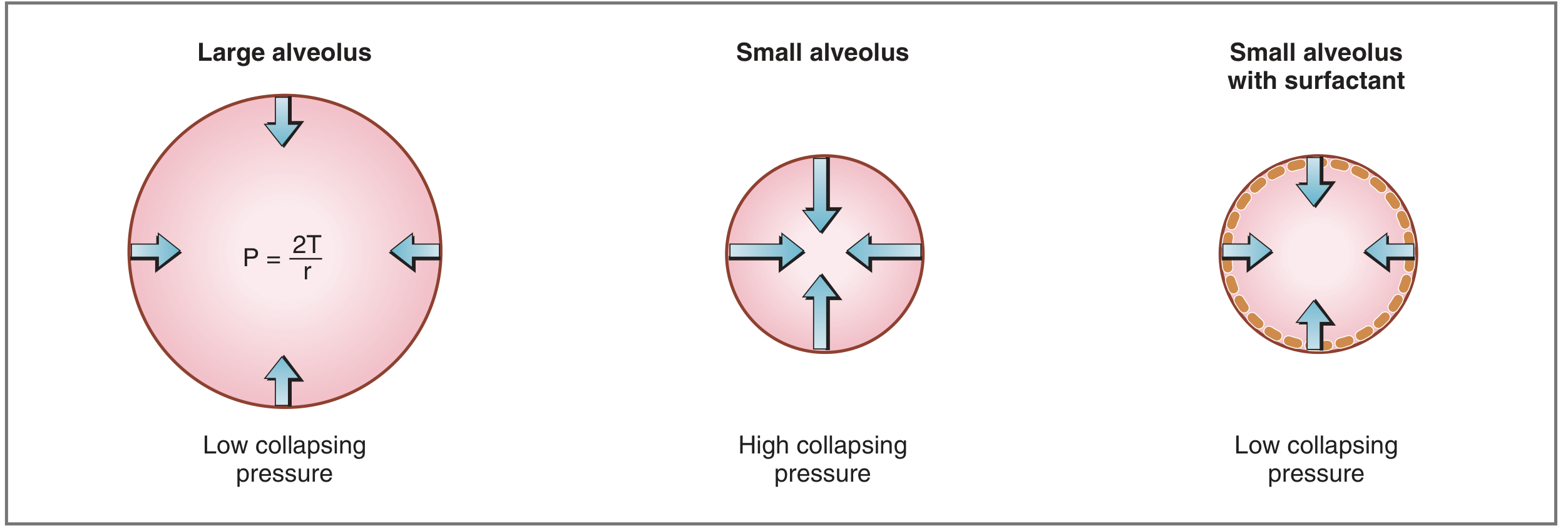
2. What Surfactant Is
Surfactant is a mixture of lipids and proteins secreted into the alveolar space by type II alveolar cells (AT2 cells). Its key components are:
- Dipalmitoylphosphatidylcholine (DPPC) - the most important component, comprising ~50-60% of surfactant lipid
- Unsaturated phosphatidylcholine, phosphatidylglycerol
- Four surfactant-associated proteins (SP-A, SP-B, SP-C, SP-D)
DPPC is amphipathic - it has a hydrophilic (water-loving) head and a hydrophobic (water-repelling) tail. DPPC molecules align themselves at the alveolar air-liquid interface, with hydrophobic portions attracted to each other and hydrophilic portions repelled from each other. This intermolecular arrangement disrupts the attractive forces between water molecules that were responsible for high surface tension. The result: surface tension and collapsing pressure are dramatically reduced.
3. What Surfactant Does - Four Key Functions
| Function | Mechanism | Consequence if absent |
|---|---|---|
| Prevents alveolar collapse | Reduces surface tension, lowering collapsing pressure | Widespread atelectasis |
| Stabilizes alveoli of different sizes | Reduces T more in smaller alveoli (denser DPPC packing at low volumes) | Small alveoli collapse into large ones |
| Increases lung compliance | Less force needed to expand alveoli during inspiration | Greatly increased work of breathing |
| Prevents alveolar edema | Low surface tension means less fluid drawn into alveoli | Fluid accumulation impairs gas exchange |
4. Why Alveolar Stability is Especially Critical
Without surfactant, not only do small alveoli collapse, but by the Law of Laplace, air from small high-pressure alveoli would be forced into large low-pressure alveoli - causing uneven ventilation. Surfactant compensates for this because DPPC molecules pack more tightly as radius decreases, so surface tension falls even further as an alveolus shrinks. This makes the collapsing pressure roughly equal across alveoli of different sizes, keeping them all open.
5. Clinical Consequences of Surfactant Deficiency
Neonatal Respiratory Distress Syndrome (NRDS / Hyaline Membrane Disease)
- Surfactant synthesis by AT2 cells begins around gestational week 24 and is reliably present by week 35
- Premature infants born before week 35 may lack surfactant
- Consequences:
- High alveolar surface tension → widespread atelectasis
- Collapsed alveoli are not ventilated → shunting → hypoxemia
- Decreased lung compliance → massive increase in work of breathing
- Retention of fluid in the lungs (immature ENaC channels fail to absorb fluid normally)
- Treatment: exogenous surfactant replacement therapy (intratracheal instillation)
Adult Respiratory Distress Syndrome (ARDS)
- Surfactant is inactivated by inflammatory mediators, proteins, and cell damage
- Results in the same physiological consequences as neonatal deficiency: diffuse atelectasis, shunting, stiff lungs, hypoxemia
Pulmonary Alveolar Proteinosis (PAP)
- Overproduction or dysregulated clearance of surfactant proteins
- Alveoli fill with surfactant material, impairing gas exchange
Summary
Surfactant is essential because without it:
- Small alveoli collapse due to unopposed surface tension (Laplace's Law)
- Lungs become stiff, requiring enormous inspiratory effort
- Ventilation-perfusion mismatch (shunting) causes hypoxemia
- Gas exchange becomes impossible in collapsed units
In essence, surfactant is what allows normal quiet breathing to be effortless and effective.
Sources: Costanzo Physiology 7th Edition, p. 210; Ganong's Review of Medical Physiology 26th Edition, p. 632; Murray & Nadel's Textbook of Respiratory Medicine
Search for recent research on surfactant replacement therapy in ARDS
Foetal haemoglobin more affinity for oxygen why?
I'll handle both questions simultaneously - searching PubMed for ARDS surfactant research while also querying the medical library for fetal hemoglobin.
Searching sources...
Searching PubMed
surfactant replacement therapy ARDS
Finding Sources
The PubMed results are all about neonatal RDS, not adult ARDS. Let me refine the ARDS search and pull fetal hemoglobin content from the best textbook sources simultaneously.
Searching PubMed
surfactant therapy adult \"acute respiratory distress syndrome\"
Finding Sources
Finding Sources
Good ARDS results found. Now pulling the key ARDS review abstract and fetal hemoglobin textbook content in parallel:
Reading Article
PMID: 41407316
Reading Article
PMID: 37497603
Finding Sources
Finding Sources
Reading Article
PMID: 38684519
Excellent - rich content on both topics. Now reading the best fetal hemoglobin textbook passages:
Reading File
Reading File
Reading File
Excellent - I now have everything needed to give comprehensive answers to both questions. Let me also quickly fetch the hemoglobin OD curve image and check for a Lippincott-level explanation of the 2,3-BPG mechanism:
Finding Sources
Finding Sources
Finding Sources
Finding Sources
I have everything I need from the textbooks already. Here are both comprehensive answers:
Part 1: Recent Research on Surfactant Replacement Therapy in ARDS
Background: Why Adult ARDS is Different from Neonatal RDS
In ARDS, surfactant dysfunction arises from a different mechanism than neonatal deficiency. Rather than absence of production, ARDS causes inactivation of existing surfactant by:
- Plasma proteins leaking into alveoli (fibrinogen, albumin compete with DPPC)
- Inflammatory mediators from recruited neutrophils and macrophages
- Destruction of type II alveolar cells (AT2) that produce surfactant
- Oxidative stress damaging surfactant lipids and proteins
This distinction is key - it explains why simply replacing surfactant in adults has been harder than in neonates.
What Recent Research Shows
1. The Core Problem: Past Trials Were Inconclusive
A major 2026 review [Lee KG et al., J Appl Physiol, PMID: 41407316] titled "Surfactant therapy for the treatment of acute respiratory distress syndrome: time to revisit?" provides the most current synthesis:
- Exogenous surfactant has not shown consistent mortality benefit in adult ARDS in past clinical trials
- However, the review argues past trial failures may reflect methodological flaws, not a lack of biological efficacy
- Key shortcomings identified:
- Wrong formulations - early trials used protein-free synthetic surfactants that lacked SP-B and SP-C (needed for resistance to inactivation)
- Inadequate dosing - insufficient phospholipid dose to overcome inhibition by plasma proteins
- Wrong delivery route - aerosol delivery failed to reach collapsed alveoli; intratracheal bolus is superior
- Heterogeneous patient populations - ARDS is not one disease; patients were not stratified by surfactant dysfunction severity
- The review concludes: "Advances in surfactant research suggest a potential role for exogenous surfactant therapy for adult patients with ARDS" - calling for new, better-designed trials
2. New Synthetic Surfactants Show Promise in Animal Models
A 2024 study [Mikolka P et al., Lung, PMID: 38684519] tested the synthetic surfactant CHF5633 (containing analogues of SP-B and SP-C) in a severe adult rabbit ARDS model:
- Results: Both CHF5633 and natural surfactant (Poractant alfa/Curosurf) produced comparable:
- Improvements in oxygenation (P/F ratio)
- Reduction in lung injury score
- Reduction in pro-inflammatory cytokines
- Decreased lung edema
- Why this matters: CHF5633 is a synthetic surfactant with modified peptide analogues designed to resist inactivation in the inflammatory ARDS environment - a key limitation of older preparations
- Conclusion: Clinical trials with newer-generation surfactants are warranted
3. Surfactant in COVID-19 ARDS
A 2023 narrative review [Khudadah K et al., Biosci Rep, PMID: 37497603] examined surfactant therapy specifically for COVID-19-associated ARDS:
- Rationale: SARS-CoV-2 spike protein directly targets AT2 cells, destroying the cells that make surfactant - causing a pattern resembling neonatal RDS more closely than classic ARDS
- Post-mortem findings: Desquamated AT2 cells with hyaline membrane formation
- Clinical findings from two trials: Inhaled/intratracheal surfactant produced marked improvement in respiratory parameters (oxygenation, compliance) but not mortality
- Surfactant also proposed as a drug delivery vehicle (liposomal carrier) for antiviral agents directly to alveoli
- Conclusion: Larger comparative trials needed on formulation and delivery method
4. Inhaled PEG-ADM: A Novel Surfactant-Related Approach (2025 RCT)
A 2025 Phase II multicenter RCT [Karagiannidis C et al., Crit Care, PMID: 41131549] tested inhaled PEG-ADM (adrenomedullin), a vasoprotective peptide that also modulates surfactant function, in ARDS patients. This represents one of the newer biological approaches to protecting alveolar function in ARDS beyond traditional surfactant replacement.
Summary Table
| Area | Current Status |
|---|---|
| Neonatal RDS | Surfactant replacement = standard of care, proven effective |
| Adult ARDS (overall) | Past trials inconclusive; new trials warranted with better formulations |
| COVID-19 ARDS | Respiratory improvement shown; mortality benefit not yet proven |
| Synthetic surfactants (CHF5633) | Promising in animal models; human trials pending |
| Key problem | Inactivation by plasma proteins; inadequate delivery in past trials |
| Future direction | Newer protein-containing surfactants + intratracheal bolus + targeted patient selection |
Key sources: Lee KG et al. (2026) [PMID: 41407316]; Mikolka P et al. (2024) [PMID: 38684519]; Khudadah K et al. (2023) [PMID: 37497603]
Part 2: Why Fetal Haemoglobin (HbF) Has Higher Oxygen Affinity
The Core Answer in One Sentence
HbF binds oxygen more avidly than adult HbA because its gamma (γ) chains interact much more weakly with 2,3-bisphosphoglycerate (2,3-BPG) - the molecule that normally reduces hemoglobin's affinity for oxygen.
Step-by-Step Explanation
Step 1: Structure of HbF vs HbA
| Hemoglobin | Globin chains | Who has it |
|---|---|---|
| HbA (adult) | α₂β₂ | Normal adults (>97%) |
| HbF (fetal) | α₂γ₂ | Fetus; <1% in normal adults |
The critical difference is the γ (gamma) chain in HbF replacing the β (beta) chain in HbA. This single structural difference has a profound physiological consequence.
Step 2: The Role of 2,3-BPG (2,3-Bisphosphoglycerate)
2,3-BPG is a negatively charged molecule produced in red blood cells by the Rapoport-Luebering shunt. It acts as a "brake" on oxygen binding:
- 2,3-BPG fits into the central cavity of deoxyhemoglobin and binds to the β chains
- This stabilizes the taut (T) conformation of hemoglobin (the low-affinity form)
- Result: hemoglobin releases oxygen more readily to tissues (rightward shift of ODC)
- Without 2,3-BPG, hemoglobin stays in the relaxed (R) conformation and holds oxygen tightly
Step 3: Why HbF Ignores 2,3-BPG
The γ chain of HbF has a serine residue at position 143, whereas the β chain of HbA has a histidine at the same position. Histidine is positively charged - it forms a salt bridge with the negatively charged 2,3-BPG. Serine is neutral - it cannot form this bond.
The result: 2,3-BPG binds weakly to HbF compared to HbA.
Because 2,3-BPG cannot effectively stabilize the T (deoxy) conformation of HbF, HbF remains in the high-affinity R conformation and holds onto oxygen more tightly.
As Lippincott's Biochemistry states: "Because 2,3-BPG reduces the affinity of hemoglobin for oxygen, the weaker interaction between 2,3-BPG and HbF results in a higher oxygen affinity for HbF relative to HbA."
Step 4: Why This Matters Physiologically - The Placenta
The fetus lives in a relatively oxygen-poor environment (fetal PaO₂ is only ~30-35 mmHg). Oxygen must be transferred from maternal blood to fetal blood across the placenta. This is only possible because of a concentration (partial pressure) gradient - but that gradient is small.
HbF's higher oxygen affinity solves this problem:
- In the placenta: At the same PO₂, HbF picks up more oxygen than maternal HbA releases - creating a net transfer from mother to fetus
- The ODC curve: HbF's oxygen-dissociation curve is shifted to the LEFT compared to HbA - meaning HbF loads oxygen at lower partial pressures
- This allows the fetus to "steal" oxygen from maternal blood even though maternal blood is not highly oxygenated
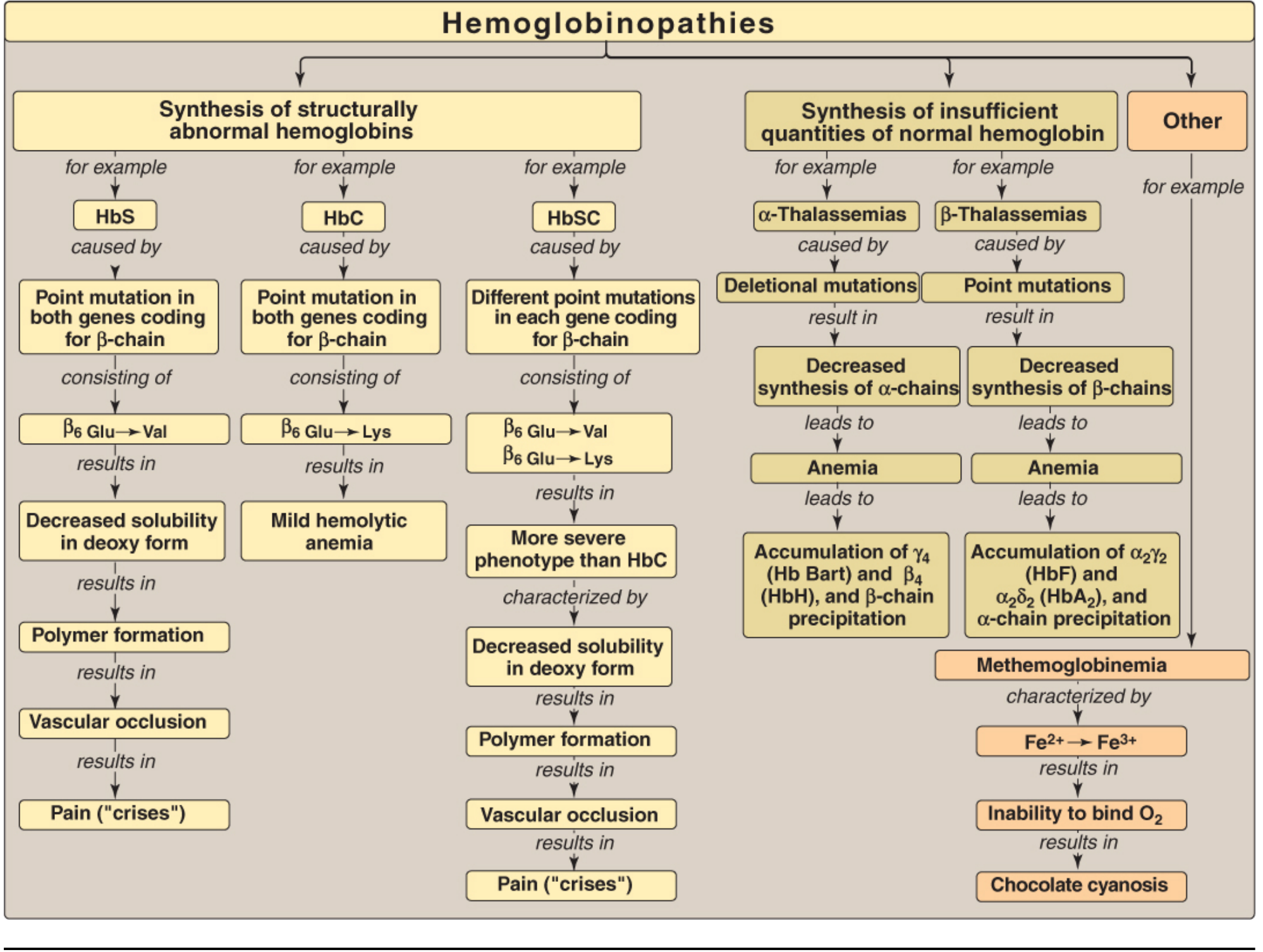
"To aid oxygen transfer, the fetal hemoglobin oxygen dissociation curve is shifted to the left such that fetal hemoglobin has a greater affinity for oxygen than does maternal hemoglobin." - Morgan & Mikhail's Clinical Anesthesiology
Step 5: Transition After Birth
At birth, the situation reverses: now the problem is delivering oxygen to fetal tissues (not loading it). High oxygen affinity means HbF reluctantly releases oxygen. The body compensates:
- Levels of 2,3-DPG rise in neonatal red cells
- The resulting mild acidosis (Bohr effect) reduces oxygen affinity
- Over 1-2 months, γ chains are gradually replaced by β chains (HbF → HbA switch)
Summary
| Feature | HbA (Adult) | HbF (Fetal) |
|---|---|---|
| Globin chains | α₂β₂ | α₂γ₂ |
| 2,3-BPG binding | Strong (His-143 on β chain) | Weak (Ser-143 on γ chain) |
| Oxygen affinity | Normal | Higher |
| ODC curve | Normal | Shifted LEFT |
| P50 (PO₂ at 50% saturation) | ~27 mmHg | ~20 mmHg |
| Physiological purpose | O₂ delivery to tissues | O₂ loading from placenta |
Sources: Mulholland & Greenfield's Surgery, p. 5282; Lippincott's Biochemistry 8th Ed, p. 135; Morgan & Mikhail's Clinical Anesthesiology 7e, p. 1583
。
It looks like you sent just a punctuation mark. Did you mean to type something? Feel free to ask your next question!
Foetal haemoglobin more affinity for oxygen why? For 3 mark ans for university exam
Here is a concise 3-mark university answer:
Fetal Haemoglobin (HbF) - Higher Oxygen Affinity
1. Structural difference (γ chains instead of β chains)
HbF has the composition α₂γ₂, unlike adult HbA (α₂β₂). The γ (gamma) chains replace the β (beta) chains.
2. Weaker binding to 2,3-BPG
The γ chain has a serine at position 143, whereas the β chain has a histidine. Histidine is positively charged and forms a strong electrostatic bond with 2,3-bisphosphoglycerate (2,3-BPG). Serine is neutral and cannot bind 2,3-BPG effectively. Since 2,3-BPG normally stabilizes the low-affinity (T/deoxy) conformation of haemoglobin, its reduced binding to HbF means HbF stays in the high-affinity (R/oxy) conformation.
3. Physiological significance
HbF's oxygen-dissociation curve is shifted to the left (lower P50 ~20 mmHg vs ~27 mmHg for HbA). This allows HbF to load oxygen at the low PO₂ of the placenta (~30-35 mmHg), facilitating oxygen transfer from maternal blood to the fetus.
One-line summary for exams: HbF has higher O₂ affinity because its γ chains bind 2,3-BPG poorly, keeping it in the relaxed (high-affinity) conformation, which left-shifts its ODC curve and enables placental O₂ uptake.
Pancreas is resistant to autodigestion why? For university 3 marke question ans
The pancreas is protected from autodigestion by several mechanisms including secreting enzymes as inactive zymogens and having inhibitors within the gland. Let me get the precise textbook answer for you.
Searching sources...
Finding Sources
Finding Sources
Reading File
Reading File
Here is the precise 3-mark university answer:
Why the Pancreas is Resistant to Autodigestion
The pancreas produces powerful proteolytic, lipolytic, and amylolytic enzymes capable of digesting tissue. Several "fail-safe" mechanisms protect it from self-destruction:
1. Enzymes are synthesized and stored as inactive zymogens
Pancreatic enzymes (trypsinogen, chymotrypsinogen, proelastase, prophospholipase, etc.) are synthesized as inactive precursors (zymogens) and stored in membrane-bound zymogen granules within acinar cells. They are only activated after entering the duodenum, where the intestinal brush-border enzyme enterokinase (enteropeptidase) converts trypsinogen → trypsin, which then activates all other zymogens. Activation within the pancreas itself is thus normally prevented.
2. Trypsin inhibitors (SPINK1)
Acinar and ductal cells also secrete pancreatic secretory trypsin inhibitor (SPINK1/PSTI), which is co-packaged in the zymogen granule. If any trypsin is prematurely activated within the gland, SPINK1 immediately neutralizes it before it can trigger a cascade of enzyme activation.
3. Trypsin self-inactivation and acinar cell resistance
Trypsin undergoes autolysis - it cleaves and inactivates itself, providing a negative feedback limit on any local trypsin activity. Additionally, acinar cells are intrinsically resistant to the action of activated enzymes such as trypsin, chymotrypsin, and phospholipase A₂, and the zymogen granule membrane is impermeable to proteins, sequestering enzymes away from the cytoplasm.
One-line clinical link: When these mechanisms fail (e.g., alcohol, gallstones triggering premature intrapancreatic trypsin activation), the result is acute pancreatitis - autodigestion of the gland itself.
Sources: Medical Physiology (Boron & Boulpaep), p. 1328; Robbins & Kumar Basic Pathology, p. 639
Enter hepatic circulation
The hepatic circulation is a dual blood supply system - the liver receives blood from both the portal vein (nutrient-rich from gut) and hepatic artery (oxygenated). Let me get the full textbook detail.
Searching sources...
Finding Sources
Finding Sources
Reading File
Finding Sources
Reading File
Loading Image
Hepatic Circulation
The liver has a unique dual blood supply - it is the only organ in the body that receives blood from two separate sources simultaneously.
1. Sources of Blood Supply
| Source | Vessel | % of Flow | % of O₂ Supply | Pressure |
|---|---|---|---|---|
| Systemic (arterial) | Hepatic artery | 25% | ~75% | ~90 mmHg |
| Portal (venous) | Portal vein | 75% | ~25% | ~10-12 mmHg |
- The hepatic artery (branch of the coeliac trunk) brings oxygenated blood from the systemic circulation
- The portal vein brings nutrient-rich but relatively deoxygenated blood from the stomach, intestines, pancreas, and spleen
- Despite carrying 75% of blood flow, portal blood has already given up much of its O₂ to the gut - so the hepatic artery, carrying only 25% of flow, provides ~75% of the liver's oxygen
2. Intrahepatic Flow - Through the Sinusoids
Both vessels enter at the portal triad (at the periphery of each liver lobule/acinus) and flow into the hepatic sinusoids:
Portal triad → Sinusoids → Central vein → Hepatic veins → Inferior vena cava
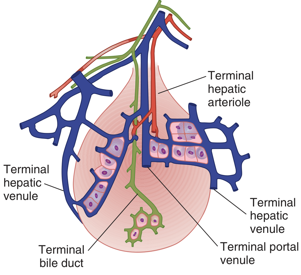
Key features of sinusoids:
- Highly permeable - endothelial cells have large fenestrations and gaps
- More permeable to proteins than capillaries anywhere else in the body
- Allow rapid exchange between blood and hepatocytes
- Kupffer cells line the sinusoids - they phagocytose bacteria and particulate matter from portal blood, acting as a first-line immunological filter
3. Pressure Relationships
| Location | Pressure |
|---|---|
| Hepatic artery | ~90 mmHg |
| Portal vein | ~10-12 mmHg |
| Sinusoids | ~8-9 mmHg |
| Hepatic vein | ~5 mmHg |
| Inferior vena cava | ~2-5 mmHg |
- The massive pressure drop from hepatic artery (90 mmHg) to sinusoids (8-9 mmHg) occurs across high-resistance pre-sinusoidal arterioles
- This is essential - if arterial pressure reached the sinusoids, blood would flow backwards into the portal vein
- The portal vein - sinusoid pressure gradient is very small (only 2-4 mmHg), so portal inflow resistance is very low
4. Reciprocal Regulation (Hepatic Arterial Buffer Response)
If portal blood flow decreases (e.g., between meals), hepatic arterial flow automatically increases to compensate, and vice versa. This is called the hepatic arterial buffer response:
- Mediated by adenosine - produced continuously around arterioles
- When portal flow is low → adenosine accumulates → dilates hepatic arterioles → increases arterial flow
- When portal flow is high → adenosine is washed away → arterioles constrict
Between meals, many sinusoids are collapsed. After eating, increased portal flow recruits reserve sinusoids, preventing a linear rise in portal pressure.
5. Functional Zones of the Liver Acinus
The liver acinus (functional unit) is divided into three zones based on proximity to blood supply:
| Zone | Location | O₂ Level | Susceptibility |
|---|---|---|---|
| Zone 1 (periportal) | Nearest portal triad | Well oxygenated | Toxic injury (first exposure to drugs/toxins) |
| Zone 2 | Middle | Moderately oxygenated | Intermediate |
| Zone 3 (centrilobular) | Nearest central vein | Least oxygenated | Ischaemic/anoxic injury (e.g., in heart failure, paracetamol toxicity) |
6. Clinical Significance
| Condition | Mechanism |
|---|---|
| Portal hypertension | Increased resistance to portal flow (e.g., cirrhosis) → portal pressure >12 mmHg |
| Ascites | Elevated sinusoidal/hepatic vein pressure → fluid transudation into peritoneal cavity |
| Oesophageal varices | Portal blood diverted through portosystemic collaterals |
| Hepatic congestion | Right heart failure → elevated IVC/hepatic vein pressure → sinusoidal distension → "nutmeg liver" |
| Centrilobular necrosis | Zone 3 is most vulnerable to ischaemia (e.g., shock, heart failure) |
Sources: Medical Physiology (Boron & Boulpaep), p. 834; Ganong's Review of Medical Physiology 26th Ed
Enterohepatic circulation
The enterohepatic circulation is the recycling loop of bile acids between the liver and intestine. Let me get the full textbook content.
Searching sources...
Finding Sources
Finding Sources
Reading File
Reading File
Reading File
Loading Image
Loading Image
Enterohepatic Circulation
Definition
The enterohepatic circulation (EHC) is the continuous recycling loop of bile salts (and other substances) between the liver and the intestine via the portal circulation. It is one of the most efficient conservation mechanisms in the body.
Anatomical Components
The circuit involves: Liver → Bile ducts → Gallbladder → Duodenum → Small intestine → Terminal ileum → Portal vein → Liver
The components act as:
- Storage chambers - gallbladder and small intestine
- Valves - sphincter of Oddi and ileocecal valve
- Chemical pumps - hepatocytes, cholangiocytes, and ileal enterocytes (ileocytes)
Step-by-Step Pathway
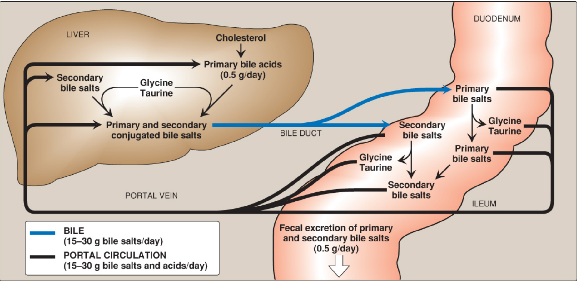
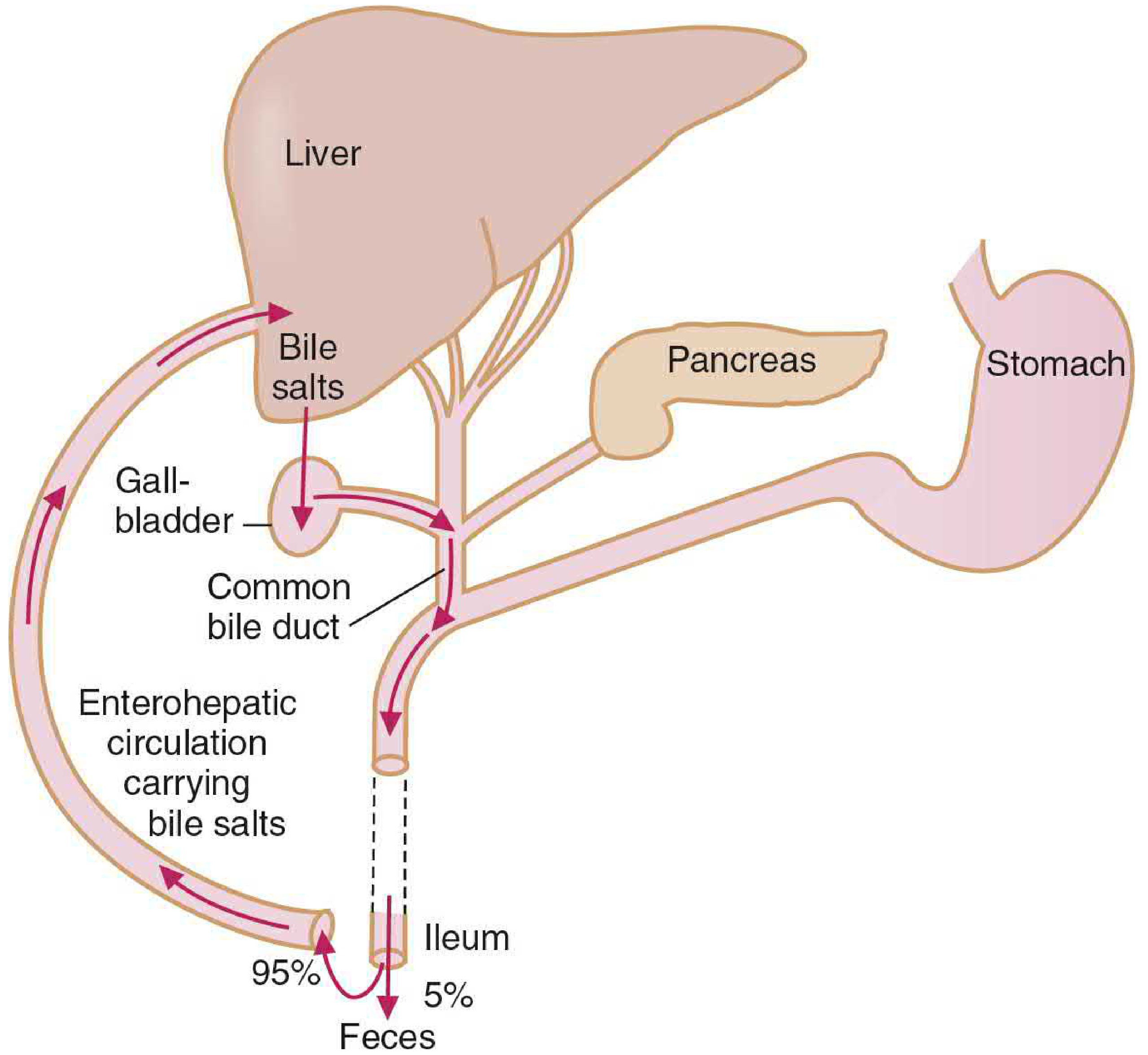
Step 1 - Synthesis in Liver
- Cholesterol is converted to primary bile acids - cholic acid and chenodeoxycholic acid - by the rate-limiting enzyme cholesterol 7α-hydroxylase
- These are conjugated with glycine or taurine to form primary bile salts (more water-soluble, less toxic)
- Secreted into bile canaliculi via the bile salt export pump (BSEP/ABCB11)
Step 2 - Storage in Gallbladder (Fasting)
- During fasting, the sphincter of Oddi contracts → bile acids back up and are concentrated ~10-fold in the gallbladder
- On eating, CCK (cholecystokinin) is released from intestinal mucosa → relaxes sphincter of Oddi + contracts gallbladder → bile squirts into the duodenum
Step 3 - Function in the Intestine
- In the duodenum and jejunum, bile salts form mixed micelles with phospholipids and cholesterol
- Micelles solubilize fat digestion products (fatty acids, monoglycerides, fat-soluble vitamins) and carry them to the brush border for absorption
- The intraluminal bile acid concentration (~5-10 mmol/L) stays well above the critical micellar concentration (~1.5 mmol/L) needed for micelle formation
Step 4 - Bacterial Modification in the Gut
- Gut bacteria deconjugate bile salts (remove glycine/taurine) and dehydroxylate at carbon 7, converting:
- Cholic acid → Deoxycholic acid (secondary bile acid)
- Chenodeoxycholic acid → Lithocholic acid (secondary bile acid)
Step 5 - Reabsorption at Terminal Ileum (Key Step)
- ~95% of bile salts are actively reabsorbed in the terminal ileum via the apical sodium-dependent bile acid transporter (ASBT / SLC10A2)
- Na⁺ co-transports bile salt into the ileal enterocyte
- They exit the basolateral membrane into the portal blood (via OST-α/β transporter)
- Bound to albumin, transported via portal vein back to the liver
Step 6 - Hepatic Re-uptake and Re-secretion
- Hepatocytes efficiently extract bile salts from portal blood via Na⁺-taurocholate cotransporting polypeptide (NTCP/SLC10A1) and OATPs (organic anion transporting polypeptides)
- Re-secreted back into bile → the cycle repeats
- The recirculated bile salts stimulate further biliary secretion (choleretic effect)
Key Quantitative Facts
| Parameter | Value |
|---|---|
| Total bile salt pool | ~2.5 g |
| Bile salts secreted per day | 15-30 g |
| Number of cycles per day | ~6-8 times (2-3 times per meal) |
| % reabsorbed in terminal ileum | >95% |
| Fecal loss per day | ~0.5 g/day |
| Daily synthesis to replace loss | ~0.5 g/day (from cholesterol) |
Regulation of Bile Acid Synthesis
The enzyme cholesterol 7α-hydroxylase is under negative feedback by returning bile salts:
- High bile salt return → enzyme inhibited → less synthesis needed
- Low bile salt return → enzyme stimulated → more synthesis
- FGF19 (fibroblast growth factor 19) secreted by ileum also suppresses hepatic bile acid synthesis via FGFR4 receptor
Clinical Significance
| Condition | Mechanism | Consequence |
|---|---|---|
| Ileal resection / Crohn's disease | EHC interrupted; bile salts not reabsorbed | Bile salt pool depleted; fat malabsorption; steatorrhoea |
| Bile salt diarrhoea | Excess bile salts enter colon (secretory diarrhoea) | Watery diarrhoea |
| Cholestyramine (bile acid sequestrant) | Binds bile salts in gut; prevents reabsorption | ↑ bile acid synthesis from cholesterol → lowers LDL cholesterol (used in hypercholesterolaemia) |
| Gallstones (cholelithiasis) | Disrupted EHC → bile supersaturated with cholesterol | Precipitation of cholesterol crystals |
| Drug recycling | Some drugs (e.g. morphine, digoxin) undergo EHC | Prolongs their duration of action |
One-liner for exams: The enterohepatic circulation recycles >95% of bile salts secreted by the liver via active reabsorption in the terminal ileum and return through the portal vein, allowing 15-30 g of bile salts to be secreted daily from a pool of only 2.5 g.
Sources: Costanzo Physiology 7th Ed, p. 379; Lippincott's Biochemistry 8th Ed, p. 633-634; Sleisenger & Fordtran's Gastrointestinal and Liver Disease, p. 1204
Basal electrical rhythm short note 5 mans
Basal electrical rhythm (BER) refers to the spontaneous rhythmic depolarizations in GI smooth muscle that set the maximum frequency of contractions. Let me get the full textbook content.
Searching sources...
Finding Sources
Finding Sources
Finding Sources
Finding Sources
Reading File
Reading File
Finding Sources
Reading File
Loading Image
Basal Electrical Rhythm (BER) - Short Note (5 Marks)
Definition
The Basal Electrical Rhythm (BER), also called slow waves or pacemaker potentials, refers to the spontaneous, rhythmic oscillations in membrane potential of gastrointestinal smooth muscle cells. These fluctuations occur between approximately -65 mV and -45 mV and are a unique electrical feature of GI smooth muscle.
Origin - Interstitial Cells of Cajal (ICC)
BER is not generated by smooth muscle itself but originates in the interstitial cells of Cajal (ICC) - stellate mesenchymal pacemaker cells located:
- In the stomach and small intestine: outer circular muscle layer, near the myenteric plexus
- In the colon: submucosal border of the circular muscle layer
The ICC are the GI equivalent of the sinoatrial node of the heart. Cyclic depolarizations from ICC spread rapidly to adjacent smooth muscle via low-resistance gap junctions. In regions with a descending frequency gradient (stomach to ileum), the pacemaker with the highest frequency dominates - analogous to cardiac pacemaker hierarchy.
Ionic Mechanism
- Depolarizing phase: Cyclic opening of Ca²⁺ channels → inward Ca²⁺ current → membrane potential rises toward threshold
- Plateau phase: Continued Ca²⁺ channel opening maintains partial depolarization
- Repolarizing phase: Opening of K⁺ channels → outward K⁺ current → membrane repolarizes
Relationship to Spike Potentials and Contraction
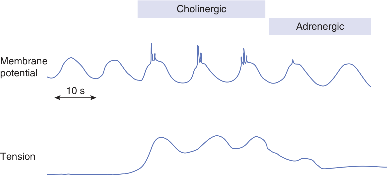
The BER alone rarely causes contraction, but sets the scene for it:
| Electrical event | Mechanical consequence |
|---|---|
| BER below threshold | Basal/tonic contraction (weak) |
| BER reaches threshold → spike potentials superimposed | Phasic (strong) contraction |
| More spikes per BER wave | Greater force of contraction |
The BER therefore sets the maximum frequency at which contractions can occur - contractions can only happen during the depolarizing portion of the BER wave.
Frequencies Along the GI Tract
| Region | BER Frequency |
|---|---|
| Stomach | ~3-4 per minute |
| Duodenum | ~12 per minute (highest) |
| Distal ileum | ~8 per minute |
| Caecum | ~2 per minute |
| Sigmoid colon | ~6 per minute |
The descending gradient from duodenum to ileum is physiologically important - the progressively slower frequency distally allows more time for absorption of fats and bile salts.
Modulation by Neural and Hormonal Factors
BER frequency is intrinsic and fixed - it is NOT altered by neural or hormonal input. However, these inputs modulate whether spike potentials are generated on top of BER waves:
| Agent | Effect on Spikes | Effect on Contraction |
|---|---|---|
| Acetylcholine (parasympathetic) | ↑ spike number | ↑ contraction strength |
| Epinephrine (sympathetic) | ↓ spike number | ↓ contraction strength |
| Gastrin, CCK, motilin | Modulate spike firing | Alter contraction patterns |
Clinical Significance
| Situation | Effect on BER |
|---|---|
| Vagotomy / stomach wall transection | Peristalsis becomes irregular and chaotic (BER loses coordination) |
| Gastroparesis | Disrupted ICC network → loss of normal BER frequency |
| Hirschsprung's disease | Abnormal enteric nervous system affecting coordination of BER-driven contractions |
Exam summary: BER (slow waves) are spontaneous rhythmic oscillations of GI smooth muscle membrane potential (-65 to -45 mV), generated by interstitial cells of Cajal. They set the maximum frequency of contractions (3/min in stomach, 12/min in duodenum). BER alone causes weak tonic contractions; when BER reaches threshold, spike potentials occur → strong phasic contractions. The frequency of BER is fixed but the occurrence of spikes is modulated by ACh (increases) and epinephrine (decreases).
Sources: Ganong's Review of Medical Physiology 26th Ed, p. 498; Costanzo Physiology 7th Ed, p. 354; Mulholland & Greenfield's Surgery 7e
Achalasia cardia
Achalasia cardia is a primary oesophageal motility disorder characterised by failure of the lower oesophageal sphincter to relax, combined with absent peristalsis. Let me get the full textbook content.
Searching sources...
Finding Sources
Finding Sources
Reading File
Reading File
Reading File
Reading File
Now let me get the radiology/investigations and image content:
Reading File
Finding Sources
I now have comprehensive content. Here is the full structured answer:
Achalasia Cardia
Definition
Achalasia (Greek: "does not relax") is a primary oesophageal motility disorder characterised by two cardinal features:
- Impaired / absent relaxation of the Lower Oesophageal Sphincter (LES) on swallowing
- Aperistalsis (absence of peristaltic contractions) in the smooth muscle oesophagus
It affects approximately 1 in 100,000 people and is the most common primary motility disorder of the oesophagus.
Pathophysiology
Normal LES Relaxation
Normally, swallowing triggers release of nitric oxide (NO) and vasoactive intestinal peptide (VIP) from inhibitory ganglion neurons in the myenteric (Auerbach) plexus → LES relaxes → food passes into the stomach.
What Goes Wrong in Achalasia
The fundamental lesion is loss of ganglion cells in the myenteric plexus of the distal oesophagus and LES. Specifically:
- Inhibitory neurons (NO and VIP secreting) are selectively destroyed first
- Without NO/VIP, the LES cannot relax after swallowing (deglutitive inhibition is lost)
- As disease progresses, excitatory (cholinergic) neurons are also affected → aperistalsis
- Muscle strips from the LES do not relax in response to ganglionic stimulation
- CCK (which normally reduces LES pressure via inhibitory neurons) paradoxically increases LES pressure in achalasia
Cause of Ganglion Cell Loss
Increasing evidence points to an autoimmune process in genetically susceptible individuals:
- Myenteric plexus infiltrated predominantly by cytotoxic T cells
- Antibodies against myenteric neurons detected in patient serum (associated with specific HLA alleles)
- Suspected trigger: Herpes simplex virus-1 (HSV-1) chronic/latent infection
- Chagas disease (Trypanosoma cruzi) causes an identical picture by destroying autonomic ganglion cells
Chicago Classification - 3 Types (High-Resolution Manometry)
| Type | Features | Response to Treatment |
|---|---|---|
| Type I (Classic) | Absent peristalsis + impaired LES relaxation; dilated oesophagus | Moderate |
| Type II (with compression) | Absent peristalsis + pan-oesophageal pressurisation; early disease | Best (~100% with pneumatic dilation) |
| Type III (Spastic) | Premature/spastic contractions + impaired LES relaxation; plexus inflammation but not destruction | Poorest; better with surgical myotomy |
Clinical Features
| Symptom | Details |
|---|---|
| Dysphagia | Most common; both solids AND liquids (distinguishes from mechanical obstruction which is solids first) |
| Regurgitation | Undigested food from hours/days before; non-bilious, non-acid, mixed with saliva |
| Chest pain | ~2/3 of patients early on; squeezing, may mimic cardiac pain |
| Weight loss | Progressive as disease advances |
| Halitosis | From stagnant food in oesophagus |
| Aspiration pneumonia | Up to 10% with advanced disease |
| Heartburn | Paradoxical complaint; actually from bacterial fermentation of retained food, not true reflux |
Investigations
1. Barium Swallow (X-ray)
- Classic finding: "Bird's beak" / "Rat tail" appearance
- Grossly dilated oesophagus tapering smoothly to a narrow segment at the gastro-oesophageal junction (GOJ)
- Barium retained >5 minutes is supportive
- Absent peristaltic waves
- Chest X-ray may show air-fluid level in a widened mediastinum
2. Endoscopy (OGD) - Mandatory
- Done in every new case to exclude pseudoachalasia (especially gastric cardia carcinoma which mimics achalasia)
- Typically shows dilated oesophagus with retained saliva/food
- The endoscope passes through the LES with a characteristic "pop"
- Pseudoachalasia suspected if: age >50, symptoms <1 year, weight loss >7 kg
3. High-Resolution Manometry (HRM) - Gold Standard
- Elevated LES resting pressure (>45 mmHg; normal ~15-30 mmHg)
- Failure of LES to relax on swallowing (integrated relaxation pressure >15 mmHg)
- Absent peristalsis in distal oesophagus
- Allows Chicago classification into types I, II, III
4. CT Chest/Abdomen
- Grossly distended oesophagus with liquid and solid material
- Helps exclude malignancy causing pseudoachalasia
Differential Diagnosis
- Pseudoachalasia - carcinoma of gastric cardia/oesophagus, lymphoma, metastases
- Chagas disease - identical picture from T. cruzi infection (check travel history, serology)
- Distal Oesophageal Spasm (DES) - spastic type III achalasia overlaps
- Scleroderma - but causes absent LES tone (patulous LES), not hypertonic LES
Treatment
The underlying neuropathology cannot be reversed. Treatment aims to reduce LES pressure to allow gravity-aided oesophageal emptying.
1. Pharmacological (Temporising only - limited efficacy)
- Nitrates (isosorbide dinitrate) - releases NO → relaxes LES
- Calcium channel blockers (nifedipine) - reduce LES pressure
- Botulinum toxin (Botox) injection into LES - blocks ACh release from excitatory neurons → reduces LES pressure; effect lasts 6-12 months; repeated injections needed
2. Pneumatic (Balloon) Dilation
- Forceful dilation of LES using a pneumatic balloon
- ~90% efficacy; risk of perforation ~1%
- Treatment of choice for Type II achalasia (100% efficacy in trials)
- May need repeat dilations
3. Surgical - Laparoscopic Heller Myotomy (LHM)
- Surgical division of the LES muscle fibres (myotomy) at the cardia
- ~90% efficacy; comparable to pneumatic dilation
- Combined with fundoplication (partial) to prevent post-op reflux
- Preferred for Type III achalasia (86% efficacy vs 40% for dilation)
4. POEM (Per-Oral Endoscopic Myotomy)
- Newer endoscopic technique: submucosal tunnelling + myotomy via flexible endoscope
- Particularly effective for Type III spastic achalasia
- Avoids external incisions
Complications of Untreated Achalasia
- Aspiration pneumonia / lung abscess
- Severe malnutrition and weight loss
- Oesophageal squamous cell carcinoma (long-standing achalasia is a risk factor - 16x increased risk due to chronic mucosal irritation from retained food)
- Mega-oesophagus (end-stage - sigmoid-shaped grossly dilated oesophagus)
Exam one-liner: Achalasia is a primary motility disorder caused by autoimmune destruction of inhibitory (NO/VIP) myenteric neurons of the oesophagus, resulting in impaired LES relaxation and aperistalsis, classically presenting with dysphagia to both solids and liquids, diagnosed by manometry (elevated LES pressure + absent peristalsis) and barium swallow (bird's beak sign), and treated by pneumatic dilation or Heller myotomy.
Sources: Sleisenger & Fordtran's Gastrointestinal and Liver Disease, pp. 729-738; Grainger & Allison's Diagnostic Radiology; Current Surgical Therapy 14e
This is a shared conversation. Sign in to Orris to start your own chat.