

IMG-20260717-WA0003.jpg
Which ointment should we prescribe for this
analyse_media
Reading File
Now let me search the medical library for granuloma annulare treatment (the most likely diagnosis based on the image analysis) and also search for molluscum/verruca treatment in parallel.
Finding Sources
Finding Sources
Finding Sources
Finding Sources
Reading File
Reading File
Let me read the treatment section for granuloma annulare from Andrews' Diseases of the Skin:
Reading File
I can see the textbook has an image of GA on the foot (Fig. 31.5). Let me get the treatment section and fetch the relevant image:
Reading File
Loading Image
Now let me read the localized GA treatment section more specifically:
Reading File
Reading File
I now have all the information needed for a thorough answer. Here is the clinical assessment and management recommendation:
Clinical Assessment
The image shows two well-demarcated, flesh-colored to slightly pinkish, smooth-surfaced papules/plaques on the lateral aspect of the foot. This morphology and location are classic for Granuloma Annulare (GA), localized type.
For comparison, here is a textbook image of GA on the foot:
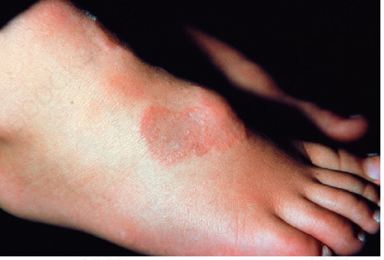
The match is strong - GA characteristically appears on the lateral or dorsal surfaces of the feet, presenting as erythematous to flesh-colored firm papules or plaques that spread peripherally. The lesions are typically asymptomatic and benign.
Treatment Recommendations
Important first point: Many mild, localized cases of GA require no treatment at all - over 50% resolve spontaneously within 2 years. Simply reassuring the patient is valid.
When treatment is desired, follow this ladder for localized GA:
First-Line Options (Topical/Intralesional)
| Treatment | Details |
|---|---|
| Intralesional triamcinolone | Most effective for localized disease; 2.5-5 mg/mL injected into the active border. Relapse common at 3-7 months. |
| Superpotent topical corticosteroids (e.g., clobetasol propionate 0.05% ointment) | Applied once or twice daily under occlusion; best for macular/early lesions |
| Topical calcineurin inhibitors (tacrolimus 0.1% ointment or pimecrolimus 1% cream) | Useful if steroid-sparing needed; suitable for longer-term use |
| Imiquimod 5% cream | Case reports show efficacy; applied 3x/week |
For Localized Disease - Recommended Starting Point
For these two localized papules on the foot, the most practical initial approach is:
- Intralesional triamcinolone acetonide (2.5-5 mg/mL) - most effective, done in clinic
- If patient prefers topical therapy: Clobetasol propionate 0.05% ointment under occlusion (applied nightly, covered with plastic wrap for 2-4 weeks)
- If steroid use is a concern (e.g., atrophic skin): Tacrolimus 0.1% ointment twice daily
Second-line (for refractory/generalized cases only)
- Hydroxychloroquine, NB-UVB phototherapy, doxycycline, dapsone - not needed for this presentation.
Differential Diagnoses to Keep in Mind
- Lichen planus (would typically be more violaceous and pruritic)
- Verruca plana (flat warts - would have a rougher surface)
- Dermatofibroma (usually firmer, dimples inward on lateral pinch)
A biopsy is not mandatory for classic localized GA but can confirm the diagnosis if any atypical features are present. The textbook notes that simply performing a biopsy sometimes leads to lesion involution.
Source: Andrews' Diseases of the Skin - Clinical Dermatology, Chapter 31 (Granuloma Annulare), p. 816-818
Disclaimer: This is clinical decision support, not a replacement for in-person examination. A dermatologist should confirm the diagnosis before treatment, particularly to rule out other granulomatous or neoplastic conditions.
IMG-20260717-WA0003.jpg
Which ointment to used for this condition
This is the same image as our previous discussion. Based on the localized granuloma annulare (GA) on the lateral foot, here is a focused answer on ointments specifically:
Topical Ointments for Localized Granuloma Annulare
First Choice - Potent/Superpotent Topical Corticosteroid
Clobetasol propionate 0.05% ointment
- Apply a thin layer to the lesion once or twice daily
- Use under occlusion (cover with plastic wrap overnight) for better penetration - this significantly improves efficacy for GA
- Duration: 2-4 weeks, then reassess
- Ointment base is preferred over cream for this condition as it penetrates thicker skin better
Alternatives in the same class:
- Betamethasone dipropionate 0.05% ointment (slightly less potent, still effective)
- Mometasone furoate 0.1% ointment (mid-high potency, good for longer use)
Second Choice - Topical Calcineurin Inhibitor (steroid-sparing)
Tacrolimus 0.1% ointment
- Apply twice daily
- Useful if you want to avoid steroid-related skin atrophy (important on the thin skin of the foot dorsum)
- Can be used for longer durations than clobetasol without risk of atrophy
Third Choice - Imiquimod 5% cream
- Applied 3 times per week
- Case reports support efficacy in GA
- Can cause local irritation reactions
Practical Recommendation
For these two small, localized lesions on the lateral foot:
Start with Clobetasol 0.05% ointment under occlusion nightly for 3-4 weeks.
If no response after 4 weeks, switch to tacrolimus 0.1% ointment twice daily for 6-8 weeks.
If topical therapy fails, the most effective next step is intralesional triamcinolone acetonide (2.5-5 mg/mL) injected directly into the lesion border in clinic.
Note: Many localized GA lesions resolve on their own within 2 years without any treatment. If the lesions are asymptomatic and the patient is not bothered cosmetically, watchful waiting is a perfectly reasonable option. Always confirm the diagnosis clinically before committing to long-term treatment.Source: Andrews' Diseases of the Skin - Clinical Dermatology, Chapter 31, p. 818
This is a shared conversation. Sign in to Orris to start your own chat.