
nephrotic syndrome
"nephrotic syndrome"[MeSH Terms] AND management
Nephrotic Syndrome
Definition
Pathophysiology
The Tetrad of Features
| Feature | Mechanism |
|---|---|
| Proteinuria | Impaired glomerular size/charge selectivity; albumin and other proteins escape |
| Hypoalbuminemia | Urinary losses exceed hepatic synthesis; compensatory increase in synthesis is blunted |
| Hyperlipidemia/Lipiduria | Increased hepatic lipoprotein synthesis, impaired lipid catabolism, abnormal transport; lipiduria from increased GBM permeability to lipoproteins |
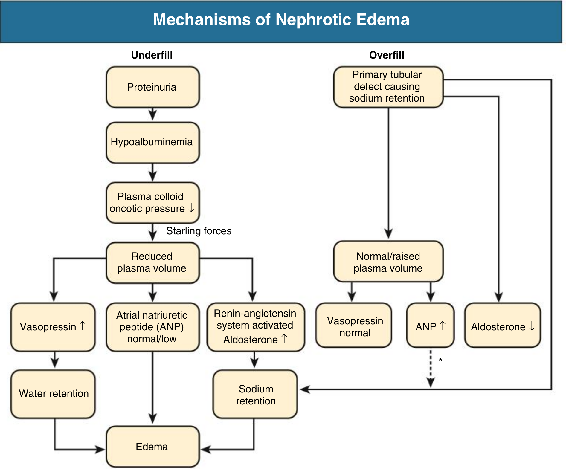
| Edema | Low plasma oncotic pressure drives fluid into the interstitium (underfill) + primary renal Na+ retention (overfill) |
Mechanisms of Nephrotic Edema
- Underfill: Hypoalbuminemia lowers plasma oncotic pressure → fluid shifts to interstitium → reduced plasma volume → RAAS activation → aldosterone-driven Na+ retention → worsens edema
- Overfill (more common): Primary defect in distal nephron Na+ excretion (likely ENaC activation by proteolytic enzymes in tubular fluid) → Na+ and water retention → expanded plasma volume, suppressed RAAS
Causes
Primary (Idiopathic) Glomerular Diseases
| Disease | Key Features | Age Group |
|---|---|---|
| Minimal Change Disease (MCD) | Normal LM; podocyte foot process effacement on EM; responds to steroids (>80% remission); relapses common | Most common in children; up to 15% of adult cases |
| Focal Segmental Glomerulosclerosis (FSGS) | Focal, segmental sclerosis of glomeruli; higher prevalence in African Americans; often steroid resistant | Adolescents/adults |
| Membranous Nephropathy (MN) | Subepithelial immune deposits; PLA2R antibody in primary form; most common adult primary nephrotic syndrome | Adults >40 yr |
| Membranoproliferative GN (MPGN) | Mesangial expansion + double contour ("tram-track") GBM | Any age |
| IgA Nephropathy | Mainly nephritic, but ~10-15% can present with nephrotic range proteinuria | Young adults |
Secondary Causes
- Diabetes mellitus - Kimmelstiel-Wilson nodular glomerulosclerosis; most common secondary cause
- Lupus (SLE) - can mirror membranous or other patterns (lupus nephritis class V for nephrotic)
- Preeclampsia - nephrotic-range proteinuria + hypertension in pregnancy
- Infections - Hepatitis B (membranous pattern), hepatitis C, HIV, malaria, infective endocarditis
- Malignancy - Hodgkin lymphoma (classically associated with MCD), carcinomas (membranous)
- Drugs - Gold, penicillamine (membranous), heroin, NSAIDs, captopril
- Amyloidosis - AL or AA amyloid deposited in glomeruli; Congo red staining with apple-green birefringence
Clinical Features
Symptoms
- Foamy urine (frothy due to proteinuria)
- Peripheral edema - often worse in the morning, periorbital edema (especially in children); may progress to anasarca (generalized severe edema including ascites, scrotal/vulvar edema, pleural effusions)
- Fatigue, dyspnea, orthopnea
- Nausea/vomiting (from bowel wall edema or ascites)
Signs
- Hypertension, pitting edema
- Ascites, pleural effusions
- Muehrcke's lines - white transverse bands on nails (hypoalbuminemia sign)
- Eruptive xanthomata, xanthelasma (hyperlipidemia)
Laboratory Findings
- Proteinuria >3.5 g/day (or spot urine protein:creatinine ratio >3.5)
- Serum albumin <2.5-3 g/dL
- Total cholesterol usually >180 mg/dL; raised LDL and triglycerides
- Lipiduria on urinalysis (fatty casts, oval fat bodies)
- Serum creatinine variable
Complications
1. Thromboembolism (up to 40% VTE risk)
- Loss of anticoagulant proteins in urine: antithrombin III, proteins C and S
- Increased hepatic synthesis of procoagulant proteins (fibrinogen, factors V, VIII)
- Hyperlipidemia causes hyperviscosity; fibrinolytic inhibitors increase clot risk
- Most common sites: renal vein thrombosis, DVT, pulmonary embolism
- Arterial thrombosis also possible
2. Infection
- Loss of IgG and complement factors in urine
- Reduced T-cell function
- Increased risk of cellulitis, spontaneous bacterial peritonitis, pneumococcal infections
- Steroid therapy adds further immunosuppression
3. Atherosclerosis / Cardiovascular Disease
- Prolonged hyperlipidemia accelerates vascular disease
4. Protein malnutrition
- Loss of vitamin D-binding protein → vitamin D deficiency, secondary hyperparathyroidism
- Loss of transferrin → iron-deficiency anemia
- Loss of hormone-binding proteins → thyroid/hormonal disturbances
Diagnosis
- Urinalysis with microscopy: proteinuria, lipiduria, fatty casts, oval fat bodies (no RBC casts - those suggest nephritic)
- 24-hour urine or spot urine protein:creatinine ratio to quantify proteinuria
- Serum: albumin, lipid panel, creatinine, BUN, electrolytes, CBC
- Complement (C3, C4, CH50) - low in MPGN, lupus nephritis
- Serologies: ANA, anti-dsDNA (SLE), anti-PLA2R (primary membranous), SPEP (amyloid), hepatitis B/C serology, HIV
- Renal biopsy - indicated in adults (most cases); children usually treated empirically for MCD first
Treatment
General / Supportive
| Goal | Intervention |
|---|---|
| Edema | Loop diuretics (furosemide); restrict dietary Na+ (<2 g/day); fluid restriction if severe hyponatremia |
| Proteinuria reduction | ACE inhibitors or ARBs - reduce intraglomerular pressure, reduce proteinuria, slow CKD progression |
| Hyperlipidemia | Statins |
| Thromboembolism prophylaxis | Anticoagulation considered if serum albumin <2.5 g/dL or other high-risk features; long-term if renal vein thrombosis occurs |
| Infections | Pneumococcal and influenza vaccination; prophylactic antibiotics in selected cases |
Disease-Specific Treatment
| Cause | First-Line | Notes |
|---|---|---|
| Minimal Change Disease | Oral corticosteroids (prednisone 1 mg/kg/day, max 80 mg) | >80% achieve remission; relapses common; steroid-dependent cases use calcineurin inhibitors (cyclosporine, tacrolimus) or rituximab |
| FSGS | Corticosteroids | Often steroid-resistant; calcineurin inhibitors, mycophenolate, rituximab for resistant cases |
| Membranous Nephropathy | Observation for mild cases; immunosuppression for high-risk | Cyclophosphamide + steroids (Ponticelli regimen) or rituximab (now preferred for primary MN) |
| Diabetic Nephropathy | RAAS blockade; SGLT2 inhibitors | Address underlying glycemic and blood pressure control |
| Lupus Nephritis (Class V) | Mycophenolate mofetil ± steroids | Belimumab and voclosporin now adjunctive options |
KDIGO Pediatric Protocol (2021)
- Initial episode: Prednisone 60 mg/m²/day (max 60 mg/day) for 4-6 weeks, then 40 mg/m² on alternate days for another 4-6 weeks
- Relapse: Prednisone until remission, then taper
- Frequent relapsers/steroid-dependent: Calcineurin inhibitors (cyclosporine, tacrolimus) or mycophenolate mofetil; rituximab for refractory cases
Key Distinctions: Nephrotic vs. Nephritic
| Feature | Nephrotic | Nephritic |
|---|---|---|
| Proteinuria | >3.5 g/day (massive) | Mild-moderate |
| Hematuria | Absent or mild | Present (RBC casts) |
| Hypertension | Variable | Prominent |
| Edema | Prominent | Mild |
| GFR | Normal or mildly reduced | Reduced (azotemia) |
| Complement | Usually normal | Often low (MPGN, post-strep, lupus) |
Recent Evidence Note
What is the role of SGLT2 inhibitors in slowing nephrotic syndrome progression?
"SGLT2 inhibitor" AND "nephrotic syndrome"
"SGLT2 inhibitor" AND proteinuria AND glomerular
PMID: 41203232
PMID: 41202026
PMID: 38061371
"SGLT2 inhibitor" AND "IgA nephropathy" OR "focal segmental glomerulosclerosis" OR "membranous nephropathy"
SGLT2 Inhibitors in Nephrotic Syndrome Progression
Background: What SGLT2 Inhibitors Do in the Kidney
Core Mechanisms of Renoprotection
| Mechanism | Detail |
|---|---|
| Tubuloglomerular feedback (TGF) restoration | By blocking proximal Na+ reabsorption, more Na+ and Cl- reach the macula densa → afferent arteriole constriction → reduced intraglomerular capillary pressure → reduced glomerular hyperfiltration |
| Reduced glomerular hyperfiltration | In diabetes, hyperfiltration damages glomeruli and drives proteinuria. SGLT2i reverses this; but the same TGF mechanism applies even in non-diabetic CKD |
| Reduction in proteinuria | Lower intraglomerular pressure directly reduces the hydraulic driving force for protein leakage through the glomerular filtration barrier |
| Anti-fibrotic / anti-inflammatory effects | Reduced tubular glucose burden lowers advanced glycation end products (AGEs) and pro-fibrotic cytokines (TGF-β, connective tissue growth factor) |
| Natriuresis and volume reduction | Reduces preload/afterload, lowers intraglomerular pressure via hemodynamic effects |
| Mitochondrial and metabolic effects | Promotes ketone body use, reduces oxidative stress in tubular cells; reduces mTOR signaling and tubular hypertrophy |
Evidence Base: From Diabetic to Non-Diabetic Proteinuric Disease
Landmark Trials
| Trial | Drug | Population | Key Kidney Outcome |
|---|---|---|---|
| CREDENCE (2019) | Canagliflozin | T2DM + diabetic nephropathy (mean UACR ~927 mg/g) | 34% relative risk reduction in renal composite endpoint |
| DAPA-CKD (2020) | Dapagliflozin | CKD (eGFR 25-75) with UACR ≥200 mg/g; ~33% non-diabetic | 39% reduction in sustained eGFR decline, ESKD, or renal death; benefit preserved in non-diabetic patients |
| EMPA-KIDNEY (2022-2024) | Empagliflozin | Broad CKD including eGFR as low as 20; many non-diabetic | Halved the chronic eGFR decline rate from -2.75 to -1.37 mL/min/1.73m²/year (-50%, 95% CI 42-58%); largest relative benefit in those with lower baseline UACR |
DAPA-CKD: FSGS Pre-specified Subgroup
Cutting-Edge 2026 Meta-Analysis Evidence
1. Neuen et al., JAMA 2026 (PMID 41203232)
- SGLT2 inhibitors reduced CKD progression risk by 38% overall (HR 0.62, 95% CI 0.57-0.68) across 70,361 participants
- Benefit was preserved regardless of baseline eGFR - including stage 4 CKD (eGFR <30): HR 0.71
- Benefit was preserved regardless of albuminuria level - including those with >300 mg/g (nephrotic-range proteinuria): HR 0.57 (95% CI 0.52-0.64)
- Annual eGFR decline was slowed across all UACR subgroups, in both diabetic and non-diabetic patients
2. Staplin et al., JAMA 2026 (PMID 41202026)
- Benefits on kidney disease progression were clear in both diabetic and non-diabetic patients (HR 0.65 vs. 0.74)
- Absolute benefits were largest in those with UACR ≥200 mg/g - the heavy proteinuria range that overlaps with nephrotic syndrome
- Benefits also extended to reduced AKI risk, hospitalizations, and mortality
Specific Relevance to Nephrotic Syndrome Causes
Diabetic Nephropathy (most common secondary NS)
IgA Nephropathy
FSGS
Minimal Change Disease / Primary NS
Practical Role in the Nephrotic Syndrome Management Algorithm
Nephrotic Syndrome
       |
       ├─ Identify underlying cause (biopsy, serologies)
       |
       ├─ ALL patients with persistent proteinuria:
       |    ├─ RAAS blockade (ACE-i or ARB) ← first line
       |    └─ SGLT2 inhibitor ← now standard adjunct if eGFR ≥20
       |         (particularly if UACR still >200 mg/g on RAAS alone)
       |
       ├─ Diabetic nephropathy: SGLT2i + RAAS + finerenone
       ├─ FSGS/IgA: SGLT2i + RAAS ± specific immunosuppression
       └─ MCD: Steroids first; SGLT2i not established
Key Practical Points
- eGFR dip: An initial acute drop of ~2-3 mL/min/1.73m² is expected and acceptable - this reflects the TGF-mediated afferent arteriole constriction and does not indicate harm. The long-term chronic slope benefit is preserved.
- SGLT2i + RAAS combination is additive - each reduces intraglomerular pressure via different mechanisms
- Volume effects: SGLT2i-driven natriuresis can exacerbate volume depletion in underfill-type nephrotic syndrome (particularly MCD) - monitor closely
- Furosemide interaction: The natriuretic effect of SGLT2i may reduce diuretic requirements; dose adjustment may be needed
- Infections: Genital mycotic infections are an adverse effect; already immunocompromised nephrotic patients require vigilance
Summary
- Strongest evidence: Diabetic nephropathy, IgA nephropathy, FSGS with CKD
- Mechanism: Primarily hemodynamic (TGF-mediated reduction in glomerular hyperfiltration) + anti-fibrotic + metabolic
- Not yet established: Primary MCD, MPGN, membranous nephropathy as dedicated indications
- Current guideline position: KDIGO 2024 recommends SGLT2 inhibitors for CKD with eGFR ≥20 and UACR ≥200 mg/g, regardless of diabetes status - which captures the majority of patients with clinically significant nephrotic syndrome
sickle cell disease
"sickle cell disease"[MeSH Terms] AND management
"sickle cell"[MeSH Terms] AND treatment
"sickle cell disease" AND (hydroxyurea OR voxelotor OR crizanlizumab OR gene therapy)
PMID: 41831848
PMID: 39118728
Sickle Cell Disease
Definition and Genetics
- Globally affects millions, particularly in sub-Saharan Africa, the Middle East, India, and the Mediterranean
- In the United States, ~8% of people of African descent are HbS carriers; ~1 in 600 have sickle cell anemia (HbSS)
- The HbS allele is maintained by selective pressure because heterozygous carriers are protected against falciparum malaria
Genotypes and Severity
| Genotype | Description | HbS% | Severity |
|---|---|---|---|
| HbSS (sickle cell anemia) | Homozygous; no HbA | >85% | Most severe |
| HbSS + α-thalassemia | Milder due to reduced MCHC | >85% | Moderate-severe |
| HbSC disease | HbS + HbC (Lys at β6) | 50% | Moderate |
| HbS β⁰-thalassemia | HbS + no β-globin production | >85% | Severe (= HbSS) |
| HbS β⁺-thalassemia | HbS + reduced β-globin | 70-95% | Mild-moderate |
| Sickle cell trait (HbAS) | Heterozygous carrier | 35-40% | Asymptomatic (usually) |
Pathophysiology
Step 1: HbS Polymerization
- Sickling is initially reversible on reoxygenation
- Repeated cycles of sickling cause cumulative membrane damage (Ca²+ influx, K+ and water loss) → irreversibly sickled cells prone to hemolysis
- High intracellular HbS concentration
- Low pH (acidosis)
- High temperature
- Slow transit through the microvasculature (delayed deoxygenation)
- Presence of HbF or HbA (dilutes HbS, weak interaction)
- Good hydration (reduces intracellular HbS concentration)
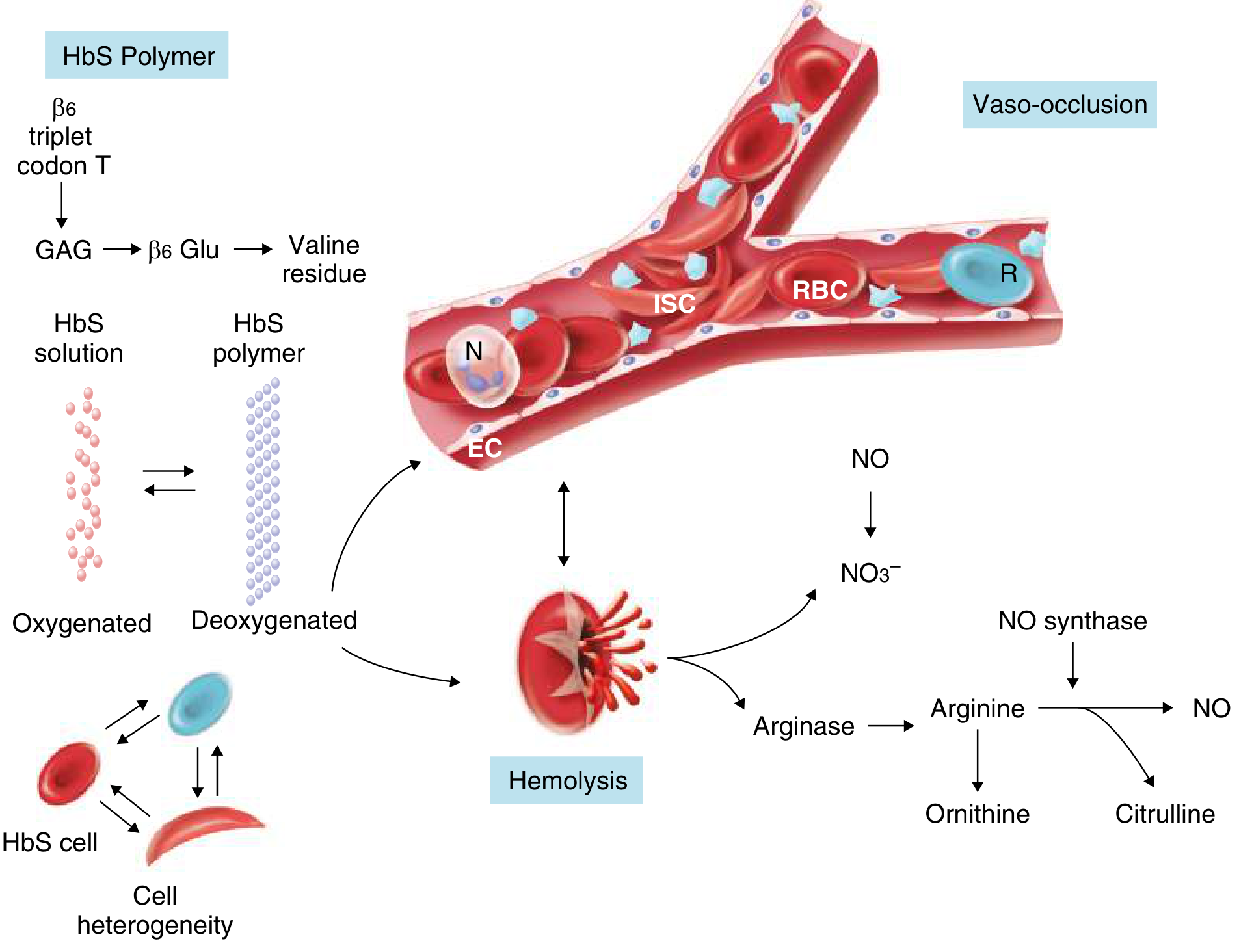
Step 2: Two Downstream Cascades
A. Hemolysis
- Extravascular: Macrophages phagocytose deformed sickled cells in the spleen and liver
- Intravascular: Direct red cell lysis releases free hemoglobin into plasma, which:
- Depletes haptoglobin
- Scavenges nitric oxide (NO) → vasoconstriction, platelet activation, endothelial inflammation
- Releases arginase → destroys L-arginine (the NO synthase substrate) → further reduces NO
- Produces reactive oxygen species
B. Vaso-Occlusion
- Microvascular obstruction → tissue ischemia
- Reperfusion injury → further endothelial activation
- Inflammation → more sickling
Clinical Manifestations
Acute Complications
1. Vaso-Occlusive Crisis (VOC) / Painful Crisis
- Most frequent acute complication; caused by microvascular occlusion
- Severe pain in bones (especially long bones, back, chest, abdomen)
- Triggers: cold, infection, dehydration, hypoxia, stress, alcohol
- Management: IV fluids, analgesia (NSAIDs + opioids), warmth, oxygen if hypoxic
2. Acute Chest Syndrome (ACS)
- New pulmonary infiltrate + fever + respiratory symptoms
- Caused by fat embolism (from bone marrow infarction), infection, or in-situ sickling in pulmonary vasculature
- Most common cause of death in SCD
- Treatment: broad-spectrum antibiotics + exchange transfusion + respiratory support; hydroxyurea for recurrent ACS
3. Stroke and Cerebrovascular Disease
- Children: ischemic stroke from large vessel intracranial stenosis
- Adults: silent infarcts, hemorrhagic stroke
- Transcranial Doppler (TCD) screening identifies high-risk children; chronic transfusion program reduces stroke risk by ~90%
- Hydroxyurea is an alternative to transfusion in lower-resource settings
4. Aplastic Crisis
- Triggered by Parvovirus B19 infection (infects erythroid precursors)
- Sudden severe drop in hemoglobin + reticulocytopenia
- Self-limited; supportive transfusion
5. Splenic Sequestration
- Rapid pooling of blood in the spleen → acute splenomegaly + severe anemia + cardiovascular collapse
- Most common in children (before functional asplenia develops)
- Emergency splenectomy or transfusion; elective splenectomy after recurrence
6. Acute Kidney Injury
- Occurs in 2-10% of acute painful crises
- Precipitated by dehydration, sepsis, drugs (NSAIDs)
Chronic Complications (Organ Damage)
| System | Complication |
|---|---|
| Spleen | Functional asplenia by age 5-6 years from repeated infarcts (autosplenectomy) → increased risk of encapsulated bacteria (pneumococcus, meningococcus, H. influenzae) |
| Bones | Avascular necrosis (AVN) of femoral/humeral heads; vertebral body H-shaped infarcts ("Lincoln log" sign); osteomyelitis (Salmonella classically) |
| Kidney | Hyposthenuria (inability to concentrate urine), microalbuminuria (20% of children, 60% of adults), proteinuria, nephrotic syndrome, renal papillary necrosis, renal medullary carcinoma, end-stage renal disease |
| Lungs | Pulmonary hypertension (30% of adults); restrictive lung disease (70% of adults); sleep-disordered breathing (40-60%) |
| Heart | High-output cardiac failure; dilated cardiomyopathy from chronic anemia |
| Eyes | Proliferative retinopathy (especially HbSC); vitreous hemorrhage; retinal detachment |
| CNS | Silent cerebral infarcts (35% of children by age 14); cognitive impairment; headaches |
| Liver | Sickle hepatopathy; intrahepatic cholestasis; pigment gallstones (from chronic hemolysis) in 50-70% |
| Skin | Leg ulcers (chronic, hard to heal; from local ischemia) |
| Genitourinary | Priapism (stuttering or fulminant) → risk of erectile dysfunction; enuresis |
| Immune | Increased COVID-19 severity; sepsis from encapsulated organisms |
Diagnosis
Blood Tests
- CBC: Hemoglobin 5-10 g/dL (HbSS); normochromic normocytic anemia; reticulocytosis (10-25%); leukocytosis; thrombocytosis
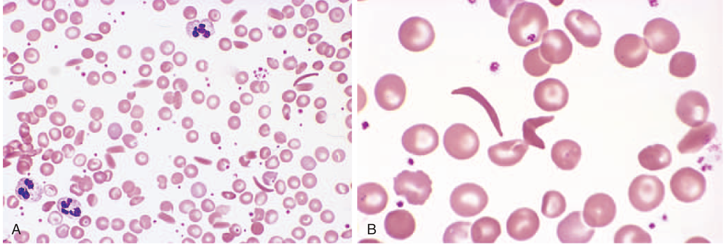
- Peripheral smear: Sickle cells, target cells, Howell-Jolly bodies (asplenia), nucleated RBCs
- Hemoglobin electrophoresis / HPLC: Gold standard for diagnosis; identifies HbS, HbA, HbF, HbA2 percentages
- Newborn screening: Now mandatory in most high-income countries (isoelectric focusing or HPLC of cord blood)
Additional Tests
- Elevated bilirubin, LDH (hemolysis markers)
- Low haptoglobin
- TCD ultrasound for stroke risk stratification
- Echocardiography (pulmonary hypertension screening via tricuspid regurgitation velocity)
Treatment
Disease-Modifying Therapy
1. Hydroxyurea (Hydroxycarbamide) - First-line
- Mechanism: Increases HbF production (inhibits ribonucleotide reductase, activates HbF gene transcription via stress erythropoiesis); reduces WBC and platelet adhesion; decreases MCHC
- Benefits: 50% reduction in painful crises, ACS, hospitalizations; improved survival; reduces renal hyperfiltration in children
- Dose: 15-35 mg/kg/day in adults; uptitrate to maximum tolerated dose
- Monitoring: CBC every 4-8 weeks (myelosuppression)
- Pregnancy: Teratogenic - contraindicated in pregnancy; use with effective contraception
- Administered under supervision of an experienced provider; now being expanded in low-resource settings
2. L-Glutamine
- FDA approved 2017; reduces oxidative stress in sickle cells; reduces VOC frequency
3. Crizanlizumab (Adakveo)
- Mechanism: Anti-P-selectin monoclonal antibody; blocks sickle cell-endothelium adhesion
- FDA approved 2019; reduces VOC frequency
- Note: In 2023, the FDA withdrew crizanlizumab's accelerated approval pending confirmatory trial data - its status as of 2026 requires current guidelines review
4. Voxelotor (Oxbryta)
- Mechanism: Allosterically stabilizes HbS in the oxygenated state, preventing polymerization; raises hemoglobin
- FDA approved 2019; also withdrawn from market in 2024 by manufacturer following safety data review - prescribers should check current status
5. Chronic Transfusion Program
- Monthly exchange or simple transfusions to maintain HbS <30%
- Indications: stroke prevention (TCD-positive children), recurrent ACS, recurrent VOC on hydroxyurea
- Risks: alloimmunization, iron overload (requires chelation), transfusion reactions
Curative Therapies
6. Allogeneic Hematopoietic Stem Cell Transplantation (HSCT)
- Only established cure prior to gene therapies
- Best results with HLA-matched sibling donor (<15% graft failure, >90% event-free survival in children)
- Expanding use of haploidentical and unrelated donors
- Risks: graft-versus-host disease, graft failure, transplant-related mortality
7. Gene Therapies (2023-2024 FDA Approvals)
| Therapy | Mechanism | Brand |
|---|---|---|
| Exagamglogene autotemcel (exa-cel) | CRISPR/Cas9 editing - reactivates BCL11A enhancer → increases HbF | Casgevy (first-ever CRISPR therapy) |
| Betibeglogene autotemcel (beti-cel) | Lentiviral vector adds functional β-globin gene (βA-T87Q) | Zynteglo |
Supportive and Preventive Care
| Intervention | Details |
|---|---|
| Penicillin prophylaxis | From 2 months to at least 5 years (prevents pneumococcal sepsis in asplenic patients) |
| Vaccinations | Pneumococcal (PCV13 + PPSV23), meningococcal, H. influenzae, influenza, COVID-19 |
| Folic acid supplementation | 5 mg/day - supports increased erythropoiesis |
| Hydration | Prevents sickling by reducing intracellular HbS concentration |
| Pain management | NSAIDs + opioids (IV morphine for severe crises); avoid ketorolac >5 days; NSAIDS avoid after 30 weeks in pregnancy |
| Oxygen | Supplement only if hypoxic; routine O₂ does not prevent sickling in normoxic patients |
| Iron chelation | For transfusion-related iron overload (deferasirox orally; deferoxamine SC infusion) |
| TCD screening | Annual from age 2-16 years; chronic transfusion if velocity >200 cm/s |
| Ophthalmic screening | Annual from age 10 for retinopathy (especially HbSC) |
Special Situations
- Increased maternal mortality (6-fold vs. controls), preterm labor, placental infarction, SGA infants
- Hydroxyurea is contraindicated (teratogenic); NSAIDs avoided after 30 weeks
- Regional anesthesia preferred over general for cesarean
- Therapeutic transfusion goal: HbS <40%, Hb ~10 g/dL
- Preoperative transfusion to Hb 10 g/dL (simple transfusion, not exchange) reduces perioperative complications
- Maintain hydration, normothermia, normoxia
Prognosis
- Median life expectancy currently in the fifth decade for both sexes in high-income countries
- Major causes of death: ACS, infection, stroke, multiorgan failure
- Quality and length of life have improved significantly with hydroxyurea, transfusion programs, and now gene therapies
- The 2026 Lancet review identifies the transition from pediatric to adult care and equitable access to gene therapies as key ongoing challenges
REFEEDING SYNDROME
"refeeding syndrome"[MeSH Terms] AND management
PMID: 40090863
PMID: 39187889
Refeeding Syndrome
Definition
Pathophysiology
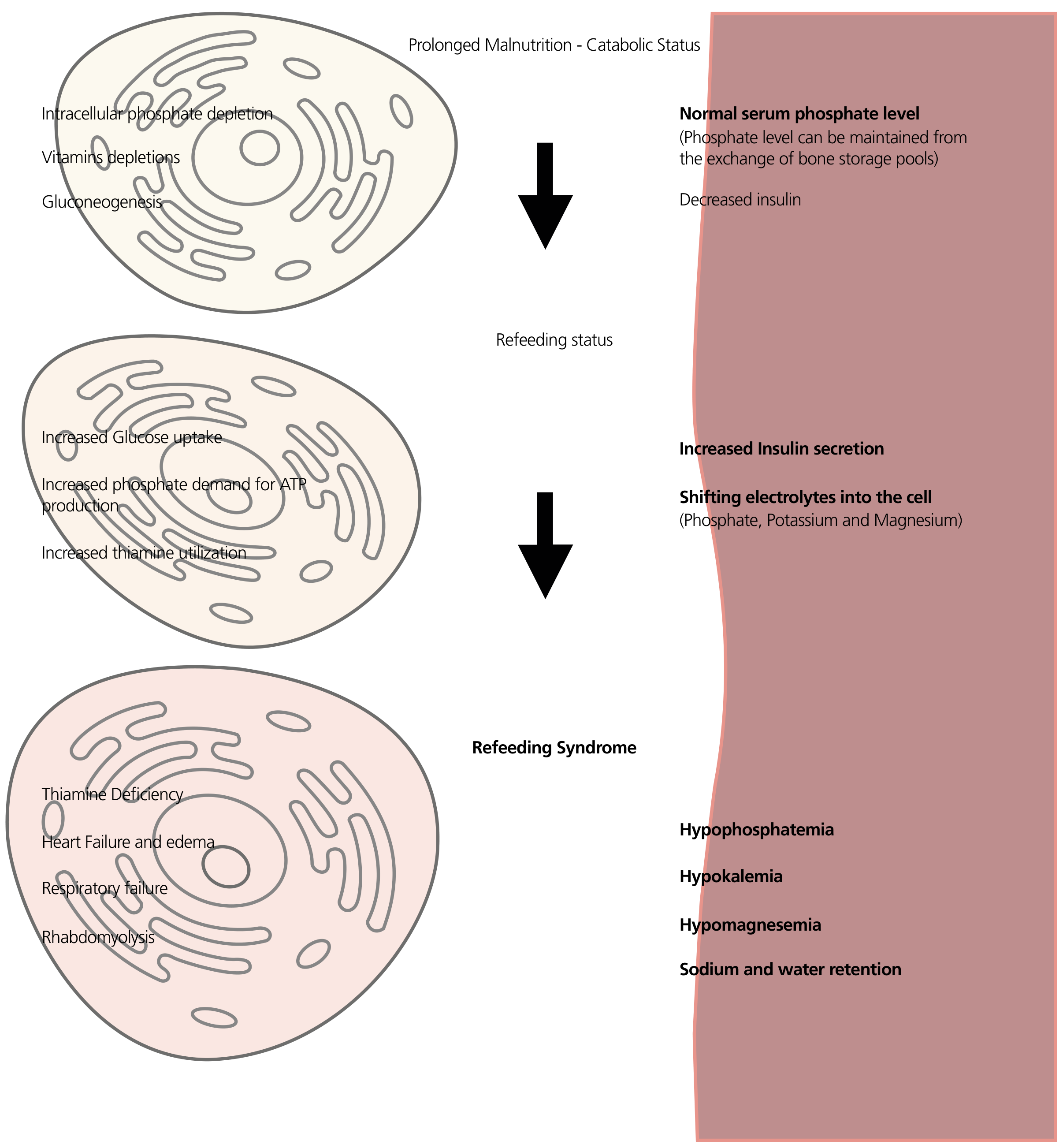
During Starvation
- Insulin secretion is low
- Intracellular electrolytes (phosphate, potassium, magnesium) become severely depleted from cells due to lack of intake and ongoing losses
- Serum levels of phosphate, potassium, and magnesium can appear normal because bone mineral stores release phosphate to maintain a near-normal extracellular concentration - this masks the true total body depletion
- Thiamine (vitamin B1) stores are also depleted
On Reintroduction of Carbohydrates
- Na+/K+ ATPase is stimulated → potassium (and sodium) are driven intracellularly → hypokalemia
- Phosphate is consumed for glycolysis (synthesis of ATP, glucose-6-phosphate, fructose-1,6-bisphosphate) and rapidly shifts intracellularly → hypophosphatemia (the hallmark)
- Magnesium shifts intracellularly via incompletely understood mechanisms; hypomagnesemia also exacerbates hypokalemia (Mg²+ is required for K+ transport)
- Insulin promotes Na+ and water retention → volume expansion → dilutional electrolyte drop + risk of pulmonary edema and heart failure
- Thiamine is consumed as a cofactor for pyruvate dehydrogenase in the reactivated Krebs cycle → if already depleted, pyruvate cannot enter the cycle → Wernicke encephalopathy
Who is at Risk? (NICE Criteria)
High Risk - One criterion is sufficient:
| Criterion | Threshold |
|---|---|
| BMI | <16 kg/m² |
| Unintentional weight loss | >15% in the last 3-6 months |
| Nutritional intake | Little or none for >10 days |
| Pre-feeding electrolytes | Low potassium, phosphate, or magnesium before feeding |
Moderate Risk - Two or more criteria needed:
| Criterion | Threshold |
|---|---|
| BMI | <18.5 kg/m² |
| Unintentional weight loss | >10% in 3-6 months |
| Nutritional intake | Little or none for >5 days |
| Medications/substances | Insulin, chemotherapy, antacids, diuretics, alcohol misuse |
Clinical Conditions Commonly Associated with High Risk
- Anorexia nervosa (prolonged severe restriction)
- Chronic alcoholism (poor intake + thiamine depletion)
- Cancer (cachexia, chemotherapy, poor intake)
- Post-bariatric surgery (malabsorption, restriction)
- Prolonged nil-by-mouth (ICU, post-surgical, dysphagia)
- Chronic malnutrition from inflammatory bowel disease, short bowel syndrome, malabsorption
- Elderly patients with poor intake or acute illness
- HIV/AIDS
Clinical Features
Electrolyte Disturbances and Their Effects
| Electrolyte | Consequence |
|---|---|
| Hypophosphatemia (hallmark) | Muscle weakness, respiratory muscle failure (diaphragm), cardiac arrhythmias, heart failure, hemolysis (depletion of 2,3-DPG), rhabdomyolysis, seizures, coma |
| Hypokalemia | Cardiac arrhythmias (VT/VF), muscle weakness, ileus, respiratory failure |
| Hypomagnesemia | Arrhythmias, tetany, seizures, exacerbates hypokalemia and hypocalcemia |
| Hypocalcemia | Tetany, seizures, prolonged QT |
Other Features
- Sodium and water retention (insulin effect) → peripheral edema, acute heart failure, pulmonary edema
- Hyperglycemia → osmotic diuresis, worsening electrolyte losses
- Thiamine (B1) deficiency → Wernicke encephalopathy (ophthalmoplegia, ataxia, confusion) or Wernicke-Korsakoff syndrome; also cardiovascular beriberi (wet beriberi - high-output cardiac failure)
- Rhabdomyolysis → acute kidney injury
- Respiratory failure → ventilator dependence in ICU patients
Diagnosis
- The patient has had adequate nutrition intake (≥50% of estimated requirements) commenced
- Electrolyte imbalances emerge after its commencement
- Clinical symptoms are present
Laboratory Monitoring
| Electrolyte | Threshold of concern |
|---|---|
| Serum phosphate | <0.6 mmol/L = severe hypophosphatemia |
| Serum potassium | <3.5 mmol/L |
| Serum magnesium | <0.7 mmol/L |
| Serum calcium | Low |
| Blood glucose | Hyperglycemia / hypoglycemia |
| Thiamine | Clinical assessment; supplement preemptively |
| ECG | Arrhythmia detection |
| Fluid balance | Daily weight, intake/output |
Prevention and Management
Step 1 - Correct Pre-Existing Deficiencies BEFORE Feeding
- Replace low phosphate, potassium, and magnesium to normal levels before starting nutrition
- Give thiamine 100-200 mg IV/IM before initiating feeds or any glucose-containing fluids - this is non-negotiable
- Continue thiamine 100 mg/day for 5-7 days (longer in alcoholics and severely starved patients)
- Give multivitamins and trace elements
Step 2 - Start Nutrition Slowly
| Risk Level | Starting Rate |
|---|---|
| Standard at-risk patients | Start at ~50% of estimated requirements on day 1 |
| Highest risk (prolonged starvation, chronic electrolyte losses) | Start at 10 kcal/kg/day maximum |
| Escalation | Increase gradually to meet full needs over 4-7 days |
Step 3 - Monitor Electrolytes Aggressively and Replace
- Check electrolytes (phosphate, potassium, magnesium, glucose) daily during the first week
- Replace electrolytes aggressively according to severity:
| Deficiency | Replacement Approach |
|---|---|
| Phosphate | Mild-moderate: oral sodium/potassium phosphate; Severe (<0.32 mmol/L): IV phosphate infusion. Select potassium-phosphate vs. sodium-phosphate based on concurrent potassium level |
| Potassium | Oral or IV; IV via central line for severe hypokalemia |
| Magnesium | Oral magnesium for mild; IV for severe; repleting Mg also helps correct refractory hypokalemia |
Step 4 - Multidisciplinary Approach
- Nutrition support team (dietitian, gastroenterologist/intensivist)
- Pharmacist (electrolyte replacement protocols)
- Nursing (monitoring, fluid balance)
- Medicine/ICU (cardiac monitoring, respiratory support if needed)
Specific Routes of Nutrition
| Route | Key Points |
|---|---|
| Enteral nutrition (EN) | Risk is the same as PN; does not confer protection. Enteral nutrition was independently associated with higher RFS risk in the Zheng 2025 systematic review - likely due to more patients receiving EN overall |
| Parenteral nutrition (PN) | Higher incidence historically (up to 1 in 3 in some series); start at 50% dextrose on day 1 |
| Oral refeeding | Still carries risk; gradual reintroduction with supplementation required |
Special Populations
Summary: The "Refeeding Checklist"
Before feeding a malnourished patient:
  1. Assess risk (NICE criteria)
  2. Check baseline electrolytes (PO4, K+, Mg2+, Ca2+)
  3. Correct deficiencies BEFORE starting
  4. Give thiamine 100-200mg IV before any glucose/feeds
  5. Start at 10 kcal/kg/day if highest risk; ~50% of needs if moderate risk
  6. Monitor daily electrolytes + glucose for first week
  7. Replace electrolytes aggressively as needed
  8. Escalate calories gradually over 4-7 days
  9. Ensure multidisciplinary oversight
oxygen administration in neonates
"oxygen" AND "neonate" AND "oxygen saturation" AND resuscitation
"neonatal resuscitation" AND oxygen AND "target saturation"
"neonatal" AND "oxygen" AND ("retinopathy of prematurity" OR "bronchopulmonary dysplasia" OR "hyperoxia")
PMID: 39481153
Oxygen Administration in Neonates
Why Oxygen Management in Neonates is Unique
Fetal-to-Neonatal Transition: Normal SpO₂ Pattern
Neonatal Resuscitation: Targeted Preductal SpO₂ Goals (NRP/AHA)
NRP Targeted SpO₂ After Birth
| Time After Birth | Target Preductal SpO₂ |
|---|---|
| 1 minute | 60% - 65% |
| 2 minutes | 65% - 70% |
| 3 minutes | 70% - 75% |
| 4 minutes | 75% - 80% |
| 5 minutes | 80% - 85% |
| 10 minutes | 85% - 95% |
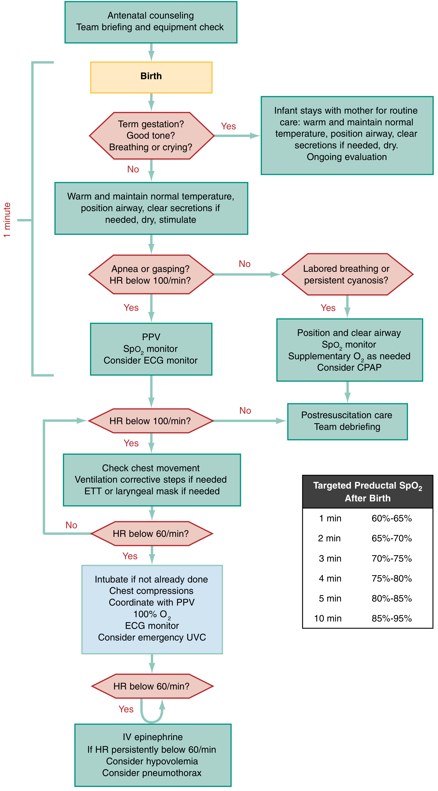
Initial FiO₂ for Resuscitation: Evidence-Based Starting Points
| Infant Category | Initial FiO₂ |
|---|---|
| Term infant (≥37 weeks) | 21% (room air) - start with room air and titrate up |
| Moderate preterm (≥35 weeks) | 21% - room air |
| Preterm (28-35 weeks) | 21-30% - begin with low FiO₂ and blend up as needed |
| Extremely preterm (<28 weeks) | May require higher initial FiO₂; titrate to SpO₂ targets |
| Bradycardia <60 bpm despite PPV | 100% O₂ - escalate to 100% FiO₂ while chest compressions are ongoing |
- Increased oxidative stress
- Direct cardiac and renal injury
- Worse neurologic outcomes (excess cerebral free radical generation)
- Increased mortality
Oxygen Delivery in the NICU Setting
SpO₂ Target Ranges by Clinical Situation
| Clinical Situation | Target SpO₂ | Rationale |
|---|---|---|
| Extremely preterm (NICU) | 90-94% (BOOST II/SUPPORT trial range) | Balance between ROP risk and mortality |
| Extremely preterm - lower target | 85-89% | Lower ROP incidence but higher mortality risk |
| Term infant with respiratory disease / pulmonary hypertension | 90-95% | Prevents hypoxic pulmonary vasoconstriction |
| Severe acidosis / therapeutic hypothermia / dark skin pigmentation | 93-98% | Pulse oximetry may underestimate true saturation |
| Cyanotic congenital heart disease (single ventricle) | 75-85% (lesion-specific) | Higher O₂ causes pulmonary over-circulation |
| Late-stage ROP (active disease) | Higher targets may limit progression | Phase 2 of ROP is oxygen-dependent for vascular regrowth |
| Intraoperative / perioperative (preterm) | 93-95% | Pragmatic balance; vigilance is key |
The SpO₂ Target Controversy in Extremely Preterm Infants
- Lower target (85-89%): Reduced severe retinopathy of prematurity (ROP) requiring treatment, but higher mortality
- Higher target (90-94%): Lower mortality, but more ROP requiring laser/anti-VEGF treatment
Complications of Oxygen Therapy in Neonates
1. Retinopathy of Prematurity (ROP)
- Phase 1 (hyperoxia-related, first weeks): Introduction of supplemental oxygen inhibits VEGF in the immature retina → suppression of normal retinal vascular development → vascular obliteration
- Phase 2 (hypoxia-driven, ~32-34 weeks corrected): The poorly vascularized retina becomes ischemic → massive VEGF upregulation → abnormal neovascularization → traction, retinal detachment, blindness
2. Bronchopulmonary Dysplasia (BPD)
- Defined as need for supplemental O₂ >21% for ≥28 days after birth
- Caused by disruption of alveolar and vascular lung development from hyperoxia, mechanical ventilation, infection, and inflammation
- Oxygen therapy is both a cause and a treatment - requiring careful balance
- Severity graded by O₂ requirement at 36 weeks corrected gestational age (mild = room air; moderate = <30% O₂; severe = ≥30% or positive pressure)
- Long-term consequences: airway hyperreactivity, pulmonary hypertension, neurodevelopmental delay
3. Hyperoxic Brain Injury
- Free radical generation from excessive O₂ in reperfusion contexts (post-asphyxia, post-resuscitation)
- Paradoxically worsens outcomes in hypoxic-ischemic encephalopathy (HIE) when used excessively
- After resuscitation from cardiac arrest or severe asphyxia: titrate to SpO₂ 94-99%; avoid hyperoxia
4. Free Radical Injury to Other Organs
- Cardiac injury (myocardial oxidative stress)
- Renal tubular injury
- Gut injury (necrotizing enterocolitis - multifactorial but hyperoxia implicated)
Practical Oxygen Delivery Methods in Neonates
| Device | FiO₂ Range | Use |
|---|---|---|
| Headbox / O₂ hood | 21-100% | Reliable FiO₂ delivery for infants not requiring positive pressure; older method, less used now |
| Nasal cannula (low-flow) | Variable (~24-40%) | Post-stabilization maintenance O₂ in NICU; FiO₂ depends on flow rate and infant's minute ventilation |
| High-flow nasal cannula (HFNC) | 21-100% at ≥2 L/min | Non-invasive respiratory support; provides positive distending pressure + O₂ |
| CPAP (nasal) | 21-100% | Prevents alveolar collapse, reduces apnea, reduces intubation rate especially in preterm; first-line in RDS |
| Bag-valve-mask (BVM) | 21-100% (blended) | Acute resuscitation; use with O₂ blender to titrate FiO₂ |
| Mechanical ventilation (ETT) | 21-100% | Severe respiratory failure, apnea, surfactant delivery |
| T-piece resuscitator | 21-100% | Preferred over self-inflating bag for preterm resuscitation - provides reliable PEEP |
Special Neonatal Situations
Respiratory Distress Syndrome (RDS) in Preterm Infants
- Primary treatment is surfactant + CPAP, not escalating O₂
- Target SpO₂ 90-94%; escalate to intubation if FiO₂ requirement rises above 0.30-0.40 on CPAP (INSURE or LISA technique)
Hypoxic-Ischemic Encephalopathy (HIE)
- Resuscitate with room air; escalate only as needed
- Post-resuscitation: maintain SpO₂ 94-99%; therapeutic hypothermia (33.5°C for 72h in term infants ≥36 weeks)
- Hyperoxia is especially harmful in this context due to reperfusion free-radical injury
Persistent Pulmonary Hypertension of the Newborn (PPHN)
- Pulmonary vasoconstriction worsened by hypoxia; maintain SpO₂ ≥95% (higher than usual)
- High-flow O₂, inhaled nitric oxide, CPAP/ventilation; avoid acidosis
- Pre-ductal and post-ductal saturation difference >3-5% suggests right-to-left ductal shunting
Meconium Aspiration Syndrome (MAS)
- Routine tracheal suctioning no longer recommended even in vigorous or depressed infants; standard PPV with titrated O₂
- If apneic/depressed with poor tone after MAS, intubate and suction before PPV
Cyanotic Congenital Heart Disease (CHD)
- Do not over-oxygenate - in duct-dependent lesions, high O₂ can close the ductus arteriosus and precipitate cardiovascular collapse
- In single-ventricle physiology (e.g. hypoplastic left heart), deliberately target lower SpO₂ 75-85% to balance pulmonary and systemic blood flow
Monitoring
- Pulse oximetry: Continuous and mandatory whenever supplemental O₂ is used or during resuscitation; right hand (preductal) placement during delivery room resuscitation
- Blood gas (arterial/capillary): PaO₂ target 50-80 mmHg in preterm; avoid PaO₂ >80 mmHg
- FiO₂ documentation: Record all FiO₂ changes in NICU; cumulative hyperoxia exposure is an independent ROP risk factor
- SpO₂ alarm limits in NICU: Typically low alarm 89%, high alarm 95% for preterm infants - tightly set to minimize time outside target range
- Transcutaneous PO₂/PCO₂ monitoring: Continuous non-invasive; useful adjunct in preterm ventilation management
Summary: Core Principles
- Start resuscitation with room air (21% FiO₂) in term and near-term infants; use 21-30% for moderately preterm; titrate to SpO₂ targets
- Normal newborn SpO₂ rises gradually over the first 10 minutes - do not treat physiologic transition as pathologic hypoxia
- Titrate oxygen using an O₂ blender - never default to 100% FiO₂ without indication
- Escalate to 100% O₂ only for chest compressions (HR <60 bpm despite adequate ventilation)
- Both hypoxia and hyperoxia cause harm - the goal is precise titration within a tight target range
- Airway and ventilation take priority over oxygen concentration in most neonatal emergencies
- Monitor preductal SpO₂ (right hand) during delivery room resuscitation
- ROP and BPD are oxygen-related complications that make tight SpO₂ targeting an ongoing NICU priority, not just a resuscitation concern