
Identity slide and 5-7 points on it
Reading File
Finding Sources
Finding Sources
Reading File
Loading Image
Slide Identity: Peripheral Nerve — H&E Section (Cross + Longitudinal)
The slide shows a peripheral nerve in H&E stain, displaying both cross-sectional and longitudinal views simultaneously. The key identifying feature is the circular perineurial ring (the dark purple ring-like structure in the center) enclosing a fascicle of nerve fibers, surrounded by collagen-rich epineurium with elongated spindle cells.
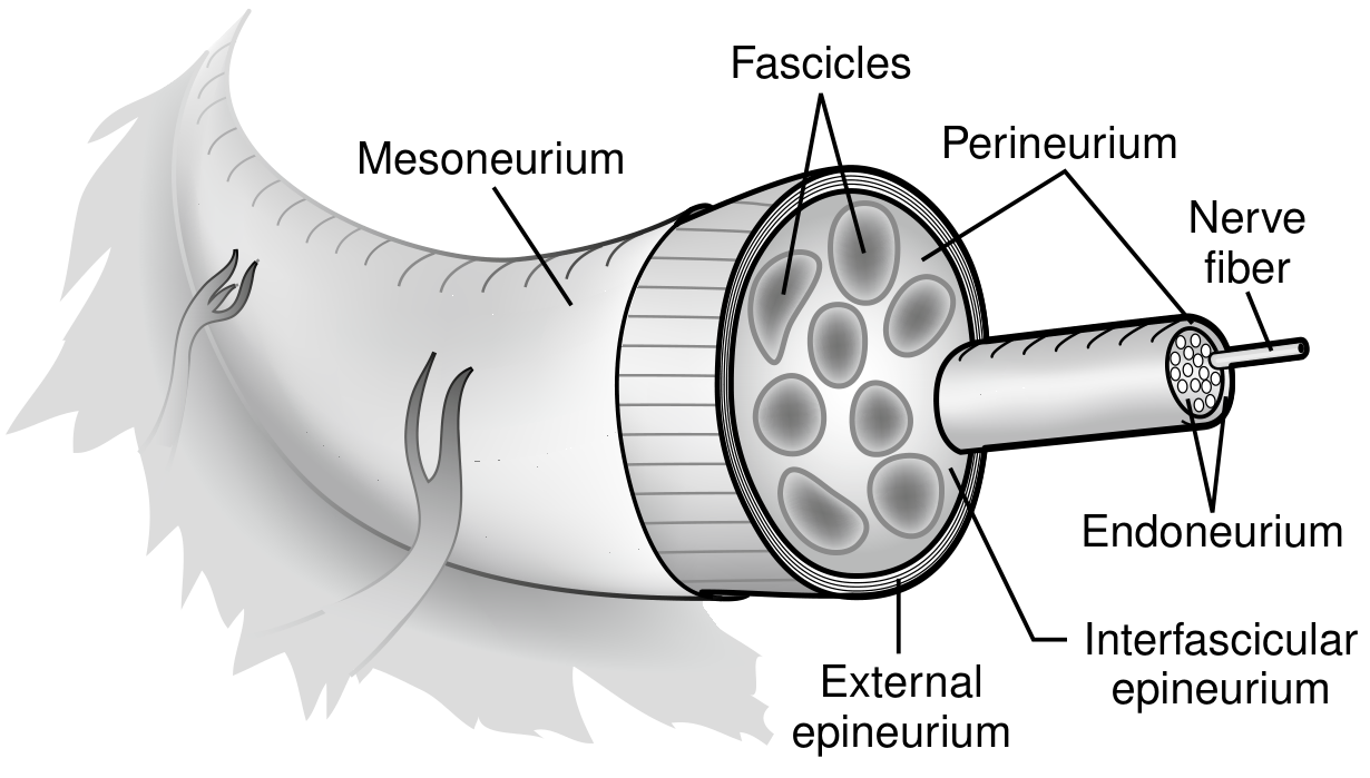
Reference diagram for orientation:
5–7 Key Points
-
Three connective tissue sheaths — The nerve is wrapped by three distinct layers: epineurium (outermost, dense irregular CT surrounding the whole nerve), perineurium (surrounds each fascicle — visible as the prominent ring), and endoneurium (innermost, reticular fibers + capillaries immediately around individual axons/Schwann cells). (Junqueira's Basic Histology, 17e)
-
Perineurium = blood-nerve barrier — The perineurium consists of 2–6 layers of flattened fibrocytes joined by tight junctions, forming a selectively permeable barrier that regulates the ionic microenvironment of axons. This is visible as the compact purple ring on the slide.
-
Fascicular organization — Axons are grouped into fascicles (bundles) by the perineurium. The number and size of fascicles vary along the nerve's length. Large nerves may contain multiple fascicles separated by interfascicular epineurium.
-
Schwann cells and wavy nerve fiber pattern — In longitudinal sections, nerve fibers display a characteristic wavy (undulating) pattern due to the arrangement of Schwann cell cytoplasm and myelin. The elongated spindle-shaped nuclei visible in the slide belong to Schwann cells and endoneurial fibroblasts.
-
Epineurium contains blood vessels — The epineurium's looser deep layer contains arterioles and venules (vasa nervorum) that supply the nerve. These may appear as small circular vascular profiles in the connective tissue around the fascicle on this slide.
-
Myelinated vs unmyelinated fibers — In H&E, myelinated fibers appear as axons surrounded by a clear halo (myelin washed out in processing), while unmyelinated fibers are smaller and more compact. Both are present in mixed peripheral nerves.
-
Clinical relevance — This architecture is directly relevant to nerve injury classification (Sunderland grades I–V / Seddon's neuropraxia, axonotmesis, neurotmesis): injury to endoneurium allows regeneration; disruption of perineurium and epineurium leads to poor functional recovery and may require surgical repair.
This is a shared conversation. Sign in to Orris to start your own chat.