
Principles of FESS - ENT with diagrams
FESS functional endoscopic sinus surgery anatomy diagram
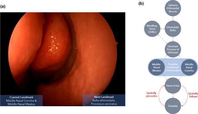
This composite educational graphic illustrates navigational landmarks and logic for Functional Endoscopic Sinus Surgery (FESS). Image (a) is a real-world clinical photograph captured during nasal endoscopy, showing a close-up view of the internal nasal anatomy. The image identifies the middle nasal concha (turbinate) and the middle nasal meatus, characterized by a healthy, reddish-orange mucosal surface with a moist, smooth texture. Image (b) is a clinical flowchart/algorithm detailing the spatial transitions between anatomical landmarks encountered during the procedure. The diagram uses a hierarchical structure to show connectivity between nodes such as the 'Outside', 'Nose Entry', 'Middle Nasal Meatus/Concha', 'Uncinate Process of Ethmoid', 'Ethmoidal Bulla', 'Maxillary Sinus Orifice', and 'Spheno-Ethmoidal Recess'. Red arrows define directional semantic relations, specifically 'spatially precedes' and 'spatially follows', which indicate the relative depth or inward/outward movement of the endoscope within the nasal cavity. This resource is designed to train surgical navigation prediction models by mapping visual anatomical landmarks to procedural pathways.
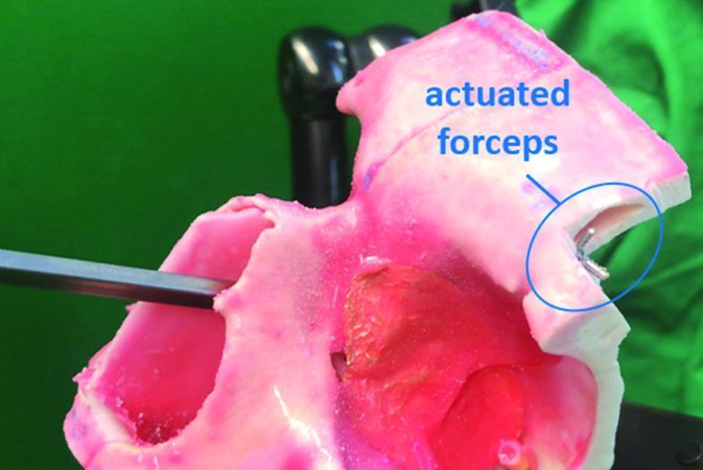
This clinical photograph displays a medical simulation setup for functional endoscopic sinus surgery (FESS), specifically demonstrating the reachability of a novel manipulator system within the lateral region of the frontal sinus. The image features a pink, semi-translucent anatomical phantom representing the human frontal sinus, including internal cavities and regions of varying density to simulate pathological tissue. A flexible surgical manipulator arm is inserted through the sinus floor (simulated Draf IIa window). At the distal end of the manipulator, a pair of metallic grasping forceps is shown in an actuated (closed) state within a lateral recess of the phantom. Blue text and a circular annotation highlight the 'actuated forceps,' indicating the stage of the procedure involving tissue manipulation or retrieval. The demonstration illustrates the surgical system's ability to navigate the complex, pyramid-shaped anatomy of the frontal sinus and access hard-to-reach lateral pathology through minimally invasive nasal access routes.
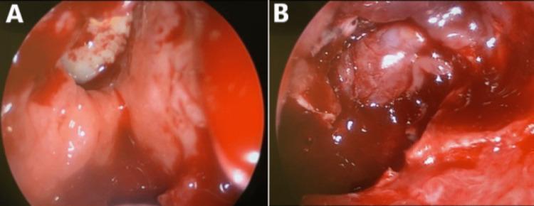
This composite intraoperative endoscopic clinical photograph illustrates a functional endoscopic sinus surgery (FESS). Image A displays the right sphenoid sinus cavity containing diffuse reddish-pink tissue, indicative of inflamed and swollen nasal mucosal polyps. A small area of whitish, denser material is visible among the irregular, vascularized tissue surfaces. Image B demonstrates a more advanced stage of the procedure, specifically showing the dehiscence of the sphenoid sinus roof. Centrally, a prominent, whitish, tubular structure is identified as the exposed optic nerve, which has become visible due to the erosion or removal of the overlying bone. The surgical field is highly vascularized with visible blood and fluid surrounding the exposed nerve and adjacent dural remnants. These images are clinically significant for demonstrating surgical management of Allergic Fungal Rhinosinusitis (AFRS) or similar inflammatory processes causing compressive optic neuropathy. The material serves as an educational resource for otorhinolaryngology and ophthalmology regarding sinus anatomy, surgical complications, and endoscopic landmarks.
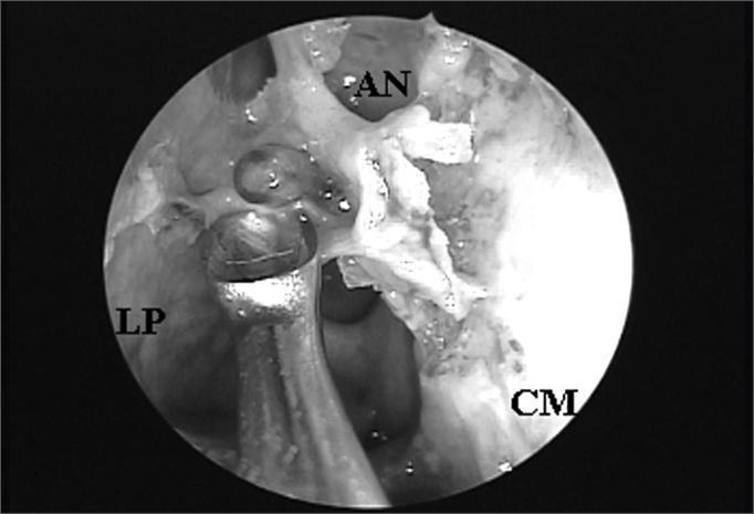
This procedural clinical photograph displays an endoscopic view of the right nasal cavity during a functional endoscopic sinus surgery (FESS). The image was captured using a 4mm 45-degree rigid endoscope. Key anatomical landmarks are labeled: the lamina papyracea (LP) on the lateral wall, the middle turbinate (CM) medially, and the agger nasi cell (AN) superiorly. The agger nasi cell is positioned such that it obstructs the direct endonasal visualization of the frontal sinus ostium, demonstrating a common anatomical variant that complicates frontal recess access. A surgical instrument is visible in the foreground, positioned near the lamina papyracea, indicating active manipulation for dissection or clearance of the obstructive ethmoidal cells. This image serves as an educational tool for identifying paranasal sinus anatomy and understanding the surgical challenges posed by agger nasi pneumatization in managing frontal sinus drainage.
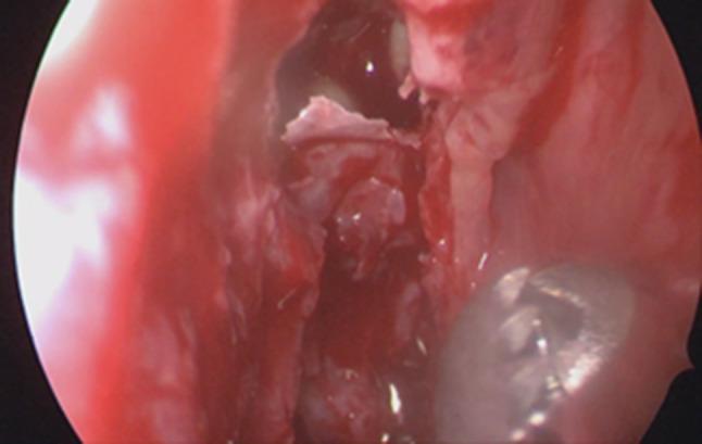
This clinical endoscopic image captures an intraoperative view during a functional endoscopic sinus surgery (FESS), specifically focusing on the right ethmoidal sinus. The visual field demonstrates an antero-posterior ethmoidectomy in progress. Several fractured ethmoidal septations are visible as irregular, whitish bony fragments. The surrounding sinonasal mucosa is notably congested, hyperemic, and edematous, exhibiting a deep red hue consistent with inflammatory changes and surgical trauma. Minimal hemorrhagic and potentially purulent fluid is interspersed among the tissues. In the lower right foreground, a metallic surgical instrument, likely a microdebrider or probe, is positioned to manipulate the bony septa. This image serves as an educational example of intranasal surgical anatomy, demonstrating the controlled breakdown of ethmoidal cells to improve sinus drainage in the context of inflammatory disease or secondary complications like empyema.
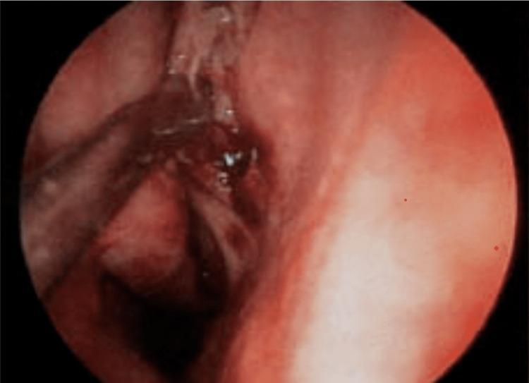
This diagnostic image is an intraoperative endoscopic view of the human nasal cavity during Functional Endoscopic Sinus Surgery (FESS). The visual field demonstrates the nasal septum and adjacent sinonasal mucosa, which exhibits a hyperemic, reddish-pink hue consistent with high vascularity or inflammatory response. Within the surgical field, thin metallic surgical instruments are visible, actively manipulating the tissue. There is evidence of dark red pooled blood and moist surfaces, characteristic of the intraoperative environment in sinus surgery. The image serves to illustrate the challenge of maintaining a clear surgical field in the presence of capillary bleeding and mucosal oozing. Key educational concepts include the visualization of endoscopic anatomy under surgical conditions and the importance of hemostasis for anatomical clarity during rhinological procedures.
paranasal sinus anatomy ostiomeatal complex endoscopic surgery
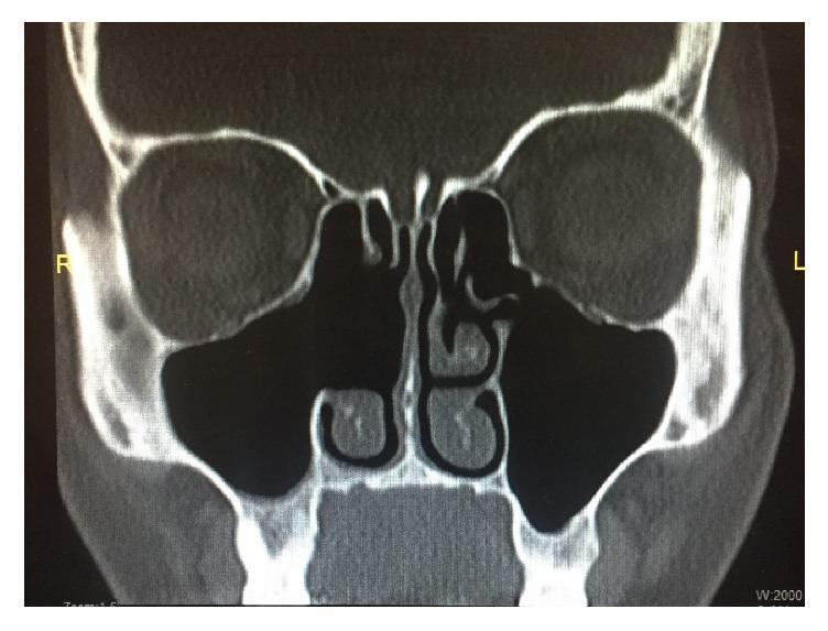
This diagnostic image is a coronal computerized tomography (CT) scan of the paranasal sinuses in bone window. The image demonstrates the postoperative status of the sinonasal cavities following right-sided Functional Endoscopic Sinus Surgery (FESS). Key visual findings include a widely patent right ostiomeatal complex and a right maxillary antrostomy. The right maxillary and ethmoid sinuses are clear and well-aerated, characterized by a lack of opacification, fluid levels, or significant mucosal thickening. Surgical changes on the right side are further evidenced by the absence of the middle turbinate, consistent with a prior middle turbinectomy. In contrast, the left nasal cavity shows intact anatomy, including the left middle and inferior turbinates and a normally aerated left maxillary sinus. The nasal septum is midline, and the orbital structures and skull base appear intact. This image serves as an educational example of a successful surgical outcome in the management of chronic or infectious sinusitis, such as sinonasal actinomycosis, showing complete resolution of previous disease and restoration of normal sinus drainage pathways.
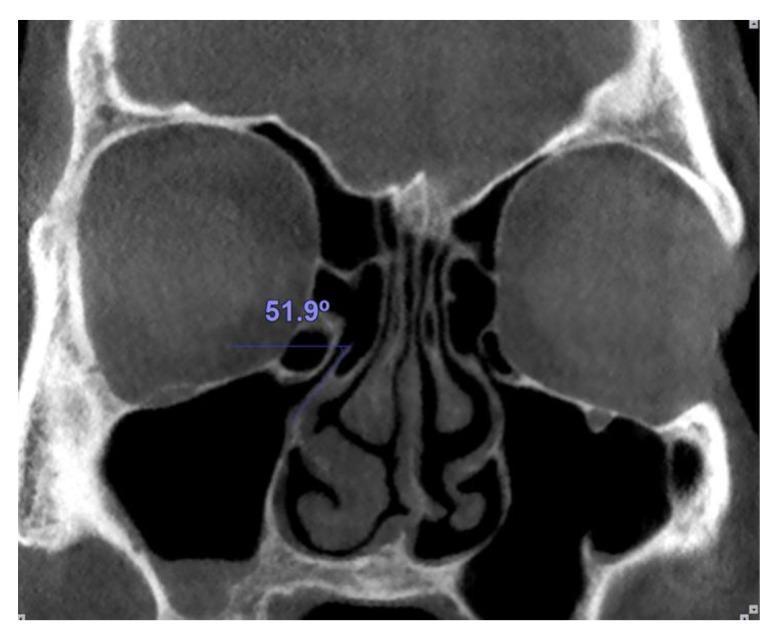
This diagnostic image is a coronal Computed Tomography (CT) scan of the paranasal sinuses and nasal cavity using a bone window setting. The view displays key sinonasal anatomy, including the maxillary sinuses, the ethmoid air cells, the nasal septum, and the inferior and middle nasal conchae (turbinates). The image specifically illustrates the morphometry of the ostiomeatal complex. An overlaid blue line measures the inclination of the uncinate process on the patient's right side. The measurement shows an angle of 51.9 degrees, calculated between a horizontal reference line originating from the superior edge of the uncinate process and its superior border. This anatomical assessment is clinically relevant for evaluating the patency of the infundibulum and planning functional endoscopic sinus surgery (FESS), as the uncinate process orientation can influence the drainage pathway of the anterior ethmoid and maxillary sinuses.
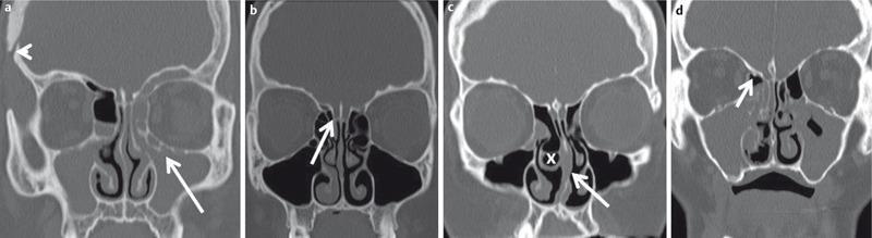
This diagnostic image set consists of four coronal low-dose CT (ldCT) scans of the paranasal sinuses, illustrating various anatomical variants and pathological findings relevant to Functional Endoscopic Sinus Surgery (FESS). Panel (a) shows bilateral complete maxillary sinus opacification and a left-sided infraorbital ethmoid cell (Haller cell) obstructing the ostiomeatal complex, with evidence of prior cranial surgery (arrowhead). Panel (b) highlights an ethmoid bulla and a Keros type II cribriform plate variant, indicating a moderate depth of the olfactory fossa. Panel (c) demonstrates a right-sided concha bullosa (pneumatized middle turbinate, marked 'x'), a deviated nasal septum to the left with a bony spur, and an opacified ethmoid bulla. Panel (d) illustrates a Keros type I cribriform plate (shallow olfactory fossa) and a small bony spur near the anterior ethmoid artery. These images serve as an educational comparison of sinonasal anatomy, highlighting variants like the Haller cell and concha bullosa that contribute to chronic rhinosinusitis and surgical risk.
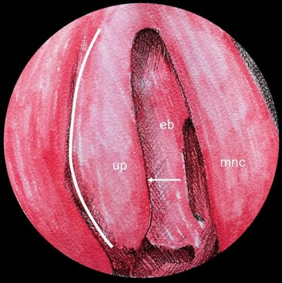
This medical illustration depicts the endoscopic anatomy of the right nasal cavity lateral wall, specifically focusing on the ostiomeatal complex (OMC). The diagram uses a color-shaded style to differentiate key surgical landmarks. Centrally, the ethmoidal bulla (eb) is shown as a prominent pneumatized structure. Anterior to it lies the uncinate process (up), a thin, hook-like bone. The space between the uncinate process and the ethmoidal bulla, known as the semilunar hiatus, is indicated by a horizontal white arrow. Medially, the middle nasal concha (mnc), or middle turbinate, is visible. For surgical context, a curved white line is superimposed on the lateral aspect of the uncinate process to mark the approximate limit for an uncinectomy, a primary step in functional endoscopic sinus surgery (FESS) to access the ethmoidal infundibulum and maxillary ostium. This educational visual is designed to assist in identifying anatomical relationships necessary for safe navigation during paranasal sinus procedures.
Functional Endoscopic Sinus Surgery (FESS) - Principles
1. Concept and Rationale
2. Indications
- Chronic rhinosinusitis (CRS) with or without nasal polyps (CRSwNP / CRSsNP) that has failed adequate medical treatment
- Recurrent acute rhinosinusitis
- Sinonasal polyposis
- Mucocele / pyocele drainage
- Orbital complications of sinusitis
- Sinonasal tumours (benign and selected malignant)
- Access surgery (e.g. dacryocystorhinostomy, pituitary tumours, skull base)
- Choanal atresia, nasal foreign body
3. Pre-operative Requirements
- Cribriform plate depth (Keros classification I/II/III)
- Uncinate process orientation and attachment
- Lamina papyracea integrity
- Agger nasi pneumatisation
- Haller cells, concha bullosa, Onodi cells
- Position of anterior ethmoidal artery
- Internal carotid artery dehiscence over sphenoid
4. Anaesthesia and Patient Preparation
- General anaesthesia preferred (occasionally local anaesthesia + sedation in fit adults)
- Hypotensive anaesthesia (MAP 60-70 mmHg) reduces bleeding and improves visibility
- Head-up tilt 15-20° reduces venous congestion
- Topical vasoconstrictors: cocaine 4-10%, or xylometazoline with/without adrenaline
- Eyes: Traditionally left open with lubrication but limited or complete taping is now acceptable, still allowing rapid access to assess the globe. Corneal abrasions from surgical drapes have led to medicolegal cases.
5. Key Anatomical Landmarks
The Ostiomeatal Complex
| Structure | Relevance |
|---|---|
| Uncinate process | Boomerang-shaped bony blade; first structure encountered |
| Ethmoidal bulla | Largest anterior ethmoid cell; sits posterosuperior to uncinate |
| Semilunar hiatus | Groove between uncinate and bulla; drainage pathway |
| Infundibulum | Funnel-shaped space receiving maxillary, frontal sinus drainage |
| Middle meatus | The working corridor for FESS |
| Agger nasi cell | Most anterior ethmoidal cell; key to frontal recess |
6. Surgical Technique - Step-by-Step
i) Uncinectomy (First Step)
- The superior uncinate is more protected by the hard bone of the frontal process of maxilla; a sickle knife is safer here
- After the inferior incision, the uncinate is removed with angled through-biting forceps, a back-biter, or a microdebrider
- The horizontal portion of the uncinate (inferior to the maxillary natural ostium) must also be addressed
- The natural maxillary ostium can be stretched open with an angled probe without removing mucosa; in advanced disease, a larger middle meatal antrostomy is created
ii) Removal of the Ethmoidal Bulla
iii) Posterior Ethmoidectomy
- The roof of the maxillary sinus guides the superior limit - anatomical studies confirm it always lies below the skull base
- Partitions between posterior cells are removed with microdebrider, Kerrison's punch, or through-biting instruments
- Onodi cell awareness: the optic nerve may traverse through an Onodi cell in the posterior ethmoid
iv) Sphenoidotomy
- Pass endoscope medial to superior turbinate to identify ostium directly
- Resect the inferior 1/3 of the superior turbinate using a back-biting forceps to enter the superior meatus
- If still inaccessible, create an artificial opening through the posterior ethmoid, then extend medially to incorporate the natural ostium
v) Frontal Sinus Surgery (Draf I/IIa/IIb/III)
- A Kerrison's punch at the axilla of the middle turbinate removes the anterior portion of the agger nasi cell
- The posterior wall and roof of the agger nasi are then removed to expose the frontal recess (Figure 98.5)
- Kuhn cells (frontal cells) obstructing the frontal ostium are removed with a Rosemann punch - circumferential mucosal injury here must be avoided to prevent scarring/stenosis
- 45° or 70° angled endoscopes are essential for visualization
7. Instruments Used in FESS
| Instrument | Purpose |
|---|---|
| 0° and 30° Hopkins rod endoscopes | Primary visualisation |
| 45° and 70° angled endoscopes | Frontal recess, high recesses |
| Freer's elevator | Identifying uncinate free edge |
| Paediatric/adult back-biting forceps | Inferior uncinectomy |
| Sickle knife | Superior uncinectomy (safer) |
| Double right-angle ball probe | Locating ethmoidal bulla ostium |
| Through-biting forceps (angled) | Removing bulla, ethmoid cells |
| Microdebrider (shaver) | Atraumatic tissue removal |
| Kerrison's punch | Removing fine bony partitions |
| Rosemann punch | Frontal recess Kuhn cells |
| 45° antral curette | Opening posterior ethmoid/sphenoid |
8. Post-operative Care
- Saline irrigations (high volume) are routinely used to clear mucopus, blood clot, and debris. Ringer's lactate solution has shown improved outcomes over isotonic or hypotonic saline in one RCT.
- Endoscopic debridement (nasal toilet) in clinic is critical - there is strong evidence supporting regular post-operative toilet
- Oral steroids: In CRSwNP, prednisolone 25 mg daily for 10 days (without tapering) is one regime. For extensive eosinophilic CRS, up to 3 weeks may be needed, with a taper to avoid adrenal insufficiency
- Post-op antibiotics: No evidence that prophylactic antibiotics improve routine outcomes. A per-operative swab is taken if mucopus is found and targeted antibiotics prescribed
- Follow-up schedule: 7-10 days post-op → weekly if concerns/fortnightly if not → 6 weeks (cavity fully healed) → 3 months review
- Long-term topical steroids and saline irrigations are required indefinitely in eosinophilic CRS to prevent polyp recurrence
9. Complications of FESS
Minor Complications
- Bleeding / epistaxis (most common)
- Adhesion / synechiae formation
- Anosmia (temporary or permanent)
- Crusting and scabbing
Major Complications
| Complication | Mechanism | Management |
|---|---|---|
| Orbital haematoma | Damage to lamina papyracea or anterior ethmoidal artery | Lateral canthotomy, decompress orbit, call ophthalmology |
| CSF leak / meningitis | Skull base injury at cribriform plate or fovea ethmoidalis | Surgical repair (free mucosal graft or fat), lumbar drain |
| Optic nerve injury | Damage in sphenoid or Onodi cell | Rare; irreversible visual loss |
| Internal carotid injury | Bony dehiscence over carotid in sphenoid | Pack, endovascular stent; immediately life-threatening |
| Anosmia | Middle turbinate sacrifice or olfactory cleft damage | Prevention by preserving olfactory mucosa |
10. Evidence Base
- A landmark RCT (90 patients) comparing FESS vs medical treatment (long-term antibiotics + intranasal steroids + saline) showed both treatments improved symptoms significantly, with no statistically significant difference between arms
- However, non-RCT data and observational studies support surgery in patients who have failed medical management
- For CRSwNP, FESS shows more evidence of benefit than for CRSsNP
- The UK Royal College of Surgeons national audit (>3000 patients, 2000) showed functional outcomes from FESS are comparable or superior to other approaches with potential for decreased morbidity
- Current consensus: FESS should be offered after adequate medical treatment has failed (typically 3 months of topical steroids ± saline irrigation ± antibiotics for acute exacerbations)
11. Image-Guided (Navigation) FESS
- Revision surgery (distorted anatomy)
- Extensive polyposis
- Anterior skull base or orbital wall involvement
- Tumour surgery
- Training